Pbx1 regulates self-renewal of long-term hematopoietic stem cells by maintaining their quiescence - PubMed (original) (raw)
Pbx1 regulates self-renewal of long-term hematopoietic stem cells by maintaining their quiescence
Francesca Ficara et al. Cell Stem Cell. 2008.
Abstract
Self-renewal is a defining characteristic of stem cells; however, the molecular pathways underlying its regulation are poorly understood. Here, we demonstrate that conditional inactivation of the Pbx1 proto-oncogene in the hematopoietic compartment results in a progressive loss of long-term hematopoietic stem cells (LT-HSCs) that is associated with concomitant reduction in their quiescence, leading to a defect in the maintenance of self-renewal as assessed by serial transplantation. Transcriptional profiling revealed that multiple stem cell maintenance factors are perturbed in Pbx1-deficient LT-HSCs, which prematurely express a large subset of genes, including cell-cycle regulators, normally expressed in non-self-renewing multipotent progenitors. A significant proportion of Pbx1-dependent genes is associated with the TGF-beta pathway, which serves a major role in maintaining HSC quiescence. Prospectively isolated, Pbx1-deficient LT-HSCs display altered transcriptional responses to TGF-beta stimulation in vitro, suggesting a possible mechanism through which Pbx1 maintenance of stem cell quiescence may in part be achieved.
Figures
Figure 1. Hematopoietic defects in Pbx1-conditional ko mice
(A) Gross appearance is shown for spleen (top left) and thymus (bottom left) of 5-week old representative Tie2Cre+.Pbx1+/f control and Tie2Cre+.Pbx1−/f mutant mice. Histograms on the right show the average number (± SD) of hematopoietic cells in the respective organs (*p<0.00001, n=14 and 27 for spleen and thymus, respectively). (B) Hypocellular BM in mutant mice is revealed by H&E staining of decalcified BM sections from 6-week old mice. Histogram on the right shows average BM cell counts as total nucleated blood cells in femurs and tibiae. Controls are Tie2Cre+.Pbx1+/f (*p=0.005, n=7). (C) Histogram represents cell numbers for the indicated populations as determined by FACS analysis on BM of 3–4 week old mice. Myeloid cells, CD11b+; ProB cells, B220+CD43+IgM−; PreB cells, B220+CD43−IgM−; Immature B cells (ImmB), B220+CD43−IgM+; T cells, TCRβ+; NK cells, TCRβ−.NK1.1+; NKT cells, TCRβ+NK1.1+ (*p<0.03; **p<0.00004, n=11).
Figure 2. Decreased BM stem and progenitor cells in Pbx1 mutant mice
(A-D) Representative FACS analyses (left) and average absolute numbers (right) of BM cells are shown for control Tie2Cre+.Pbx1+/f and mutant Tie2Cre+.Pbx1−/f mice. Numbers in the FACS plots indicate percentages among total BM cells. (A) CLPs are defined as Lin−IL7R+Sca1locKitlo (Kondo et al., 1997). Depicted contour plots are referred to the Lin−IL7R+ gate (*p<0.02, n=5). (B) FACS plots are referred to the Lin−cKithiSca1− gate, in which CMPs are defined as CD34+/loCD16/32int, GMPs as CD34+CD16/32+, and MEPs as CD34−CD16/32− (*p<0.005, n=17). (C) KSL are Lin−IL7R−Sca1+cKit+ (*p<0.03, n=7). (D) LT-HSCs are defined as KSL, CD150+CD41−CD48−. FACS plots are referred to the KSL gate (*p<0.0001, n=10). (E-F) Experimental outline, representative FACS analyses, and average absolute numbers of LT-HSCs in control and MxCre+.Pbx1−/f mice. Indicated mice received 7 poly(I:C) injections every other day. FACS plots are referred to the KSL gate, percentages among total BM cells are indicated (*p=0.02, n=24). In F, 1×10⁶ donor cells either from mutant or control (MxCre−.Pbx1f/f) mice were transplanted into lethally irradiated Ly5.1 recipients prior to poly(I:C) injections. KSL cells were all donor-derived (*p=0.01, n=4).
Figure 3. Pbx1-deficient stem cells display increased cycling and reduced quiescence
(A) Control or mutant mice were injected ip with BrdU and sacrificed 2 hrs later. Representative FACS analyses on the left show BrdU and 7-AAD incorporation in KSL cells. Graphs on the right represent the % of BrdU+ cells in LT-HSCs, ST-HSCs and MPP cells in three independent experiments (*p=0.06, **p<0.04). (B) Left: representative FACS analysis of PY/Hoechst staining on KSL cells (top) or LT-HSC (bottom). Right: % of KSL cells (top) or LT−. HSCs (bottom) in G0, G1, and S phase (n=2, with bars representing the range).
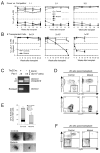
Figure 4. Defective stem cell function in Pbx1 mutant mice
(A) Total BM cells from either control (white) or mutant (black) Ly5.2 mice were transplanted into lethally-irradiated Ly5.1 recipients in competition with BM from Ly5.1/Ly5.2 mice, at different ratios of donor versus competitor, as indicated at the top of the panels. The average percentages of myeloid (circles), B (squares) and T (triangles) cells of donor origin in the PB of recipient mice are shown for different time points after transplant. The percentages of Ly5.2+ cells were measured by FACS in the CD11b+, B220+ and TCRβ+ fractions of red blood cell (RBC) lysed PB. The data are from two independent experiments (4–5 recipients/group). (B) Different doses of total BM cells (indicated at top of panels) from either control or mutant Ly5.2 mice were transplanted into lethally-irradiated Ly5.1 recipients in the absence of competition. The percentage of donor myeloid cells in the PB of recipient mice was determined at different time points after transplant. (C) Representative PCR performed on the PB of recipients of non-competitive transplants shows that reconstitution was mediated by cells with complete Pbx1 deletion in mice transplanted with mutant BM. (D) Representative FACS analyses are shown for HSCs at wk 20 post-transplant of 5×10⁵ donor whole BM cells using two different combinations of antibodies. Top panels: LT-HSCs are defined as KSL,CD150+CD48−; bottom panels: LT-HSCs are defined as KSL,CD34−.CD135−.. Plots and percentages are referred to the donor KSL gate. (E) Methylcellulose colony assays were performed at the time of sacrifice 20 weeks post-transplant using BM cells of donor origin from mice that received non-competitive transplants. (F) Representative FACS analyses are shown for B cells of donor origin (Ly5.2 gate) in the BM at wk 20 following non-competitive transplantation.
Figure 5. Pbx1-deficient cells are unable to engraft secondary recipients
(A) Survival curves represent results of secondary transplants performed with donor BM cells sorted from primary recipient mice (20 weeks after primary transplant) successfully reconstituted by control or mutant cells. Data are derived from 2 independent experiments containing a total of 9 secondary transplanted mice in each cohort. (B) Representative FACS analysis of PB at 4 weeks post-transplant is shown for secondary recipients transplanted with control (left) or mutant (right, two examples) BM cells. Top, percent of donor cells; bottom, percent of myeloid and T cells in the donor gate. Double negative cells are B220+ B cells (not shown). (C) Summary of transplantation experiments. (D) Survival curves after multiple 5-FU injections (indicated by arrows) of poly(I:C)-treated mutant (MxCre+.Pbx1−/f or MxCre−.Pbx1f/f, n=4) and control (MxCre−.Pbx1f/f, MxCre−.Pbx1−/f, or MxCre−..Pbx1−/f, n=5) mice.
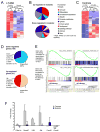
Figure 6. Gene expression profile of Pbx1-deficient HSCs
(A) Heat map shows the expression of the 241 Pbx1-regulated genes in LT-HSCs from poly (I:C) treated control (MxCre−.Pbx1f/f, C1 and C2) or mutant (MxCre+.Pbx1f/f, M1-M3) individual mice. Up-regulated and down-regulated genes are presented in red and blue, respectively. (B) Pie charts show the distribution of the 61 up-regulated (top) and the 180 down-regulated (bottom) genes in Pbx1-deficient LT-HSCs into functional groups. (C) Heat map displays the differentially expressed transcripts in LT- and ST-HSCs from poly (I:C) treated control mice. (D) Pie charts represent the overlap of the mutant LT-HSC gene signature with genes found in the current study to be differentially expressed in the LT-to ST-HSC transition in control mice (17% and 37% for the down-regulated and up-regulated genes, respectively), as well as with genes differentially expressed in the LT-HSC to MPP transition not included in the LT/ST-HSC overlap (14% and 11%). (E) Enrichment plots of selected gene sets from the GSEA in Table 1. (F) Histogram shows fold-induction for the indicated trancripts in sorted LT-HSCs upon 4 h of TGF-b exposure, as measured by qRT-PCR (**p<0.0001, *p≤0.01; p57: n=7 mutants and 8 controls; Smad7: n=3; Klf4, Ski: n=4 mutants and 2 controls; Ccnd2, Ccna2: n=4 and 3 mutants, respectively, and one control).
Similar articles
- Pbx1 restrains myeloid maturation while preserving lymphoid potential in hematopoietic progenitors.
Ficara F, Crisafulli L, Lin C, Iwasaki M, Smith KS, Zammataro L, Cleary ML. Ficara F, et al. J Cell Sci. 2013 Jul 15;126(Pt 14):3181-91. doi: 10.1242/jcs.125435. Epub 2013 May 9. J Cell Sci. 2013. PMID: 23660001 Free PMC article. - MicroRNA-127-3p controls murine hematopoietic stem cell maintenance by limiting differentiation.
Crisafulli L, Muggeo S, Uva P, Wang Y, Iwasaki M, Locatelli S, Anselmo A, Colombo FS, Carlo-Stella C, Cleary ML, Villa A, Gentner B, Ficara F. Crisafulli L, et al. Haematologica. 2019 Sep;104(9):1744-1755. doi: 10.3324/haematol.2018.198499. Epub 2019 Feb 21. Haematologica. 2019. PMID: 30792210 Free PMC article. - Prdm16 is a critical regulator of adult long-term hematopoietic stem cell quiescence.
Gudmundsson KO, Nguyen N, Oakley K, Han Y, Gudmundsdottir B, Liu P, Tessarollo L, Jenkins NA, Copeland NG, Du Y. Gudmundsson KO, et al. Proc Natl Acad Sci U S A. 2020 Dec 15;117(50):31945-31953. doi: 10.1073/pnas.2017626117. Epub 2020 Dec 2. Proc Natl Acad Sci U S A. 2020. PMID: 33268499 Free PMC article. - Leukemogenesis via aberrant self-renewal by the MLL/AEP-mediated transcriptional activation system.
Yokoyama A. Yokoyama A. Cancer Sci. 2021 Oct;112(10):3935-3944. doi: 10.1111/cas.15054. Epub 2021 Aug 2. Cancer Sci. 2021. PMID: 34251718 Free PMC article. Review. - Balancing dormant and self-renewing hematopoietic stem cells.
Wilson A, Laurenti E, Trumpp A. Wilson A, et al. Curr Opin Genet Dev. 2009 Oct;19(5):461-8. doi: 10.1016/j.gde.2009.08.005. Epub 2009 Oct 5. Curr Opin Genet Dev. 2009. PMID: 19811902 Review.
Cited by
- Meis1 preserves hematopoietic stem cells in mice by limiting oxidative stress.
Unnisa Z, Clark JP, Roychoudhury J, Thomas E, Tessarollo L, Copeland NG, Jenkins NA, Grimes HL, Kumar AR. Unnisa Z, et al. Blood. 2012 Dec 13;120(25):4973-81. doi: 10.1182/blood-2012-06-435800. Epub 2012 Oct 22. Blood. 2012. PMID: 23091297 Free PMC article. - Meis1 regulates the metabolic phenotype and oxidant defense of hematopoietic stem cells.
Kocabas F, Zheng J, Thet S, Copeland NG, Jenkins NA, DeBerardinis RJ, Zhang C, Sadek HA. Kocabas F, et al. Blood. 2012 Dec 13;120(25):4963-72. doi: 10.1182/blood-2012-05-432260. Epub 2012 Sep 20. Blood. 2012. PMID: 22995899 Free PMC article. - Prenatal inflammation perturbs murine fetal hematopoietic development and causes persistent changes to postnatal immunity.
López DA, Apostol AC, Lebish EJ, Valencia CH, Romero-Mulero MC, Pavlovich PV, Hernandez GE, Forsberg EC, Cabezas-Wallscheid N, Beaudin AE. López DA, et al. Cell Rep. 2022 Nov 22;41(8):111677. doi: 10.1016/j.celrep.2022.111677. Cell Rep. 2022. PMID: 36417858 Free PMC article. - Ovarian Cancer Chemoresistance Relies on the Stem Cell Reprogramming Factor PBX1.
Jung JG, Shih IM, Park JT, Gerry E, Kim TH, Ayhan A, Handschuh K, Davidson B, Fader AN, Selleri L, Wang TL. Jung JG, et al. Cancer Res. 2016 Nov 1;76(21):6351-6361. doi: 10.1158/0008-5472.CAN-16-0980. Epub 2016 Sep 2. Cancer Res. 2016. PMID: 27590741 Free PMC article. - Oncogenic heterogeneous nuclear ribonucleoprotein D-like modulates the growth and imatinib response of human chronic myeloid leukemia CD34+ cells via pre-B-cell leukemia homeobox 1.
Ji D, Zhang P, Ma W, Fei Y, Xue W, Wang Y, Zhang X, Zhou H, Zhao Y. Ji D, et al. Oncogene. 2020 Jan;39(2):443-453. doi: 10.1038/s41388-019-0998-9. Epub 2019 Sep 5. Oncogene. 2020. PMID: 31488872
References
- Akala OO, Clarke MF. Hematopoietic stem cell self-renewal. Curr Opin Genet Dev. 2006;16:496–501. - PubMed
- Berkes CA, Bergstrom DA, Penn BH, Seaver KJ, Knoepfler PS, Tapscott SJ. Pbx marks genes for activation by MyoD indicating a role for a homeodomain protein in establishing myogenic potential. Mol Cell. 2004;14:465–477. - PubMed
- Blank U, Karlsson G, Moody JL, Utsugisawa T, Magnusson M, Singbrant S, Larsson J, Karlsson S. Smad7 promotes self-renewal of hematopoietic stem cells. Blood. 2006;108:4246–54. - PubMed
Publication types
MeSH terms
Substances
LinkOut - more resources
Full Text Sources
Other Literature Sources
Medical
Molecular Biology Databases
Research Materials


