A proteomic comparison of immature and mature mouse gonadotrophs reveals novel differentially expressed nuclear proteins that regulate gonadotropin gene transcription and RNA splicing - PubMed (original) (raw)
A proteomic comparison of immature and mature mouse gonadotrophs reveals novel differentially expressed nuclear proteins that regulate gonadotropin gene transcription and RNA splicing
Jiajun Feng et al. Biol Reprod. 2008 Sep.
Abstract
The alphaT3-1 and LbetaT2 gonadotroph cell lines contain all the known factors required for expression of gonadotropin genes, yet only the LbetaT2 cells express the beta subunits. We hypothesized that comparison of their nuclear proteomes would reveal novel proteins and/or modifications that regulate expression of these genes. We identified nine proteins with different expression profiles in the two cell lines, of which several were chosen for further functional studies. Of those found at higher levels in alphaT3-1 nuclei, 1110005A23RIK was found associated with the Fshb gene promoter and repressed its expression. Transgelin 3 overexpression reduced transcript levels of Fshb, and its knockdown elevated Lhb and Cga transcript levels, indicating an ongoing repressive effect on these more highly expressed genes, possibly through altering levels of phosphorylated mitogen-activated protein kinase. Heterogeneous nuclear ribonucleoprotein A2/B1 repressed splicing of the Fshb primary transcript, which it binds in the first intron. Proteins at higher levels in LbetaT2 nuclei included prohibitin, the overexpression of which reduced promoter activity of all three gonadotropin subunits, and appeared to mediate the differential effect of GnRH on proliferation of the two cell lines; its knockdown also altered cell morphology. Two other splicing factors were also found at higher levels in LbetaT2 nuclei: the knockdown of PRPF19 or EIF4A3 decreased splicing of Lhb, or of both beta subunit transcripts, respectively. The levels of Eif4a3 mRNA were increased by activin, and both factors increased Fshb splicing. This study has revealed a number of novel factors that alter gonadotropin expression and gonadotroph function, and likely mediate or moderate effects of the regulatory hormones.
Figures
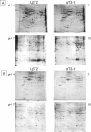
FIG. 1.
Two-dimensional gel electrophoresis of proteins from isolated nuclei (A) or solubilized nuclear proteins (B) of untreated mature LβT2 and immature αT3–1 gonadotrophs over the pI ranges 4–7 or 7–10. Nuclei were prepared as described in the Materials and Methods, before resolving over first and second dimensions. Numerous pairs of samples were run and compared, and representative pairs are shown. Protein spots showing differential expression patterns that were isolated for identification are numbered; protein 8 appears in two distinct spots on the gel (as revealed by its subsequent identification).
FIG. 2.
Identification and verification of differentially expressed proteins. A) The differentially expressed proteins marked in Figure 1 are shown magnified; an asterisk marks the spot of higher intensity for each pair. B) After the proteins were excised, in-gel digested, and subjected to mass spectrometry, a putative identity was obtained and, where antisera were available, Western analysis was carried out to verify the identification and examine differential expression in nuclear extracts of the two cell lines, with ACTB as internal control. The expression levels of these proteins were assessed, and are shown, after normalization, relative to levels in LβT2 cells. C) RT-PCR was also carried out to determine whether the differential protein levels relate to differential gene expression, with Actb as internal control. The 1110005A23RIK is abbreviated to RIK in all figures.
FIG. 3.
1110005A23RIK represses expression of the Fshb gene. A) An 1110005A23RIK expression vector (RIK) was transfected into LβT2 cells, and RT-PCR carried out for 1110005A23Rik, Fshb, and Lhb mRNAs, and the levels of 1110005A23RIK overexpression were evaluated using antisera to the HA tag in Western analysis. The effect of 1110005A23RIK overexpression was tested in LβT2 cells on transiently transfected mouse Fshb (intron and promoter (IP): includes first intron), Lhb, and Cga promoter-luciferase reporter genes with pRL-SV40 as internal control (B), or endogenous mRNA levels by real-time PCR, using Actb as internal control (C). For both types of experiment, levels are expressed relative to those in control cells after normalization with levels of the internal controls. Mean ± SEM; n = 3–4. Student _t_-test compared the promoter activity of each gene with or without 1110005A23RIK overexpression; *P < 0.05; ***_P_ < 0.001, NS: _P_ > 0.05. D) An siRNA construct targeting 1110005A23Rik (siRik) was transfected into αT3–1 cells together with each of the subunit promoter-luciferase constructs. Luciferase activity was measured after 48 h and is presented as in B. The effect of the knockdown was verified by RT-PCR and quantified relative to the controls after normalization with the levels of Actb. E) The 1110005A23RIK contains three domains, two of which were removed individually and in combination, and truncated fusion proteins created fused to LGALS4 DBD (pM vector), as shown before testing their effects on a SV40-LGALS4 reporter gene; mean ± SEM; n = 4–6. ANOVA compared means; those that are not significantly different (P > 0.05) share the same letter. F) The effect of the wild-type or C-terminus-truncated 1110005A23RIK construct on the Fshb promoter activity was tested similarly in LβT2 cells; mean ± SEM; n = 4–6. Statistical analysis is as in (E). G) The 1110005A23RIK was overexpressed in LβT2 cells before ChIP using antisera to the HA tag to detect association with the Fshb proximal promoter. Also shown are the input samples before precipitation, and the negative Actb control, which was not precipitated by the antisera. H) The effect of GnRH (10 nM, 24 h) on the 1110005A23Rik expression level in LβT2 cells was tested using RT-PCR. I) The ChIP analysis was repeated as in G after GnRH treatment (10 nM, 4 h), and the association of Fshb and Lhb proximal promoters was assessed.
FIG. 4.
Prohibitin represses promoter activity of all three gonadotropin subunits, and moderates cell numbers differently in the two cell lines. A) PHB or its 3′UTR were overexpressed in LβT2 cells, and the effects on levels of transcripts of the three gonadotropin subunits were assessed by RT-PCR and quantitated after normalization to levels of Actb. A sample gel is shown, as well as the quantitated densitometry readings relative to levels of Actb; mean ± SEM; n = 3. Student _t_-test compared means from the untreated control with activity of the same promoter in PHB- or 3′UTR-transfected cells; NS: P > 0.05; *P < 0.05; **P < 0.01. The effect of PHB overexpression on the three gonadotropin subunits was also tested by real-time PCR (B) and promoter assays (C), as in Figure 3B. Mean ± SEM; n = 3–4. Statistical analysis was as in (A); ***P < 0.001. D) Western analysis confirms the level of overexpression of PHB following transfection of the expression vector in the two cell lines. E) MTT cell growth assays were carried out in LβT2 and αT3–1 cells to evaluate the effect of PHB or its 3′UTR overexpression on cells in SFM with or without GnRH treatment (10 nM) for 48 h. Mean ± SEM; n = 6. Statistical analysis was as described in (A) and (C), with additional comparison between untransfected controls with and without GnRH treatment. F) The effect of overexpression of the 3′UTR on PHB protein levels was assessed by Western analysis on cytoplasmic or nuclear fractions from LβT2 cells.
FIG. 5.
Prohibitin prevents GnRH-induced cell proliferation and alters cell morphology. A) LβT2 cells were stably transfected with siRNA to knockdown PHB expression, and Western analysis used to confirm the degree of knockdown in two of the clones (siPHB1 and siPHB2). The degree of knockdown was quantified relative to the controls after normalization with levels of ACTB in the same samples. B) The same two clones, as well as nontransfected αT3–1 and LβT2 cells, were cultured, and some of the cells exposed to GnRH (10 nM, 16 h) before carrying out a BrdU assay to evaluate cell proliferation. BrdU incorporation is expressed as a ratio to that in untreated cells of the same type. Mean ± SEM; n = 6. Statistical analysis was as described in Figure 4. C) Wild-type αT3–1 and LβT2 cells, as well as the LβT2, PHB1, and PHB2 knockdown cells, were photographed at two magnifications showing their different morphologies.
FIG. 6.
Ribonucleoprotein A2/B1 represses Fshb first intron splicing_._ A) After overexpression of HNRNPA2B1 in LβT2 cells, RT-PCR was carried out to evaluate the spliced and unspliced Fshb mRNA using primers on the first and second exons. The relative levels of unspliced:spliced transcripts were measured by densitometry readings, and are shown as fold difference of the level in control cells. The level of HNRNPA2B1 overexpression, assessed by western analysis, is also shown. B) RIP was carried out in αT3–1 cells using antisera to HNRNPA2B1 and primers that span the first intron of Fshb. C) SiRNA constructs targeting Hnrnpa2b1 were transfected into LβT2 cells, and Western blots confirm the reduced levels of HNRNPA2B1; also shown are the quantified levels relative to those in controls, after normalization to glyceraldehyde phosphate dehydrogenase (a sum of both isoforms). The effects of these constructs on each of the gonadotropin subunits were assessed by RT-PCR using primers that span an intron, and quantified as in A. The band for the Cga transcript is the spliced form (due to the larger intron and the short amplification time used in the PCR).
FIG. 7.
EIF4A3 and PRPF19 alter splicing of the Fshb and/or Lhb transcripts_._ A) SiRNA constructs targeting Eif4a3 or Prpf19 transcripts were transfected into LβT2 cells, and the respective protein levels assessed by Western analysis and quantified as in Figure 6C. B) The effect of these siRNA constructs on the Fshb and Lhb transcripts, as well as those of the targets, were measured by RT-PCR. C) The effect of overexpression of EIF4A3 on the Fshb transcript (representing spliced and unspliced forms, the primers amplify only exon 3, and do not span an intron) was measured similarly by RT-PCR in αT3–1 cells; the degree of overexpression was assessed by Western analysis (lower panel). D) The effect of activin (100 ng/ml, 24 h) on Eif4a3 mRNA levels and on the splicing of the Fshb primary transcript in LβT2 cells was assessed by RT-PCR using primers spanning the first intron, and relative levels of unspliced:spliced transcripts measured as in Figure 6A.
FIG. 8.
TAGLN3 represses Fshb and Lhb transcript levels in LβT2 cells. The effect of TAGLN3 overexpression (+TG3) in LβT2 cells on gonadotropin promoter activity (A) and endogenous mRNA levels (B) was assayed using reporter gene assays and real-time PCR, as in Figure 3; mean ± SEM, n = 3–4. C) The degree of TAGLN3 overexpression (top) or its knockdown using an siRNA construct (siTG; bottom) in LβT2 cells was assessed by Western analysis, both with ACTB as internal control. The efficiency of the knockdown was assessed as in Figure 5A. D) The effects of the siRNA construct targeting Tagln3 on all three gonadotropin subunits was similarly tested by real-time PCR, as in Figure 3; mean ± SEM, n = 3. The effects of TAGLN3 overexpression or knockdown on pMAPK levels were also assessed in untreated (E) or untreated and GnRH-treated (10 nM, 5 min) (F) LβT2 cells using 60 μg (E) or 30 μg (F) protein. G) To determine whether the effects of TAGLN3 related to MAPK inactivation, promoter assays were carried out (as in Fig. 3) in LβT2 cells, some of which were incubated with a MEK inhibitor, PD98059, with or without transfection of the siRNA TAGLN3 construct. Luciferase assays were carried out, and data are presented as in Figure 3. A Western blot, revealing the effect of the siTAGLN3 construct transfected with or without the MEK inhibitor (PD [PD98059]), is shown on the right.
Similar articles
- Chromatin status and transcription factor binding to gonadotropin promoters in gonadotrope cell lines.
Xie H, Hoffmann HM, Iyer AK, Brayman MJ, Ngo C, Sunshine MJ, Mellon PL. Xie H, et al. Reprod Biol Endocrinol. 2017 Oct 24;15(1):86. doi: 10.1186/s12958-017-0304-z. Reprod Biol Endocrinol. 2017. PMID: 29065928 Free PMC article. - Pulse frequency-dependent gonadotropin gene expression by adenylate cyclase-activating polypeptide 1 in perifused mouse pituitary gonadotroph LbetaT2 cells.
Kanasaki H, Mutiara S, Oride A, Purwana IN, Miyazaki K. Kanasaki H, et al. Biol Reprod. 2009 Sep;81(3):465-72. doi: 10.1095/biolreprod.108.074765. Epub 2009 May 20. Biol Reprod. 2009. PMID: 19458315 - NR5A2 regulates Lhb and Fshb transcription in gonadotrope-like cells in vitro, but is dispensable for gonadotropin synthesis and fertility in vivo.
Fortin J, Kumar V, Zhou X, Wang Y, Auwerx J, Schoonjans K, Boehm U, Boerboom D, Bernard DJ. Fortin J, et al. PLoS One. 2013;8(3):e59058. doi: 10.1371/journal.pone.0059058. Epub 2013 Mar 11. PLoS One. 2013. PMID: 23536856 Free PMC article. - Mechanisms for pulsatile regulation of the gonadotropin subunit genes by GNRH1.
Ferris HA, Shupnik MA. Ferris HA, et al. Biol Reprod. 2006 Jun;74(6):993-8. doi: 10.1095/biolreprod.105.049049. Epub 2006 Feb 15. Biol Reprod. 2006. PMID: 16481592 Review. - Welcoming beta-catenin to the gonadotropin-releasing hormone transcriptional network in gonadotropes.
Salisbury TB, Binder AK, Nilson JH. Salisbury TB, et al. Mol Endocrinol. 2008 Jun;22(6):1295-303. doi: 10.1210/me.2007-0515. Epub 2008 Jan 24. Mol Endocrinol. 2008. PMID: 18218726 Free PMC article. Review.
Cited by
- Arc Regulates Transcription of Genes for Plasticity, Excitability and Alzheimer's Disease.
Leung HW, Foo G, VanDongen A. Leung HW, et al. Biomedicines. 2022 Aug 11;10(8):1946. doi: 10.3390/biomedicines10081946. Biomedicines. 2022. PMID: 36009494 Free PMC article. - Mechanoregulation and function of calponin and transgelin.
Rasmussen M, Jin JP. Rasmussen M, et al. Biophys Rev (Melville). 2024 Mar 19;5(1):011302. doi: 10.1063/5.0176784. eCollection 2024 Mar. Biophys Rev (Melville). 2024. PMID: 38515654 Review. - An epigenetic switch repressing Tet1 in gonadotropes activates the reproductive axis.
Yosefzon Y, David C, Tsukerman A, Pnueli L, Qiao S, Boehm U, Melamed P. Yosefzon Y, et al. Proc Natl Acad Sci U S A. 2017 Sep 19;114(38):10131-10136. doi: 10.1073/pnas.1704393114. Epub 2017 Aug 30. Proc Natl Acad Sci U S A. 2017. PMID: 28855337 Free PMC article. - Mitogen- and stress-activated protein kinase 1 is required for gonadotropin-releasing hormone-mediated activation of gonadotropin α-subunit expression.
Haj M, Wijeweera A, Rudnizky S, Taunton J, Pnueli L, Melamed P. Haj M, et al. J Biol Chem. 2017 Dec 15;292(50):20720-20731. doi: 10.1074/jbc.M117.797845. Epub 2017 Oct 20. J Biol Chem. 2017. PMID: 29054929 Free PMC article. - Transcriptome analysis of the hypothalamus and pituitary of turkey hens with low and high egg production.
Brady K, Liu HC, Hicks JA, Long JA, Porter TE. Brady K, et al. BMC Genomics. 2020 Sep 21;21(1):647. doi: 10.1186/s12864-020-07075-y. BMC Genomics. 2020. PMID: 32957911 Free PMC article.
References
- Alarid ET, Windle JJ, Whyte DB, Mellon PL.Immortalization of pituitary cells at discrete stages of development by directed oncogenesis in transgenic mice. Development 1996; 122: 3319–3329. - PubMed
- Jorgensen JS, Quirk CC, Nilson JH.Multiple and overlapping combinatorial codes orchestrate hormonal responsiveness and dictate cell-specific expression of the genes encoding luteinizing hormone. Endocr Rev 2004; 25: 521–542. - PubMed
- Melamed P, Abdul Kadir MN, Wijeweera A, Seah S.Transcription of gonadotropin β subunit genes involves cross-talk between the transcription factors and co-regulators that mediate actions of the regulatory hormones. Mol Cell Endocrinol 2006; 252: 167–183. - PubMed
- Lim S, Koh M, Luo M, Yang M, Abdul Kadir MN, Tan JH, Ye Z, Wang W, Melamed P.Distinct mechanisms involving diverse histone deacetylases repress expression of the two gonadotropin β-subunit genes in immature gonadotropes, and their actions are overcome by GnRH. Mol Cell Biol 2007; 27: 4105–4120. - PMC - PubMed
- Graham KE, Nusser KD, Low MJ.LβT2 gonadotroph cells secrete follicle stimulating hormone (FSH) in response to activinA. J Endocrinol 1999; 162: R1–R5. - PubMed
Publication types
MeSH terms
Substances
Grants and funding
- U54 HD012303-25A1S1/HD/NICHD NIH HHS/United States
- R01 HD43758/HD/NICHD NIH HHS/United States
- K02 HD040803-05/HD/NICHD NIH HHS/United States
- U54 HD012303-25A1/HD/NICHD NIH HHS/United States
- R01 HD037568/HD/NICHD NIH HHS/United States
- K02 HD40803/HD/NICHD NIH HHS/United States
- U54 HD012303-25A10011/HD/NICHD NIH HHS/United States
- U54 HD012303-270011/HD/NICHD NIH HHS/United States
- P50 HD012303/HD/NICHD NIH HHS/United States
- K02 HD040803/HD/NICHD NIH HHS/United States
- R01 HD037568-08/HD/NICHD NIH HHS/United States
- U54 HD012303/HD/NICHD NIH HHS/United States
LinkOut - more resources
Full Text Sources
Molecular Biology Databases
Research Materials
Miscellaneous


