Heterogeneous Ca2+ influx along the adult calyx of Held: a structural and computational study - PubMed (original) (raw)
Heterogeneous Ca2+ influx along the adult calyx of Held: a structural and computational study
G A Spirou et al. Neuroscience. 2008.
Abstract
The calyx of Held is a morphologically complex nerve terminal containing hundreds to thousands of active zones. The calyx must support high rates of transient, sound-evoked vesicular release superimposed on a background of sustained release, due to the high spontaneous rates of some afferent fibers. One means of distributing vesicle release in space and time is to have heterogeneous release probabilities (Pr) at distinct active zones, which has been observed at several CNS synapses including the calyx of Held. Pr may be modulated by vesicle proximity to Ca2+ channels, by Ca2+ buffers, by changes in phosphorylation state of proteins involved in the release process, or by local variations in Ca2+ influx. In this study, we explore the idea that the complex geometry of the calyx also contributes to heterogeneous Pr by impeding equal propagation of action potentials through all calyx compartments. Given the difficulty of probing ion channel distribution and recording from adult calyces, we undertook a structural and modeling approach based on computerized reconstructions of calyces labeled in adult cats. We were thus able to manipulate placement of conductances and test their effects on Ca2+ concentration in all regions of the calyx following an evoked action potential in the calyceal axon. Our results indicate that with a non-uniform distribution of Na+ and K+ channels, action potentials do not propagate uniformly into the calyx, Ca2+ influx varies across different release sites, and latency for these events varies among calyx compartments. We suggest that the electrotonic structure of the calyx of Held, which our modeling efforts indicate is very sensitive to the axial resistivity of cytoplasm, may contribute to variations in release probability within the calyx.
Figures
Figure 1
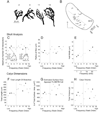
Structural Analysis of the Calyx of Held. A. Computerized tracings of four BDA-labeled calyces. Calyces are numbered by their position (rank order) along the tonotopic axis of the MNTB. Calyx 11 extended along the long axis of the postsynaptic cell; calyx 2 extended along the short axis of the cell. The cells contacted by calyces 3 and 13 were not stained. B. Locations of this population of 16 calyces projected onto a normalized section through the MNTB. Low frequencies located dorsolateral, high frequencies ventromedial. Each calyx (represented as an enclosure of its extremities) and the proximal length of axon contained within the tissue section are shown. Calyces were rank-ordered by position along the frequency axis. Dashed line separates MNTB into halves along the frequency axis. D, dorsal; L, lateral. C–E. Modified Sholl analysis. Vertical dashed lines, also in panels F–H, correspond to dashed line in panel B. Filled circles indicate the four calyces depicted in panel A. C. Calyces were similar in the maximum number of branches (peak value in the Sholl plots shown as insets). Sholl plots of number of branches vs distance from the base are shown for calyces #3 and #13. Plots are fit by splines and half width (horizontal lines) measured from the fitted plot. D. The distance from the base of the calyx at which the maximum number of intersections occurred varied over a factor of 2 but showed a slight frequency dependence. E. High frequency calyces maintained a high branch number over longer distances from the calyx base, as measured by the half width of the Sholl plot. A frequency scale, derived from Guinan et al. (1972), is applied in this panel to the normalized plot of calyx position in panel B as an approximation of the characteristic frequency for each calyx. F. High frequency calyces exhibited longer total branch length. G, H. Due to variation in the composition of calyces (number and size of stalks, branches and swellings), surface area apposed to the MNTB cell and calyx volume showed less dependence on frequency.
Figure 2
Spatial distribution of voltage drop in the calyx structure under standard model conditions, measured at both DC and 500 Hz using NEURON’s Impedance Tool. These plots of voltage throughout the structure were normalized to the voltage at the injection site, located at the junction between the axon and the calyx proper (indicated by the arrow on the inset schematic of the calyx). There are two tiers of voltage drops within the calyx, indicating that the calyx has electrotonically distinct compartments. This voltage drop is larger for higher frequencies (500 Hz, gray trace), reflecting the lower impedance of the calyx as seen by an action potential.
Figure 3
Spatially Heterogeneous “Standard” Model. A. Action potential height (left; gray open circles) and half-width (right, black filled squares) as a function of axial resistance, Ra. Each data point is the average over all swellings in the calyx. glk was also varied in these simulations over the range 1–10 µS/cm2, but changes due to glk are small and not visible. B. Action potential waveform where it invades the base of the calyx from the axon (thin black trace, labeled ax), and at three of the swellings (thick black, thick gray and thin gray traces, indicated in the inset in this Figure and Figure 4–Figure 6 by the Roman numerals I–III). The action potential is delayed, and becomes wider and smaller as it propagates into the calyx. Solid lines are from simulations for Ra = 100 Ω·cm, dashed lines for Ra = 200 Ω·cm. Traces for locations I and II nearly overlap. C. Peak Ca2+ current density for each of the 55 swellings in the calyx (note that some swellings are represented by more than one point when they were associated with a branch point; these symbols overlap), for Ra = 100 (filled circles) and 200 (open circles) Ω·cm. Two tiers of values are generated for each Ra value. ICa shows wider variation when Ra is 100 Ω·cm. D. Ca2+ current density time course for the same locations as in B, C; trace color, thickness and style corresponds to data in B. Note that the Ca2+ current is much smaller in the terminal swelling (location III), especially when Ra = 200 Ω·cm. E. Estimated peak intracellular Ca2+ concentration for the same conditions as in B–D. Each Ra value generates two tiers of peak Ca2+ concentration values. F. Estimated time course of Ca2+ concentration in the terminal.
Figure 4
Spatially Homogeneous “Uniform” Model. This model was generated by setting the Na+ and K+ currents, as measured in voltage clamp, to the same values as in Figure 3, while distributing the channels at a uniform density throughout the calyx. A. Action potential height and half-width is independent of Ra. B. Action potential time course is similar in all calyx compartments but independent of Ra, although small latency shifts remain. Measurements taken from same calyx compartments as in Figure 3. C. Ca2+ current density is uniform throughout the calyx. D. The Ca2+ current shows an inflection on the rising phase as the action potential voltage approaches ECa. E, F. The time course of the Ca2+ influx is uniform throughout the calyx, with only slight latency shifts across the structure.
Figure 5
Standard Model with Increased Ca2+ channel density. A 2-fold increase in the calyx Ca2+ channel density widens the action potential and the duration of Ca2+ current (compare with Fig. 3). A. Average action potential peak value (open circles) is slightly higher, and the half-width is greater than for the standard model. B. Action potentials are wider with a distinct hump on the falling phase with elevated Ca2+ channel density. Measurements taken from same calyx compartments as in Figure 3. C. The distribution of Ca2+ current amplitudes is compressed when Ra = 100 Ω·cm, but still shows a two-tiered separation in different parts of the calyx when Ra = 200 Ω·cm. D. The duration of the Ca2+ current is longer with elevated channel density. E. Peak Ca2+ concentration is nearly uniform across the calyx when Ra = 100 Ω·cm; however, as with the Ca2+ current, there are still 2 tiers of values when Ra = 200 Ω·cm. F. The Ca2+ concentration shows latency shifts similar to those in the standard model.
Figure 6
Standard Model with addition of Na+ channels in the proximal stalks. Na+ channel density was adjusted so that peak Na+ currents were the same as in the standard model of Figure 3. All other parameters and conventions are the same as for Figure 3. A. Action potential amplitude is significantly higher than in the standard model, although the width remains the same. B. The action potential shapes are similar to the standard case, except that with higher Ra, the most distal swellings have a slight hump on repolarization (thin gray dashed line for swelling III). Measurements taken from same calyx compartments as in Figure 3. C. Ca2+ current density in the swellings is higher than the standard model, and the decrease with distance from the axon is less than in the standard model. Two tiers of Ca2+ influx remain. D. The Ca2+ current has a brief time course, although there is a slight inflection on the rising phase of the current in the proximal regions of the calyx. E. The distribution of Ca2+ concentration exhibits two tiers. However, the decrease with distance is less than in the standard case, such that the Ca2+ influx in the more distant portions of the terminal is higher. F. Ca2+ concentration shows higher values in the more proximal regions of the calyx due to a larger action potential but a similar time course as the standard case.
Similar articles
- Calcium secretion coupling at calyx of Held governed by nonuniform channel-vesicle topography.
Meinrenken CJ, Borst JG, Sakmann B. Meinrenken CJ, et al. J Neurosci. 2002 Mar 1;22(5):1648-67. doi: 10.1523/JNEUROSCI.22-05-01648.2002. J Neurosci. 2002. PMID: 11880495 Free PMC article. - Calcium channel types with distinct presynaptic localization couple differentially to transmitter release in single calyx-type synapses.
Wu LG, Westenbroek RE, Borst JG, Catterall WA, Sakmann B. Wu LG, et al. J Neurosci. 1999 Jan 15;19(2):726-36. doi: 10.1523/JNEUROSCI.19-02-00726.1999. J Neurosci. 1999. PMID: 9880593 Free PMC article. - Exocytotic dynamics and calcium cooperativity effects in the calyx of Held synapse: a modelling study.
Gil A, González-Vélez V. Gil A, et al. J Comput Neurosci. 2010 Feb;28(1):65-76. doi: 10.1007/s10827-009-0187-x. Epub 2009 Oct 2. J Comput Neurosci. 2010. PMID: 19798561 - Spontaneous neurotransmitter release and Ca2+--how spontaneous is spontaneous neurotransmitter release?
Glitsch MD. Glitsch MD. Cell Calcium. 2008 Jan;43(1):9-15. doi: 10.1016/j.ceca.2007.02.008. Epub 2007 Mar 26. Cell Calcium. 2008. PMID: 17382386 Review. - Presynaptic calcium and control of vesicle fusion.
Schneggenburger R, Neher E. Schneggenburger R, et al. Curr Opin Neurobiol. 2005 Jun;15(3):266-74. doi: 10.1016/j.conb.2005.05.006. Curr Opin Neurobiol. 2005. PMID: 15919191 Review.
Cited by
- Presynaptic Diversity Revealed by Ca2+-Permeable AMPA Receptors at the Calyx of Held Synapse.
Lujan B, Dagostin A, von Gersdorff H. Lujan B, et al. J Neurosci. 2019 Apr 17;39(16):2981-2994. doi: 10.1523/JNEUROSCI.2565-18.2019. Epub 2019 Jan 24. J Neurosci. 2019. PMID: 30679394 Free PMC article. - Short-term synaptic depression and recovery at the mature mammalian endbulb of Held synapse in mice.
Wang Y, Manis PB. Wang Y, et al. J Neurophysiol. 2008 Sep;100(3):1255-64. doi: 10.1152/jn.90715.2008. Epub 2008 Jul 16. J Neurophysiol. 2008. PMID: 18632895 Free PMC article. - Allegro giusto: piccolo, bassoon and clarinet set the tempo of vesicle pool replenishment.
Dagostin A, Kushmerick C, von Gersdorff H. Dagostin A, et al. J Physiol. 2018 Apr 15;596(8):1315-1316. doi: 10.1113/JP275704. Epub 2018 Mar 25. J Physiol. 2018. PMID: 29446080 Free PMC article. No abstract available. - Underpinning heterogeneity in synaptic transmission by presynaptic ensembles of distinct morphological modules.
Fekete A, Nakamura Y, Yang YM, Herlitze S, Mark MD, DiGregorio DA, Wang LY. Fekete A, et al. Nat Commun. 2019 Feb 18;10(1):826. doi: 10.1038/s41467-019-08452-2. Nat Commun. 2019. PMID: 30778063 Free PMC article. - High-resolution volumetric imaging constrains compartmental models to explore synaptic integration and temporal processing by cochlear nucleus globular bushy cells.
Spirou GA, Kersting M, Carr S, Razzaq B, Yamamoto Alves Pinto C, Dawson M, Ellisman MH, Manis PB. Spirou GA, et al. Elife. 2023 Jun 8;12:e83393. doi: 10.7554/eLife.83393. Elife. 2023. PMID: 37288824 Free PMC article.
References
- Blackburn CC, Sachs MB. The representations of the steady-state vowel sound /e/ in the discharge patterns of cat anteroventral cochlear nucleus neurons. J Neurophysiol. 1990;63:1191–1212. - PubMed
- Bollmann JH, Sakmann B, Borst JG. Calcium sensitivity of glutamate release in a calyx-type terminal. Science. 2000;289:953–957. - PubMed
- Bollmann JH, Sakmann B. Control of synaptic strength and timing by the release-site Ca2+ signal. Nat Neurosci. 2005;8:426–434. - PubMed
- Borst JG, Sakmann B. Calcium influx and transmitter release in a fast CNS synapse. Nature. 1996;383:431–434. - PubMed
Publication types
MeSH terms
Substances
Grants and funding
- P20 RR015574-09/RR/NCRR NIH HHS/United States
- R01 DC004551/DC/NIDCD NIH HHS/United States
- R01 DC005035/DC/NIDCD NIH HHS/United States
- R01 DC004274/DC/NIDCD NIH HHS/United States
- P20 RR015574/RR/NCRR NIH HHS/United States
- R01 DC005035-05/DC/NIDCD NIH HHS/United States
- RR15574/RR/NCRR NIH HHS/United States
- R01 DC004551-08/DC/NIDCD NIH HHS/United States
- R01 DC004274-09/DC/NIDCD NIH HHS/United States
- R01 DC004551-07/DC/NIDCD NIH HHS/United States
LinkOut - more resources
Full Text Sources
Research Materials
Miscellaneous


