White matter in learning, cognition and psychiatric disorders - PubMed (original) (raw)
Review
White matter in learning, cognition and psychiatric disorders
R Douglas Fields. Trends Neurosci. 2008 Jul.
Abstract
White matter is the brain region underlying the gray matter cortex, composed of neuronal fibers coated with electrical insulation called myelin. Previously of interest in demyelinating diseases such as multiple sclerosis, myelin is attracting new interest as an unexpected contributor to a wide range of psychiatric disorders, including depression and schizophrenia. This is stimulating research into myelin involvement in normal cognitive function, learning and IQ. Myelination continues for decades in the human brain; it is modifiable by experience, and it affects information processing by regulating the velocity and synchrony of impulse conduction between distant cortical regions. Cell-culture studies have identified molecular mechanisms regulating myelination by electrical activity, and myelin also limits the critical period for learning through inhibitory proteins that suppress axon sprouting and synaptogenesis.
Figures
Figure 1
Myelin is the multilayered compacted cell membrane wrapped around axons by glial cells to form electrical insulation that speeds conduction of nerve impulses. (a) In the brain, myelin is wrapped around axons by oligodendrocytes, which have 20 or more cellular processes to insulate multiple axons. (b) An oligodendrocyte (green) is shown at the initial stage of wrapping myelin membrane around several axons (red), in cell cultures equipped with electrodes to stimulate axons for investigations of the role of impulse activity in regulating myelination [87]. (c) An electron micrograph of an axon from the corpus callosum of rat brain is shown in cross-section to reveal the multiple layers of myelin membrane surrounding the axon. Up to 150 layers of myelin are formed on large-diameter axons. Image in (b) courtesy of Varsha Shukla, NICHD and in (c) courtesy of Andrea Nans, NYU School of Medicine. Myelin basic protein (green); neurofilament protein (red). Figure modified from Fields [1].
Figure 2
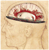
Neuroimaging reveals changes in white matter structure in the human brain. White matter (white) comprises half of the human brain and consists of bundles of myelinated axons connecting neurons in different brain regions. Gray matter (pink) is composed of neuronal cell bodies and dendrites concentrated in the outer layers of the cortex. Microstructural changes in white matter can be revealed by specialized MRI brain imaging techniques such as diffusion tensor imaging (DTI). This method analyzes the fractional anisotropy (FA) of proton diffusion in tissue, which is more restricted in white matter than in gray matter. The anisotropy increases with increased myelination, fiber diameter and axon compaction. The degree of anisotropy is represented on a pseudo color scale as shown in the human brain scan in the horizontal plane of the image above, where the major white matter tracts are revealed against a black background of low fractional anisotropy [107]. These data can be used to calculate the probable anatomy of white matter fiber bundles in living brain, a process called tractography. An example from human brain imaging is shown above as red filamentous bundles radiating out from the corpus callosum. Fiber orientation is calculated from the eigenvectors defining proton diffusion in three dimensions in each voxel. Using algorithms, the principal eigenvalue vector is connected to the next voxel to trace the fiber structure and orientation in white matter tracts [108]. Changes in white matter structure are seen by DTI in association with many neuropsychiatric disorders, cognitive function and during learning. FA image courtesy of Carlo Pierpaoli, NICHD, NIH, and DTI tractography, courtesy of Derek K. Jones, School of Psychiatry, Cardiff University. Illustration by Lydia Kibiuk, Medical Arts, NIH.
Figure 3
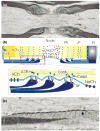
Myelin speeds impulse conduction velocity through nerve fibers by fundamentally changing the way impulses are propagated. Rather than a continuous wave of depolarization, the nerve impulse is generated by sodium and potassium currents at isolated points on the axon called nodes of Ranvier, and each node acts as a repeater. (a) An electron micrograph of a node of Ranvier in long section from spinal dorsal root nerve of rat showing the node of Ranvier flanked by intermodal segments insulated by layers of compact myelin. Each layer of myelin terminates in a series of loops adjacent to the node of Ranvier (the paranodal loops shown in [b] and [c]). (b) Three axonal domains are defined by axon interactions with myelinating glia: the Na+ channel-enriched node of Ranvier, the adjacent paranode (PN) where the loops of myelin adhere to the axon through cell-adhesion molecules linked to the axon cytoskeleton, the juxtaparanodal region (JP) which contains delayed rectifier K+ channels and the internode (IN) sealed by compacted layers of myelin membrane to restrict transmembrane ion currents to the nodal region. These domains are formed and maintained by adhesive interactions and soluble signals from myelinating glia. KCh = K+ channel; NaCh = Na+ channel; Cont = contactin; Caspr = contactin-associated protein; NF = neurofascin 155, 4.1B protein. (c) High-magnification electron micrograph of paranodal loops in a node of Ranvier from mouse spinal root nerve preserved by high-pressure freezing. Note the dense adhesive junctions between each paranodal loop and the axon. (a,c) Courtesy of Gina Sosinsky, Thomas Deerinck, Ying Jones and Mark Ellisman, UCSD, National Center for Microscopy and Imaging Research, San Diego. (b) Modified from Fields and Stevens-Graham [103].
Similar articles
- Grey matter myelination.
Timmler S, Simons M. Timmler S, et al. Glia. 2019 Nov;67(11):2063-2070. doi: 10.1002/glia.23614. Epub 2019 Mar 12. Glia. 2019. PMID: 30860619 Review. - Is psychosis a dysmyelination-related information-processing disorder?
Giotakos O. Giotakos O. Psychiatriki. 2019 Jul-Sep;30(3):245-255. doi: 10.22365/jpsych.2019.303.245. Psychiatriki. 2019. PMID: 31685456 Review. - Motor learning requires myelination to reduce asynchrony and spontaneity in neural activity.
Kato D, Wake H, Lee PR, Tachibana Y, Ono R, Sugio S, Tsuji Y, Tanaka YH, Tanaka YR, Masamizu Y, Hira R, Moorhouse AJ, Tamamaki N, Ikenaka K, Matsukawa N, Fields RD, Nabekura J, Matsuzaki M. Kato D, et al. Glia. 2020 Jan;68(1):193-210. doi: 10.1002/glia.23713. Epub 2019 Aug 29. Glia. 2020. PMID: 31465122 Free PMC article. - Myelin development in cerebral gray and white matter during adolescence and late childhood.
Corrigan NM, Yarnykh VL, Hippe DS, Owen JP, Huber E, Zhao TC, Kuhl PK. Corrigan NM, et al. Neuroimage. 2021 Feb 15;227:117678. doi: 10.1016/j.neuroimage.2020.117678. Epub 2020 Dec 29. Neuroimage. 2021. PMID: 33359342 Free PMC article. - Social Experience-Dependent Myelination: An Implication for Psychiatric Disorders.
Toritsuka M, Makinodan M, Kishimoto T. Toritsuka M, et al. Neural Plast. 2015;2015:465345. doi: 10.1155/2015/465345. Epub 2015 May 19. Neural Plast. 2015. PMID: 26078885 Free PMC article. Review.
Cited by
- Breastfeeding and early white matter development: A cross-sectional study.
Deoni SC, Dean DC 3rd, Piryatinsky I, O'Muircheartaigh J, Waskiewicz N, Lehman K, Han M, Dirks H. Deoni SC, et al. Neuroimage. 2013 Nov 15;82:77-86. doi: 10.1016/j.neuroimage.2013.05.090. Epub 2013 May 28. Neuroimage. 2013. PMID: 23721722 Free PMC article. - Protracted abstinence from chronic ethanol exposure alters the structure of neurons and expression of oligodendrocytes and myelin in the medial prefrontal cortex.
Navarro AI, Mandyam CD. Navarro AI, et al. Neuroscience. 2015 May 7;293:35-44. doi: 10.1016/j.neuroscience.2015.02.043. Epub 2015 Feb 28. Neuroscience. 2015. PMID: 25732140 Free PMC article. - Reversal of the Detrimental Effects of Post-Stroke Social Isolation by Pair-Housing is Mediated by Activation of BDNF-MAPK/ERK in Aged Mice.
Verma R, Harris NM, Friedler BD, Crapser J, Patel AR, Venna V, McCullough LD. Verma R, et al. Sci Rep. 2016 Apr 29;6:25176. doi: 10.1038/srep25176. Sci Rep. 2016. PMID: 27125783 Free PMC article. - The Impact of Cognitive Training on Cerebral White Matter in Community-Dwelling Elderly: One-Year Prospective Longitudinal Diffusion Tensor Imaging Study.
Cao X, Yao Y, Li T, Cheng Y, Feng W, Shen Y, Li Q, Jiang L, Wu W, Wang J, Sheng J, Feng J, Li C. Cao X, et al. Sci Rep. 2016 Sep 15;6:33212. doi: 10.1038/srep33212. Sci Rep. 2016. PMID: 27628682 Free PMC article. - Effects of Chronic Scopolamine Treatment on Cognitive Impairments and Myelin Basic Protein Expression in the Mouse Hippocampus.
Park JH, Choi HY, Cho JH, Kim IH, Lee TK, Lee JC, Won MH, Chen BH, Shin BN, Ahn JH, Tae HJ, Choi JH, Chung JY, Lee CH, Cho JH, Kang IJ, Kim JD. Park JH, et al. J Mol Neurosci. 2016 Aug;59(4):579-89. doi: 10.1007/s12031-016-0780-1. Epub 2016 Jun 24. J Mol Neurosci. 2016. PMID: 27343058
References
- Fields RD. White matter matters. Sci Am. 2008;298:42–49. - PubMed
- Bullock TH, et al. Evolution of myelin sheaths: both lamprey and hagfish lack myelin. Neurosci Lett. 1984;48:145–148. - PubMed
- Tkachev D, et al. Oligodendrocyte dysfunction in schizophrenia and bipolar disorder. Lancet. 2003;362:798–805. - PubMed
- Stewart SE, et al. A genetic family-based association study of OLIG2 in obsessive-compulsive disorder. Arch Gen Psychiatry. 2007;64:209–214. - PubMed
Publication types
MeSH terms
LinkOut - more resources
Full Text Sources
Other Literature Sources
Medical


