Exploiting the anisotropy of anomalous scattering boosts the phasing power of SAD and MAD experiments - PubMed (original) (raw)
Exploiting the anisotropy of anomalous scattering boosts the phasing power of SAD and MAD experiments
Marc Schiltz et al. Acta Crystallogr D Biol Crystallogr. 2008 Jul.
Abstract
The X-ray polarization anisotropy of anomalous scattering in crystals of brominated nucleic acids and selenated proteins is shown to have significant effects on the diffraction data collected at an absorption edge. For conventionally collected single- or multi-wavelength anomalous diffraction data, the main manifestation of the anisotropy of anomalous scattering is the breakage of the equivalence between symmetry-related reflections, inducing intensity differences between them that can be exploited to yield extra phase information in the structure-solution process. A new formalism for describing the anisotropy of anomalous scattering which allows these effects to be incorporated into the general scheme of experimental phasing methods using an extended Harker construction is introduced. This requires a paradigm shift in the data-processing strategy, since the usual separation of the data-merging and phasing steps is abandoned. The data are kept unmerged down to the Harker construction, where the symmetry-breaking is explicitly modelled and refined and becomes a source of supplementary phase information. These ideas have been implemented in the phasing program SHARP. Refinements using actual data show that exploitation of the anisotropy of anomalous scattering can deliver substantial extra phasing power compared with conventional approaches using the same raw data. Examples are given that show improvements in the phases which are typically of the same order of magnitude as those obtained in a conventional approach by adding a second-wavelength data set to a SAD experiment. It is argued that such gains, which come essentially for free, i.e. without the collection of new data, are highly significant, since radiation damage can frequently preclude the collection of a second-wavelength data set. Finally, further developments in synchrotron instrumentation and in the design of data-collection strategies that could help to maximize these gains are outlined.
Figures
Figure 1
Anomalous scattering factors _f_′ and _f_′′ for Se in selenomethionine residues. The curves represent the anomalous scattering factors when the polarization direction of the incident X-ray beam is aligned with one of the principal molecular directions in a C—Se—C moiety. Black curves: along the direction u (perpendicular to the plane containing the C—Se—C bonds). Green curves: along the direction w (bisecting the C—Se—C angle). Red curves: along the direction v (perpendicular to u and w). Data from Bricogne et al. (2005 ▶).
Figure 2
Anomalous scattering factors _f_′ and _f_′′ for Br in brominated nucleotides. The curves represent the anomalous scattering factors when the polarization direction of the incident X-ray beam is aligned with one of the principal molecular directions in a brominated nucleobase. Black curves: along the direction u (parallel to the C—Br bond). Red curves: along the direction v (perpendicular to the C—Br bond and parallel to the plane of the nucleobase ring). Green curves: along the direction w (perpendicular to the nucleobase ring). Data from Sanishvili et al. (2007 ▶).
Figure 3
AAS-induced symmetry breaking in _p_-bromobenzamide. The ORTEP plot in the upper part of the figure displays the packing of _p_-bromobenzamide molecules in the monoclinic crystal form viewed down the a axis. The glide plane is perpendicular to the b axis and the translational component is along c. Two _p_-bromobenzamide molecules which are related by the glide-plane symmetry operation are highlighted. The direction of linear polarization of the incident X-ray beam is also indicated for two experiments (I) and (II) that were carried out successively. In experiment (I), the C—Br bonds of the two symmetry-related molecules experience the polarization direction at different angles. Thus, in the vicinity of the Br K edge, these two Br atoms display different anomalous scattering factors and are no longer equivalent. This symmetry-breaking effect of AAS leads to the appearance of the glide-plane forbidden reflections [(h_0_l), l = odd] as is shown in the lower left part of the picture, which shows the reconstruction of the (h_0_l) layer from experimental data. In experiment (II), the direction of linear polarization of the incident X-ray beam was oriented parallel to the glide plane. The C—Br bonds of the two symmetry-related molecules therefore experienced the polarization direction at identical angles. Thus, in this particular configuration, the symmetry-equivalence of the two Br atoms is restored and the glide-plane forbidden reflections [(h_0_l), l = odd] are truly absent as is shown in the lower right part of the picture, which shows the reconstruction of the (h_0_l) layer from experimental data.
Figure 4
Packing of d(CGCG[BrU]G) molecules viewed down the crystal c axis. The eight C—Br moieties in the unit cell are displayed, with the Br atoms highlighted as green spheres. Owing to the orientation of the helical DNA duplexes in the crystal, all C—Br bonds are oriented almost perpendicular to [001]. Also displayed is the in-plane component of the X-ray polarization direction for data sets (I) and (II). {For data set (III), the X-ray polarization direction was almost parallel to [001] and is therefore not displayed here.}
Figure 5
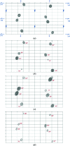
Anomalous difference Fourier maps for d(CGCG[BrU]G) computed to 1.1 Å resolution. The maps are projected down the crystal c axis. For each map, the origin is located at the upper left corner and the a axis is along the vertical direction. Contours are at intervals of 0.4 e− Å−3. (a) Map computed from data set (II) merged in point group 222. The symmetry elements of space group _P_212121 are displayed in blue. (b) Map computed from data set (I) merged in point group 1. (c) Map computed from data set (II) merged in point group 1. (d) Map computed from data set (III) merged in point group 1. The figures printed in red next to each peak indicate the angle between the direction of X-ray polarization and the C—Br bond direction of the corresponding Br site.
Figure 6
Refined f j,_s_′′ parameters of the Br atoms in the unit cell of d(CGCG[BrU]G) crystals (data from Table 3 ▶) plotted against cos2(α), where α is the angle between the C—Br direction and the X-ray polarization direction p.
Figure 7
Quality of the phases obtained by exploiting the AAS-induced symmetry-breaking effects in d(CGCG[BrU]G). The plots represent the correlation coefficients, as a function of resolution, of maps computed from experimental phases with a map computed from the final refined structure of d(CGCG[BrU]G). All three data sets (crystal orientations) have been used for phasing. Black, SAD phases computed from merged data with conventional isotropic _f_′′ factors. Red, phases computed from unmerged data using a tensorial description (f′′) for the imaginary anomalous scattering factors (see Table 2 ▶). Green, phases computed from unmerged data using distinct _f_′′ factors for ‘symmetry-unrolled’ sites and for each data set (see Table 3 ▶).
Figure 8
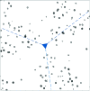
Anomalous difference Fourier map for PPAT computed to 2.2 Å resolution. The map is projected down the [111] axis. The origin is located in the centre of the map and the location of the threefold symmetry axis is displayed in blue. Contours are at intervals of 0.1 e− Å−3. The map was computed from the data merged in point group 1. It can be seen that the threefold symmetry is broken.
Figure 9
Anomalous difference Fourier maps for PPAT computed to 2.2 Å resolution from the data merged in point group 1. The maps are projected down the axes [100] (a), [010] (b) and [001] (c). For each map, the origin is located at the upper left corner and the location of the twofold symmetry axis is displayed in blue. Contours are at intervals of 0.1 e− Å−3.
Figure 10
Quality of the phases obtained by exploiting the AAS-induced symmetry-breaking effects in PPAT. The plots represent the correlation coefficients, as a function of resolution, of maps computed from experimental phases with a map computed from the final refined structure of PPAT. Black, SAD phases computed from merged data with conventional isotropic _f_′ and _f_′′ factors. Red, SAD phases computed from unmerged data, using individual _f_′′ factors for symmetry-related sites.
Figure 11
Quality of the phases obtained by exploiting the AAS-induced symmetry-breaking effects in IMPDH. The plots represent the correlation coefficients, as a function of resolution, of maps computed from experimental phases with a map computed from the final refined structure of IMPDH. Black, SAD phases computed from merged data with conventional isotropic _f_′ and _f_′′ factors. Red, SAD phases computed from unmerged data using a tensorial parametrization ( +
+  ) to describe AAS. Green, two-wavelength MAD (peak + inflection point) phases computed from merged data with conventional isotropic _f_′ and _f_′′ factors. Blue, three-wavelength MAD phases computed from merged data with conventional isotropic _f_′ and _f_′′ factors. Orange, three-wavelength MAD phases computed from unmerged data using a tensorial parametrization (
 +
+  ) to describe AAS.
Similar articles
- 'Broken symmetries' in macromolecular crystallography: phasing from unmerged data.
Schiltz M, Bricogne G. Schiltz M, et al. Acta Crystallogr D Biol Crystallogr. 2010 Apr;66(Pt 4):447-57. doi: 10.1107/S0907444909053578. Epub 2010 Mar 24. Acta Crystallogr D Biol Crystallogr. 2010. PMID: 20382998 Free PMC article. - Modelling and refining site-specific radiation damage in SAD/MAD phasing.
Schiltz M, Bricogne G. Schiltz M, et al. J Synchrotron Radiat. 2007 Jan;14(Pt 1):34-42. doi: 10.1107/S0909049506038970. Epub 2006 Dec 15. J Synchrotron Radiat. 2007. PMID: 17211070 - Single-wavelength anomalous diffraction phasing revisited.
Rice LM, Earnest TN, Brunger AT. Rice LM, et al. Acta Crystallogr D Biol Crystallogr. 2000 Nov;56(Pt 11):1413-20. doi: 10.1107/s0907444900010039. Acta Crystallogr D Biol Crystallogr. 2000. PMID: 11053839 - Anomalous X-ray diffraction with soft X-ray synchrotron radiation.
Carpentier P, Berthet-Colominas C, Capitan M, Chesne ML, Fanchon E, Lequien S, Stuhrmann H, Thiaudière D, Vicat J, Zielinski P, Kahn R. Carpentier P, et al. Cell Mol Biol (Noisy-le-grand). 2000 Jul;46(5):915-35. Cell Mol Biol (Noisy-le-grand). 2000. PMID: 10976874 Review. - Contemporary Use of Anomalous Diffraction in Biomolecular Structure Analysis.
Liu Q, Hendrickson WA. Liu Q, et al. Methods Mol Biol. 2017;1607:377-399. doi: 10.1007/978-1-4939-7000-1_16. Methods Mol Biol. 2017. PMID: 28573582 Free PMC article. Review.
Cited by
- Anomalous diffraction in crystallographic phase evaluation.
Hendrickson WA. Hendrickson WA. Q Rev Biophys. 2014 Feb;47(1):49-93. doi: 10.1017/S0033583514000018. Q Rev Biophys. 2014. PMID: 24726017 Free PMC article. Review. - Translation calibration of inverse-kappa goniometers in macromolecular crystallography.
Brockhauser S, White KI, McCarthy AA, Ravelli RB. Brockhauser S, et al. Acta Crystallogr A. 2011 May;67(Pt 3):219-28. doi: 10.1107/S0108767311004831. Epub 2011 Mar 15. Acta Crystallogr A. 2011. PMID: 21487180 Free PMC article. - PRIGo: a new multi-axis goniometer for macromolecular crystallography.
Waltersperger S, Olieric V, Pradervand C, Glettig W, Salathe M, Fuchs MR, Curtin A, Wang X, Ebner S, Panepucci E, Weinert T, Schulze-Briese C, Wang M. Waltersperger S, et al. J Synchrotron Radiat. 2015 Jul;22(4):895-900. doi: 10.1107/S1600577515005354. Epub 2015 May 9. J Synchrotron Radiat. 2015. PMID: 26134792 Free PMC article. - Recent developments in phasing and structure refinement for macromolecular crystallography.
Adams PD, Afonine PV, Grosse-Kunstleve RW, Read RJ, Richardson JS, Richardson DC, Terwilliger TC. Adams PD, et al. Curr Opin Struct Biol. 2009 Oct;19(5):566-72. doi: 10.1016/j.sbi.2009.07.014. Epub 2009 Aug 21. Curr Opin Struct Biol. 2009. PMID: 19700309 Free PMC article. Review. - PROXIMA-1 beamline for macromolecular crystallography measurements at Synchrotron SOLEIL.
Chavas LMG, Gourhant P, Guimaraes BG, Isabet T, Legrand P, Lener R, Montaville P, Sirigu S, Thompson A. Chavas LMG, et al. J Synchrotron Radiat. 2021 May 1;28(Pt 3):970-976. doi: 10.1107/S1600577521002605. Epub 2021 Apr 29. J Synchrotron Radiat. 2021. PMID: 33950005 Free PMC article.
References
- Bricogne, G. (2000). Proceedings of the Workshop on Advanced Special Functions and Applications, Melfi (PZ), Italy, 9–12 May 1999, edited by D. Cocolicchio, G. Dattoli & H. M. Srivastava, pp. 315–321. Rome: Aracne Editrice.
- Bricogne, G., Capelli, S. C., Evans, G., Mitschler, A., Pattison, P., Roversi, P. & Schiltz, M. (2005). J. Appl. Cryst.38, 168–182.
- Bricogne, G. & Schiltz, M. (2000). Communication at the International Workshop on X-ray Gyrotropy and Synchrotron-based Chiroptical Spectroscopies, 21–23 September 2000. ESRF, Grenoble, France.
- Bricogne, G., Vonrhein, C., Flensburg, C., Schiltz, M. & Paciorek, W. (2003). Acta Cryst. D59, 2023–2030. - PubMed
- Collaborative Computational Project, Number 4 (1994). Acta Cryst. D50, 760–763. - PubMed
Publication types
MeSH terms
Substances
LinkOut - more resources
Full Text Sources


