A molecule targeting VHL-deficient renal cell carcinoma that induces autophagy - PubMed (original) (raw)
A molecule targeting VHL-deficient renal cell carcinoma that induces autophagy
Sandra Turcotte et al. Cancer Cell. 2008.
Abstract
Renal cell carcinomas (RCCs) are refractory to standard therapies. The von Hippel-Lindau (VHL) tumor suppressor gene is inactivated in 75% of RCCs. By screening for small molecules selectively targeting VHL-deficient RCC cells, we identified STF-62247. STF-62247 induces cytotoxicity and reduces tumor growth of VHL-deficient RCC cells compared to genetically matched cells with wild-type VHL. STF-62247-stimulated toxicity occurs in a HIF-independent manner through autophagy. Reduction of protein levels of essential autophagy pathway components reduces sensitivity of VHL-deficient cells to STF-62247. Using a yeast deletion pool, we show that loss of proteins involved in Golgi trafficking increases killing by STF-62247. Thus, we have found a small molecule that selectively induces cell death in VHL-deficient cells, representing a paradigm shift for targeted therapy.
Figures
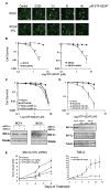
Figure 1. STF-62247 induces cytotoxicity and reduces tumor growth in VHL-deficient cells in HIF-independent manner
A. RCC4 and RCC4/VHL cells were labeled with EYFP and treated with increasing concentration of STF-62247 for 4 days. Each cell type was monitored by fluorescence separately. Cells are pseudocolored green. Scale bar, 10 μm. B. Clonogenic assay in RCC4, RCC4/VHL, SN12C, SN12C-VHL shRNA cells in the presence of STF-62247 (0–30 μM). Each point of the cell survival is calculated by the average of three different experiments in triplicate as the percent of treated on untreated plate. C. Upper Panel, clonogenic assay in RCC4 infected with HIF-1α or HIF-2α shRNA by retroviral infection in presence of drug in same conditions described above. Bottom panel, HIF-1α or HIF-2α protein expression evaluated by western blot in RCC4 cells with HIF-1α or HIF-2α shRNA. Tubulin is used as loading control. D. Upper Panel, clonogenic assay with STF-62247 in RCC4 where HIF-2α was stably introduced into VHL-positive cells by retroviral infection. Bottom panel, western blot for HIF-1α or HIF-2α in RCC4 VHL-positive cells. E. SN12C-VHL shRNA (2–3 × 10⁶ cells) and 786-O (5 × 10⁶ cells) were injected into SCID mice. SN12C shVHL tumor bearing mice were treated daily with vehicle, 2.7 mg/kg, or 8 mg/kg of STF-62247 by intraperitoneal injection. 786-O tumor bearing mice were treated with vehicle or 8 mg/kg. Five tumors per condition were analyzed where * p<0.05, **p<0.01 and ***p<0.005. All error bars represented the standard error of the mean (SEM).
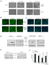
Figure 2. Presence of autophagic vacuoles with STF-62247 in RCCs
A. Phase contrast pictures of VHL-deficient RCC4 and SN12C-VHL shRNA cells and VHL-positive cells RCC4 and SN12C after 20 hrs of 1.25 μM STF-62247. B. Immunostaining for LC3 following STF-62247 treatment at 1.25 μM for 20 hrs with or without 2 mM of 3-MA in RCC4 with and without VHL cells. After treatment, cells were fixed, permeabilized and processed for the detection of punctuate staining by indirect immunofluorescence. C. Autophagic vacuoles stained with monodansylcadaverine (MDC). After STF-62247 treatment in the same conditions, MDC was added for the last 10 minutes. Cells were fixed, washed with PBS and observed directly under microscope. D. Western blot for LC3 in RCC4, RCC4/VHL, SN12C, and SN12C-VHL shRNA cells after STF-62247 treatment with or without 3-MA. Nutrient starvation (EBSS) is used as positive control. E. Western blot for LC3 in RCC4 and RCC4/VHL with different concentrations of STF-62247. Tubulin was used as loading control. F. Cell viability assay in VHL-deficient RCC4 and wild-type VHL cells treated with 1 mM of 3-MA, 1.25 μM of STF-62247 alone or in combination for 24 hrs. Viability is measured by trypan blue exclusion assay and is represented by the average of three different experiments in duplicate as the percent of treated on untreated cells. All error bars represent SEM. All scale bars, 10 μm.
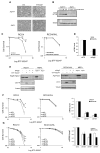
Figure 3. Atg5 is involved in autophagic cell death of STF-62247
A. MEFs Atg5+/+ and Atg5−/− were treated in presence of STF-62247 and examined under light microscopy 20 hrs later. Scale bar, 10 μm. B. LC3 processing evaluated by Western blot following 24 hours of drug treatment in MEFs. C. Cell Survival measured by clonogenic assay with STF-62247 in RCC4 and RCC4/VHL cells transiently transfected with siRNA to ATG5. Colony formation was evaluated as described before. ***p<0.005 represents the difference between RCC4 and RCC4 with siATG5 cells in presence of STF-62247. D. mRNA level of ATG5 quantified by qRT-PCR in RCC4 and RCC4/VHL cells transfected with siRNA to ATG5 for 48 hours. E. Western blot for LC3 and ATG5 in RCC4 and RCC4/VHL cells transiently transfected with siRNA against ATG5. Tubulin was used as loading control. F, G. Left and middle panels, Clonogenic assay with STF-62247 in cells transfected with ATG7 and ATG9 siRNA in VHL-deficient RCC4, RCC10 and wild-type VHL cells. Colony formation was evaluated as described before. **p<0.01 represents the difference between siControl cells and siATG cells in response to STF-62247. Right panels, mRNA levels of ATG7 and ATG9 (and ATG5 in RCC10 cells) quantified by qRT-PCR after 48 hours of transfection using siRNA to ATG7 and ATG9 (and ATG5 in RCC10 cells) in RCC4 and RCC10 cells with or without VHL. All error bars represent SEM.
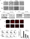
Figure 4. Golgi trafficking and PI3K involved in STF-62247 signaling pathway and acidification of vesicle after STF-62247 treatment
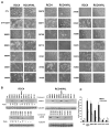
A. VHL-deficient RCC4 and wild-type VHL were incubated in presence of 10 μM LY294002, or 1 μg/ml Brefeldin A with 1.25 μM of STF-62247 for 20 hrs and the presence of vacuoles was examined under light microscopy. B,C. LC3 processing was evaluated by Western blot after the treatment with Brefeldin A or LY294002 in presence or absence of STF-62247 as described above. Tubulin was used as loading control. D. Cells were treated with 1.25 μM of STF-62247 with or without 2 mM of 3-MA, 1 μg/ml of Brefeldin A for 20 hrs. For the last 15 min, acidic components were stained with 1 μg/ml of acridine orange and visualized under fluorescence microscope. E. FACS analysis to measure the acidic vesicle organelles (AVO) in cells treated in D. y axis represents the concentrated dye in the acidic vesicles, whereas the cytoplasm and the nucleus showed green fluorescence in x axis. F. Quantification of the FACS analysis. The data are calculated as the average of three different experiments as relative units of treated over untreated cells. All error bars represent the SEM. Scale bar, 10 μm.
Figure 5. Evaluation of autophagy in RCC treated with analogs of the STF-62247
A. RCC4 and RCC4/VHL were treated with 1.25 μM of each 19 analogs for 20 hours and the presence of vacuoles was examined under light microscopy. Scale bar, 10 μm. B. Western blot for LC3 after treatment with each compound. Tubulin was used as loading control. Ratio is the relative from the IC50 in RCC4/VHL cells/IC50 in RCC4 cells. C. Acidic vesicle organelles (AVO) measured by FACS analysis in cells treated with 1.25 μM of STF-62247 or with compounds 30603, 30651, 31184, 31187 and 31270 and stained with acridine orange. All error bars represent SEM.
Figure 6. Golgi trafficking proteins are sensitive and a target for the STF-62247
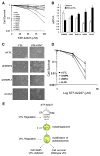
A. Killing curves for 8 deletion yeast strains. Wild-type, ALG5, APS1, CLA4, DRS2, OSH3, VPS20, VPS25, YPT31 were incubated with STF-62247 (0–100 μM) for 1 hour and colony formation was evaluated after two days. Each point of the survival is calculated by the average of two different experiments in triplicate and as the percent of treated on untreated plate. B. Quantitative real time PCR for CHMP6, PAK2, RAB11a, OSBPL3 and ALG5 in RCC4 and RCC4/VHL. Histogram is calculated by the average of three different experiments in triplicate as the percent of RCC4/VHL on RCC4 cells. The error bars are represented as SEM and *p<0.05, **p<0.01. C. VHL cells were transiently transfected with siRNA for CHMP6, OSBPL3 and ALG5 for 72 hours. The last 24 hours, 1.25 μM of STF-62247 was added and the vacuole formation was examined under light microscopy. Scale bar, 10 μm. D. Clonogenic assay with STF-62247 (0–30 μM) in RCC4/VHL cells transiently transfected in the same condition than in C. Colony formation was evaluated as described before. All error bars represent SEM. *p<0.05 represents the difference between VHL cells and VHL with siRNA in presence of 5 μM of STF-62247. E. Representative schema of the effect of STF-62247 in RCC through autophagy. STF-62247 disrupts ER-Golgi trafficking through CHMP6, OSBPL3 and ALG5 and induces autophagy. Lipidation of LC3 associated with the double membrane of autophagosome is observed in an ATG5-dependent manner. A higher acidification of the autolysosomes is associated with autophagic cell death in VHL-deficient cells whereas wild-type VHL cells are protected against acidification and survive. A regulation of CHMP6, OSBPL3 and ALG5 proteins by VHL is also observed and remains to be investigated.
Similar articles
- Targeted therapy for the loss of von Hippel-Lindau in renal cell carcinoma: a novel molecule that induces autophagic cell death.
Turcotte S, Sutphin PD, Giaccia AJ. Turcotte S, et al. Autophagy. 2008 Oct;4(7):944-6. doi: 10.4161/auto.6785. Epub 2008 Oct 13. Autophagy. 2008. PMID: 18769110 Free PMC article. - Targeting lysosome function causes selective cytotoxicity in VHL-inactivated renal cell carcinomas.
Bouhamdani N, Comeau D, Coholan A, Cormier K, Turcotte S. Bouhamdani N, et al. Carcinogenesis. 2020 Jul 10;41(6):828-840. doi: 10.1093/carcin/bgz161. Carcinogenesis. 2020. PMID: 31556451 Free PMC article. - Quantitative proteomics to study a small molecule targeting the loss of von Hippel-Lindau in renal cell carcinomas.
Bouhamdani N, Joy A, Barnett D, Cormier K, Léger D, Chute IC, Lamarre S, Ouellette R, Turcotte S. Bouhamdani N, et al. Int J Cancer. 2017 Aug 15;141(4):778-790. doi: 10.1002/ijc.30774. Epub 2017 May 25. Int J Cancer. 2017. PMID: 28486780 - MicroRNAs Associated with Von Hippel-Lindau Pathway in Renal Cell Carcinoma: A Comprehensive Review.
Schanza LM, Seles M, Stotz M, Fosselteder J, Hutterer GC, Pichler M, Stiegelbauer V. Schanza LM, et al. Int J Mol Sci. 2017 Nov 22;18(11):2495. doi: 10.3390/ijms18112495. Int J Mol Sci. 2017. PMID: 29165391 Free PMC article. Review. - Significance of PI3K signalling pathway in clear cell renal cell carcinoma in relation to VHL and HIF status.
Tumkur Sitaram R, Landström M, Roos G, Ljungberg B. Tumkur Sitaram R, et al. J Clin Pathol. 2021 Apr;74(4):216-222. doi: 10.1136/jclinpath-2020-206693. Epub 2020 May 28. J Clin Pathol. 2021. PMID: 32467322 Review.
Cited by
- VHL, the story of a tumour suppressor gene.
Gossage L, Eisen T, Maher ER. Gossage L, et al. Nat Rev Cancer. 2015 Jan;15(1):55-64. doi: 10.1038/nrc3844. Nat Rev Cancer. 2015. PMID: 25533676 Review. - Dietary downregulation of mutant p53 levels via glucose restriction: mechanisms and implications for tumor therapy.
Rodriguez OC, Choudhury S, Kolukula V, Vietsch EE, Catania J, Preet A, Reynoso K, Bargonetti J, Wellstein A, Albanese C, Avantaggiati ML. Rodriguez OC, et al. Cell Cycle. 2012 Dec 1;11(23):4436-46. doi: 10.4161/cc.22778. Epub 2012 Nov 14. Cell Cycle. 2012. PMID: 23151455 Free PMC article. - Loperamide, pimozide, and STF-62247 trigger autophagy-dependent cell death in glioblastoma cells.
Zielke S, Meyer N, Mari M, Abou-El-Ardat K, Reggiori F, van Wijk SJL, Kögel D, Fulda S. Zielke S, et al. Cell Death Dis. 2018 Sep 24;9(10):994. doi: 10.1038/s41419-018-1003-1. Cell Death Dis. 2018. PMID: 30250198 Free PMC article. - A novel sphingosine kinase inhibitor induces autophagy in tumor cells.
Beljanski V, Knaak C, Smith CD. Beljanski V, et al. J Pharmacol Exp Ther. 2010 May;333(2):454-64. doi: 10.1124/jpet.109.163337. Epub 2010 Feb 23. J Pharmacol Exp Ther. 2010. PMID: 20179157 Free PMC article. - Oxidative stress and autophagy in cardiac disease, neurological disorders, aging and cancer.
Essick EE, Sam F. Essick EE, et al. Oxid Med Cell Longev. 2010 May-Jun;3(3):168-77. doi: 10.4161/oxim.3.3.12106. Oxid Med Cell Longev. 2010. PMID: 20716941 Free PMC article. Review.
References
- Aita VM, Liang XH, Murty VV, Pincus DL, Yu W, Cayanis E, Kalachikov S, Gilliam TC, Levine B. Cloning and genomic organization of beclin 1, a candidate tumor suppressor gene on chromosome 17q21. Genomics. 1999;59:59–65. - PubMed
- An J, Fisher M, Rettig MB. VHL expression in renal cell carcinoma sensitizes to bortezomib (PS-341) through an NF-kappaB-dependent mechanism. Oncogene. 2005;24:1563–1570. - PubMed
- Bursch W, Ellinger A, Kienzl H, Torok L, Pandey S, Sikorska M, Walker R, Hermann RS. Active cell death induced by the anti-estrogens tamoxifen and ICI 164 384 in human mammary carcinoma cells (MCF-7) in culture: the role of autophagy. Carcinogenesis. 1996;17:1595–1607. - PubMed
- Chan DA, Sutphin PD, Denko NC, Giaccia AJ. Role of prolyl hydroxylation in oncogenically stabilized hypoxia-inducible factor-1alpha. J Biol Chem. 2002;277:40112–40117. - PubMed
Publication types
MeSH terms
Substances
Grants and funding
- NCI-CA-123823/CA/NCI NIH HHS/United States
- P01 CA082566/CA/NCI NIH HHS/United States
- P01 CA082566-070004/CA/NCI NIH HHS/United States
- NCI-CA-082566/CA/NCI NIH HHS/United States
- F32 CA123823/CA/NCI NIH HHS/United States
LinkOut - more resources
Full Text Sources
Other Literature Sources
Medical
Molecular Biology Databases
