The structure of an archaeal pilus - PubMed (original) (raw)
The structure of an archaeal pilus
Ying A Wang et al. J Mol Biol. 2008.
Abstract
Bacterial pili are involved in a host of activities, including motility, adhesion, transformation, and immune escape. Structural studies of these pili have shown that several distinctly different classes exist, with no common origin. Remarkably, it is now known that the archaeal flagellar filament appears to have a common origin with the bacterial type IV pilus, and assembly in both systems involves hydrophobic N-terminal alpha-helices that form three-stranded coils in the center of these filaments. Recent work has identified further genes in archaea as being similar to bacterial type IV pilins, but the function or structures formed by such gene products was unknown. Using electron cryo-microscopy, we show that an archaeal pilus from Methanococcus maripaludis has a structure entirely different from that of any of the known bacterial pili. Two subunit packing arrangements were identified: one has rings of four subunits spaced by approximately 44 A and the other has a one-start helical symmetry with approximately 2.6 subunits per turn of a approximately 30 A pitch helix. Remarkably, these schemes appear to coexist within the same filaments. For the segments composed of rings, the twist between adjacent rings is quite variable, while for the segments having a one-start helix there is a large variability in both the axial rise and the twist per subunit. Since this pilus appears to be assembled from a type IV pilin-like protein with a hydrophobic N-terminal helix, it provides yet another example of how different quaternary structures can be formed from similar building blocks. This result has many implications for understanding the evolutionary divergence of bacteria and archaea.
Figures
Figure 1. Electron micrographs and helical symmetry estimates
Electron micrograph of negatively stained (a) and frozen-hydrated archaeal pili (b). The space bar (a) is 250 Å. An averaged power spectrum (c), generated from 1,907 non-overlapping segments (each 300 pixels or 1,260 Å in length) of negatively-stained pili shows three layer lines (black arrows): 1/(17 Å), 1/(44 Å) and 1/(58 Å). The log of the intensities has been taken to reduce the dynamic range and allow all three layer lines to be visible simultaneously. Mass per unit length measurements yielded an average of 1,618.4 ± 8.8 (SEM) Da/ Å (d).
Figure 2. Sorting based on C4 symmetry and one-start helical symmetry
The 28,577 segments from the cryo-EM images were sorted into two groups by models with either C4 symmetry or one-start helical symmetry. 54% of the segments were sorted to have point group symmetry while the remaining 46 % were sorted to have a one-start helix. The averaged power spectrum for the segments sorted as having C4 symmetry (a) has a meridional layer line (red arrow) at 1/(44 Å). The averaged power spectrum for the segments sorted to have one-start helical symmetry (b) is much poorer, which suggests this group is more heterogeneous than the other group. The broad yet weak layer line at 1/(56 Å) (red arrow) can be interpreted as either n=2 or n=3, based upon the distance of the first peak from the meridian and the diameter of the filaments..
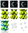
Figure 3. Sorting of the group with C4 symmetry
The 15,297 segments of the first class were sorted into five subgroups by differences in the angular rotation between adjacent rings of subunits. The averaged power spectra from the three most populated subgroups (a–c, left halves) are much improved compared to the averaged power spectrum before sorting (Fig. 2a), suggesting reduced heterogeneity in each subgroup. The meridional layer line is fixed while the n=+4 and n=−4 layer lines shift as expected in the three subgroups (a–c, red lines). The first subgroup was sorted to have a twist of ~ 64.0° and an axial rise of ~ 43.8 Å. The averaged power spectrum (a, left half) can be interpreted as having an n=0 layer line at 1/(44 Å), n=+4 layer line at 1/(60 Å) and n=−4 layer line at 1/(144 Å). The reconstruction of this subgroup (d) has a C4 symmetry with an axial rise of 43.9 Å and a twist of 65.0°. The second subgroup was sorted to have a twist of ~72.0° and an axial rise of ~43.8 Å. The averaged power spectrum (b, left half) can be interpreted as having an n=0 layer line at 1/(44 Å), n=+4 layer line at 1/(53 Å) and n=−4 at 1/(204 Å). The reconstruction of this subgroup (e) has a C4 symmetry with an axial rise of 43.8 Å and a twist of 72.0°. The third subgroup was sorted to have a twist of ~80.0° and an axial rise of ~43.8 Å. The averaged power spectrum (c, left half) can be interpreted as having an n=0 layer line at 1/(44 Å), n=+4 layer line at 1/(48 Å) and n=+3 at 1/(350 Å). The reconstruction of this subgroup (g) has a C4 symmetry with an axial rise of 43.5 Å and a twist of 80.1°. The power spectra from the projections of each reconstruction (a–c, right halves) match the corresponding averaged power spectrum of each subgroup (a–c, left halves). The reconstructions from the three subgroups are very similar, except for the difference in twist (d–g). The surface thresholds have been chosen to enclose 100% of the expected molecular volume. In all three reconstructions the main connectivity between asymmetric subunits is along a left-handed four-start helix (e, red line). A superposition of the two reconstructions (g) in which one subunit in each has been aligned (black arrow) shows a 15.1° difference in subunit twist at a subunit ~ 44 Å away (red arrow). The yellow reconstruction has a twist of 65.0° and the cyan one has a twist of 80.1°. Two cross-sectional contour plots (h,i) that are spaced 24 Å away from each other have different outer diameters and different size lumens.
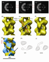
Figure 4. Sorting of the group with one-start helical symmetry
The 13,280 segments of the second group were sorted into nine subgroups by differences in the angular rotation and axial rise per subunit. Nine arbitrarily chosen symmetries, with three different angles (211°, 216°, and 221°) and three different axial rises (10.7, 11.7, and 12.7 A°) were used. The averaged power spectra of three representative subgroups (a–c, left halves) are much improved compared to the averaged global power spectrum before sorting (Fig. 2b), suggesting reduced heterogeneity in each subgroup. The layer lines shift in different subgroups (a–c, red lines) as expected. The first subgroup was sorted to have a twist of ~221.0° and an axial rise of ~10.7 Å. The averaged power spectrum (a, left half) can be interpreted as having an n=−2 layer line at 1/(47 Å) and n=+3 at 1/(66 Å). The reconstruction of this subgroup (d) has an axial rise of 10.8 Å and a twist of 220.9°. The second subgroup was sorted to have a twist of ~221.0° and an axial rise of ~11.7 Å. The averaged power spectrum (b, left half) can be interpreted as having an n=−2 layer line at 1/(50 Å) and n=+3 at 1/(72 Å). The reconstruction of this subgroup (e) has an axial rise of 11.7 Å and a twist of 221.1°. The third subgroup was sorted to have a twist of ~ 221.0° and an axial rise of ~ 12.7 Å. The averaged power spectrum (c, left half) can be interpreted as having an n=−2 layer line at 1/(56 Å) and n=+3 at 1/(82 Å). The reconstruction of this subgroup (g) has an axial rise of 13.0 Å and a twist of 221.1°. The power spectra from projections of each reconstruction (a–c, right halves) match the averaged power spectrum of each class (a–c, left halves). The surface threshold for each of the reconstructions has been chosen to enclose 100% of the expected molecular mass. The reconstructions from the three subgroups are very similar, except the difference in axial rise (d–g). In all the three reconstructions, the connectivity between subunits is along a right-handed two-start helix (e, cyan line) and a left-handed three-start helix (e, red line). A superposition (g) of two reconstructions (d,f) in which one subunit in each has been aligned (black arrow) shows a 13.2 Å difference in axial rise at a subunit ~ 70 Å away (red arrow). The yellow reconstruction has an axial rise of 10.8 Å and the cyan one has an axial rise of 13.0 Å. Two cross-sectional contour plots (h,i) spaced 9.6 Å away from each other show similar outer diameters and lumen size.
Figure 5. Comparison between the two subunit packing schemes
A superposition of the reconstructions with C4 symmetry and one-start helical symmetry (a) in which one subunit in each has been aligned (black arrow) shows the similarity of the structure of each single subunit but huge difference in subunit packing. The yellow surface has a one-start helical symmetry with a twist of 221.0° and an axial rise of 11.7 Å. (Fig. 3e), while the cyan surface has C4 symmetry with an axial rise of 43.8 Å and a twist of 72.0° (Fig. 4e). The aligned subunit in the cyan structure is shown as mesh for clarity. A helical net (b,c) shows the lattice of subunits on the surface of a cylinder, using the standard convention that the cylindrical surface has been cut open and we are looking at the inside of the surface. Two helical families are labeled in the helical net for the C4 helix (b). Within segments having this symmetry, the strongest observed connectivity between subunits is along the left-handed four-start helices. Three helical families are labeled in the helical net for the one-start helix (c). A left-handed one-start helix having ~ 2.6 subunits per 30 Å pitch turn is labeled. The strongest observed connectivity between subunits within segments having a one-start symmetry is along the left-handed three-start helices and the right-handed two-start helices.
Similar articles
- The structure of F-pili.
Wang YA, Yu X, Silverman PM, Harris RL, Egelman EH. Wang YA, et al. J Mol Biol. 2009 Jan 9;385(1):22-9. doi: 10.1016/j.jmb.2008.10.054. Epub 2008 Oct 25. J Mol Biol. 2009. PMID: 18992755 Free PMC article. - Two distinct archaeal type IV pili structures formed by proteins with identical sequence.
Liu J, Eastep GN, Cvirkaite-Krupovic V, Rich-New ST, Kreutzberger MAB, Egelman EH, Krupovic M, Wang F. Liu J, et al. Nat Commun. 2024 Jun 14;15(1):5049. doi: 10.1038/s41467-024-45062-z. Nat Commun. 2024. PMID: 38877064 Free PMC article. - Filaments from Ignicoccus hospitalis show diversity of packing in proteins containing N-terminal type IV pilin helices.
Yu X, Goforth C, Meyer C, Rachel R, Wirth R, Schröder GF, Egelman EH. Yu X, et al. J Mol Biol. 2012 Sep 14;422(2):274-81. doi: 10.1016/j.jmb.2012.05.031. Epub 2012 May 30. J Mol Biol. 2012. PMID: 22659006 Free PMC article. - Recent advances in the structure and assembly of the archaeal flagellum.
Bardy SL, Ng SY, Jarrell KF. Bardy SL, et al. J Mol Microbiol Biotechnol. 2004;7(1-2):41-51. doi: 10.1159/000077868. J Mol Microbiol Biotechnol. 2004. PMID: 15170402 Review. - Diversity, assembly and regulation of archaeal type IV pili-like and non-type-IV pili-like surface structures.
Lassak K, Ghosh A, Albers SV. Lassak K, et al. Res Microbiol. 2012 Nov-Dec;163(9-10):630-44. doi: 10.1016/j.resmic.2012.10.024. Epub 2012 Nov 9. Res Microbiol. 2012. PMID: 23146836 Review.
Cited by
- Diversity and subcellular distribution of archaeal secreted proteins.
Szabo Z, Pohlschroder M. Szabo Z, et al. Front Microbiol. 2012 Jul 2;3:207. doi: 10.3389/fmicb.2012.00207. eCollection 2012. Front Microbiol. 2012. PMID: 22783239 Free PMC article. - A helical processing pipeline for EM structure determination of membrane proteins.
Fisher LS, Ward A, Milligan RA, Unwin N, Potter CS, Carragher B. Fisher LS, et al. Methods. 2011 Dec;55(4):350-62. doi: 10.1016/j.ymeth.2011.09.013. Epub 2011 Sep 20. Methods. 2011. PMID: 21964395 Free PMC article. Review. - The Iho670 fibers of Ignicoccus hospitalis are anchored in the cell by a spherical structure located beneath the inner membrane.
Meyer C, Heimerl T, Wirth R, Klingl A, Rachel R. Meyer C, et al. J Bacteriol. 2014 Nov;196(21):3807-15. doi: 10.1128/JB.01861-14. Epub 2014 Aug 25. J Bacteriol. 2014. PMID: 25157085 Free PMC article. - Editorial.
Filloux A. Filloux A. FEMS Microbiol Rev. 2015 Jan;39(1):1. doi: 10.1093/femsre/fuu005. FEMS Microbiol Rev. 2015. PMID: 25793962 No abstract available. - Cell surface structures of archaea.
Ng SY, Zolghadr B, Driessen AJ, Albers SV, Jarrell KF. Ng SY, et al. J Bacteriol. 2008 Sep;190(18):6039-47. doi: 10.1128/JB.00546-08. Epub 2008 Jul 11. J Bacteriol. 2008. PMID: 18621894 Free PMC article. Review. No abstract available.
References
- Albers SV, Szabo Z, Driessen AJM. Protein secretion in the Archaea: multiple paths towards a unique cell surface. Nature Reviews Microbiology. 2006;4:537–547. - PubMed
- Alam M, Oesterhelt D. Morphology, Function and Isolation of Halobacterial Flagella. Journal of Molecular Biology. 1984;176:459–475. - PubMed
- Cohen-Krausz S, Trachtenberg S. The structure of the archeabacterial flagellar filament of the extreme halophile Halobacterium salinarum R1M1 and its relation to eubacterial flagellar filaments and type IV pili. Journal of Molecular Biology. 2002;321:383–395. - PubMed
- Cohen-Krausz S, Trachtenberg S. The flagellar filament structure of the extreme acidothermophile Sulfolobus shibatae B12 suggests that archaeabacterial flagella have a unique and common symmetry and design. Journal of Molecular Biology. 2008;375:1113–1124. - PubMed
- Gerl L, Sumper M. Halobacterial Flagellins Are Encoded by A Multigene Family - Characterization of 5 Flagellin Genes. Journal of Biological Chemistry. 1988;263:13246–13251. - PubMed
Publication types
MeSH terms
Substances
LinkOut - more resources
Full Text Sources
Other Literature Sources


