Isolation and biological analysis of tumor stem cells from pancreatic adenocarcinoma - PubMed (original) (raw)
Isolation and biological analysis of tumor stem cells from pancreatic adenocarcinoma
Peng Huang et al. World J Gastroenterol. 2008.
Abstract
Aim: To explore the method of isolation and biological analysis of tumor stem cells from pancreatic adenocarcinoma cell line PANC-1.
Methods: The PANC-1 cells were cultured in Dulbecco modified eagle medium F12 (1:1 volume) (DMEM-F12) supplemented with 20% fetal bovine serum (FBS). Subpopulation cells with properties of tumor stem cells were isolated from pancreatic adenocarcinoma cell line PANC-1 according to the cell surface markers CD44 and CD24 by flow cytometry. The proliferative capability of these cells in vitro were estimated by 3-[4,5-dimehyl-2-thiazolyl]-2, 5-diphenyl-2H-tetrazolium bromide (MTT) method. And the tumor growth of different subpopulation cells which were injected into the hypodermisof right and left armpit of nude mice was studied, and expression of CD44 and CD24 of the CD44(+)CD24(+) cell-formed nodules and PANC-1 cells were detected by avidin-biotin-peroxidase complex (ABC) immunohistochemical staining.
Results: The 5.1%-17.5% of sorted PANC-1 cells expressed the cell surface marker CD44, 57.8% -70.1% expressed CD24, only 2.1%-3.5% of cells were CD44(+) CD24(+). Compared with CD44(-)CD24(-) cells, CD44(+)CD24(+) cells had a lower growth rate in vitro. Implantation of 10(4) CD44(-)CD24(-) cells in nude mice showed no evident tumor growth at wk 12. In contrast, large tumors were found in nude mice implanted with 10(3) CD44(+)CD24(+) cells at wk 4 (2/8), a 20-fold increase in tumorigenic potential (P < 0.05 or P < 0.01). There was no obvious histological difference between the cells of the CD44(+)CD24(+) cell-formed nodules and PANC-1 cells.
Conclusion: CD44 and CD24 may be used as the cell surface markers for isolation of pancreatic cancer stem cells from pancreatic adenocarcinoma cell line PANC-1. Subpopulation cells CD44(+)CD24(+) have properties of tumor stem cells. Because cancer stem cells are thought to be responsible for tumor initiation and its recurrence after an initial response to chemotherapy, it may be a very promising target for new drug development.
Figures
Figure 1
Analysis of Panc-1 pancreatic cancer cells by FACS.
Figure 2
Growth curve of tumors cells in vitro.
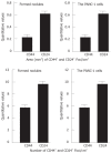
Figure 3
Quantitative values of CD44+ and CD24+ cell foci in the formed nodules and PANC-1 cells. There was no significant difference between the formed nodules and PANC-1 cells.
Similar articles
- Expression of CD44, CD24 and ESA in pancreatic adenocarcinoma cell lines varies with local microenvironment.
Wei HJ, Yin T, Zhu Z, Shi PF, Tian Y, Wang CY. Wei HJ, et al. Hepatobiliary Pancreat Dis Int. 2011 Aug;10(4):428-34. doi: 10.1016/s1499-3872(11)60073-8. Hepatobiliary Pancreat Dis Int. 2011. PMID: 21813394 - Identification of pancreatic cancer stem cells.
Li C, Heidt DG, Dalerba P, Burant CF, Zhang L, Adsay V, Wicha M, Clarke MF, Simeone DM. Li C, et al. Cancer Res. 2007 Feb 1;67(3):1030-7. doi: 10.1158/0008-5472.CAN-06-2030. Cancer Res. 2007. PMID: 17283135 - ALDH activity selectively defines an enhanced tumor-initiating cell population relative to CD133 expression in human pancreatic adenocarcinoma.
Kim MP, Fleming JB, Wang H, Abbruzzese JL, Choi W, Kopetz S, McConkey DJ, Evans DB, Gallick GE. Kim MP, et al. PLoS One. 2011;6(6):e20636. doi: 10.1371/journal.pone.0020636. Epub 2011 Jun 13. PLoS One. 2011. PMID: 21695188 Free PMC article. - Significance of CD44 and CD24 as cancer stem cell markers: an enduring ambiguity.
Jaggupilli A, Elkord E. Jaggupilli A, et al. Clin Dev Immunol. 2012;2012:708036. doi: 10.1155/2012/708036. Epub 2012 May 30. Clin Dev Immunol. 2012. PMID: 22693526 Free PMC article. Review. - More than markers: biological significance of cancer stem cell-defining molecules.
Keysar SB, Jimeno A. Keysar SB, et al. Mol Cancer Ther. 2010 Sep;9(9):2450-7. doi: 10.1158/1535-7163.MCT-10-0530. Epub 2010 Aug 17. Mol Cancer Ther. 2010. PMID: 20716638 Free PMC article. Review.
Cited by
- CD44/CD24 immunophenotypes on clinicopathologic features of salivary glands malignant neoplasms.
Soave DF, Oliveira da Costa JP, da Silveira GG, Ianez RC, de Oliveira LR, Lourenço SV, Ribeiro-Silva A. Soave DF, et al. Diagn Pathol. 2013 Feb 18;8:29. doi: 10.1186/1746-1596-8-29. Diagn Pathol. 2013. PMID: 23419168 Free PMC article. - MiR-200a inhibits epithelial-mesenchymal transition of pancreatic cancer stem cell.
Lu Y, Lu J, Li X, Zhu H, Fan X, Zhu S, Wang Y, Guo Q, Wang L, Huang Y, Zhu M, Wang Z. Lu Y, et al. BMC Cancer. 2014 Feb 12;14:85. doi: 10.1186/1471-2407-14-85. BMC Cancer. 2014. PMID: 24521357 Free PMC article. - Cancer stem cells: involvement in pancreatic cancer pathogenesis and perspectives on cancer therapeutics.
Tanase CP, Neagu AI, Necula LG, Mambet C, Enciu AM, Calenic B, Cruceru ML, Albulescu R. Tanase CP, et al. World J Gastroenterol. 2014 Aug 21;20(31):10790-801. doi: 10.3748/wjg.v20.i31.10790. World J Gastroenterol. 2014. PMID: 25152582 Free PMC article. Review. - Stem Cells in the Exocrine Pancreas during Homeostasis, Injury, and Cancer.
Lodestijn SC, van Neerven SM, Vermeulen L, Bijlsma MF. Lodestijn SC, et al. Cancers (Basel). 2021 Jun 30;13(13):3295. doi: 10.3390/cancers13133295. Cancers (Basel). 2021. PMID: 34209288 Free PMC article. Review. - Fzd7/Wnt7b signaling contributes to stemness and chemoresistance in pancreatic cancer.
Zhang Z, Xu Y, Zhao C. Zhang Z, et al. Cancer Med. 2021 May;10(10):3332-3345. doi: 10.1002/cam4.3819. Epub 2021 May 2. Cancer Med. 2021. PMID: 33934523 Free PMC article.
References
- Bardeesy N, DePinho RA. Pancreatic cancer biology and genetics. Nat Rev Cancer. 2002;2:897–909. - PubMed
- Murphy SL. Deaths: final data for 1998. Natl Vital Stat Rep. 2000;48:1–105. - PubMed
- Cameron JL, Crist DW, Sitzmann JV, Hruban RH, Boitnott JK, Seidler AJ, Coleman J. Factors influencing survival after pancreaticoduodenectomy for pancreatic cancer. Am J Surg. 1991;161:120–124; discussion 124-125. - PubMed
- Niederhuber JE, Brennan MF, Menck HR. The National Cancer Data Base report on pancreatic cancer. Cancer. 1995;76:1671–1677. - PubMed
- Jemal A, Thomas A, Murray T, Thun M. Cancer statistics, 2002. CA Cancer J Clin. 2002;52:23–47. - PubMed
Publication types
MeSH terms
Substances
LinkOut - more resources
Full Text Sources
Medical
Miscellaneous


