The angio-fibrotic switch of VEGF and CTGF in proliferative diabetic retinopathy - PubMed (original) (raw)
The angio-fibrotic switch of VEGF and CTGF in proliferative diabetic retinopathy
Esther J Kuiper et al. PLoS One. 2008.
Abstract
Background: In proliferative diabetic retinopathy (PDR), vascular endothelial growth factor (VEGF) and connective tissue growth factor (CTGF) cause blindness by neovascularization and subsequent fibrosis, but their relative contribution to both processes is unknown. We hypothesize that the balance between levels of pro-angiogenic VEGF and pro-fibrotic CTGF regulates angiogenesis, the angio-fibrotic switch, and the resulting fibrosis and scarring.
Methods/principal findings: VEGF and CTGF were measured by ELISA in 68 vitreous samples of patients with proliferative DR (PDR, N = 32), macular hole (N = 13) or macular pucker (N = 23) and were related to clinical data, including degree of intra-ocular neovascularization and fibrosis. In addition, clinical cases of PDR (n = 4) were studied before and after pan-retinal photocoagulation and intra-vitreal injections with bevacizumab, an antibody against VEGF. Neovascularization and fibrosis in various degrees occurred almost exclusively in PDR patients. In PDR patients, vitreous CTGF levels were significantly associated with degree of fibrosis and with VEGF levels, but not with neovascularization, whereas VEGF levels were associated only with neovascularization. The ratio of CTGF and VEGF was the strongest predictor of degree of fibrosis. As predicted by these findings, patients with PDR demonstrated a temporary increase in intra-ocular fibrosis after anti-VEGF treatment or laser treatment.
Conclusions/significance: CTGF is primarily a pro-fibrotic factor in the eye, and a shift in the balance between CTGF and VEGF is associated with the switch from angiogenesis to fibrosis in proliferative retinopathy.
Conflict of interest statement
Competing Interests: Noelynn Oliver is an employee of Fibrogen Inc, Roel Goldschmeding has received research support grants from Fibrogen Inc.
Figures
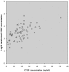
Figure 1. Correlation between the levels of CTGF and log10(VEGF) in the vitreous of all 68 patients.
A significant (p = 0.01) Spearman's rank correlation (ρ = 0.4) within all samples was found.
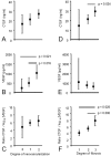
Figure 2. Mean levels of CTFG (A, D), geometric mean levels of VEGF (B, E), and mean ratio CTGF/log10(VEGF) (C, F) in relation with degree of neovascularization (A–C) and degree of fibrosis (D–F) in the vitreous of 32 PDR patients.
Vertical bars represent 95% confidence intervals. Significant differences between groups are indicated.
Figure 3. Fundus photographs of a patient with proliferative diabetic retinopathy and new vessels (nv) along the lower vascular arcade, before (A) and 8 months after (B) an injection with bevacizumab followed by pan-retinal photocoagulation.
Note the increase in fibrosis (f) after anti-VEGF and laser treatment (B).
Figure 4. Fundus photographs (A, D) and fluorescein angiographic imaging (B, C) of a patient with branch retinal vein occlusion before (A–C) and after (D) treatment with pan-retinal photocoagulation.
Note the leaky vessels consistent with angiogenesis(A, C), and the quiet aspect of the vessels after treatment without formation of fibrosis (D). n, normal; nv, neovascularization.
Similar articles
- A shift in the balance of vascular endothelial growth factor and connective tissue growth factor by bevacizumab causes the angiofibrotic switch in proliferative diabetic retinopathy.
Van Geest RJ, Lesnik-Oberstein SY, Tan HS, Mura M, Goldschmeding R, Van Noorden CJ, Klaassen I, Schlingemann RO. Van Geest RJ, et al. Br J Ophthalmol. 2012 Apr;96(4):587-90. doi: 10.1136/bjophthalmol-2011-301005. Epub 2012 Jan 29. Br J Ophthalmol. 2012. PMID: 22289291 Free PMC article. - Vitreous TIMP-1 levels associate with neovascularization and TGF-β2 levels but not with fibrosis in the clinical course of proliferative diabetic retinopathy.
Van Geest RJ, Klaassen I, Lesnik-Oberstein SY, Tan HS, Mura M, Goldschmeding R, Van Noorden CJ, Schlingemann RO. Van Geest RJ, et al. J Cell Commun Signal. 2013 Mar;7(1):1-9. doi: 10.1007/s12079-012-0178-y. Epub 2012 Oct 2. J Cell Commun Signal. 2013. PMID: 23054594 Free PMC article. - The role of CTGF in diabetic retinopathy.
Klaassen I, van Geest RJ, Kuiper EJ, van Noorden CJ, Schlingemann RO. Klaassen I, et al. Exp Eye Res. 2015 Apr;133:37-48. doi: 10.1016/j.exer.2014.10.016. Exp Eye Res. 2015. PMID: 25819453 Review. - Accumulation of NH2-terminal fragment of connective tissue growth factor in the vitreous of patients with proliferative diabetic retinopathy.
Hinton DR, Spee C, He S, Weitz S, Usinger W, LaBree L, Oliver N, Lim JI. Hinton DR, et al. Diabetes Care. 2004 Mar;27(3):758-64. doi: 10.2337/diacare.27.3.758. Diabetes Care. 2004. PMID: 14988298 - Pan-retinal photocoagulation and other forms of laser treatment and drug therapies for non-proliferative diabetic retinopathy: systematic review and economic evaluation.
Royle P, Mistry H, Auguste P, Shyangdan D, Freeman K, Lois N, Waugh N. Royle P, et al. Health Technol Assess. 2015 Jul;19(51):v-xxviii, 1-247. doi: 10.3310/hta19510. Health Technol Assess. 2015. PMID: 26173799 Free PMC article. Review.
Cited by
- Mediators of ocular angiogenesis.
Qazi Y, Maddula S, Ambati BK. Qazi Y, et al. J Genet. 2009 Dec;88(4):495-515. doi: 10.1007/s12041-009-0068-0. J Genet. 2009. PMID: 20090210 Free PMC article. Review. - Trial by CCN2: a standardized test for fibroproliferative disease?
Leask A. Leask A. J Cell Commun Signal. 2009 Mar;3(1):87-8. doi: 10.1007/s12079-009-0041-y. Epub 2009 Mar 7. J Cell Commun Signal. 2009. PMID: 19266315 Free PMC article. - APOPTOSIS AND ANGIOFIBROSIS IN DIABETIC TRACTIONAL MEMBRANES AFTER VASCULAR ENDOTHELIAL GROWTH FACTOR INHIBITION: Results of a Prospective Trial. Report No. 2.
Jiao C, Eliott D, Spee C, He S, Wang K, Mullins RF, Hinton DR, Sohn EH. Jiao C, et al. Retina. 2019 Feb;39(2):265-273. doi: 10.1097/IAE.0000000000001952. Retina. 2019. PMID: 29190236 Free PMC article. Clinical Trial. - Involvement of Müller Glial Autoinduction of TGF-β in Diabetic Fibrovascular Proliferation Via Glial-Mesenchymal Transition.
Wu D, Kanda A, Liu Y, Noda K, Murata M, Ishida S. Wu D, et al. Invest Ophthalmol Vis Sci. 2020 Dec 1;61(14):29. doi: 10.1167/iovs.61.14.29. Invest Ophthalmol Vis Sci. 2020. PMID: 33369638 Free PMC article. - Recombinant thrombomodulin domain 1 rescues pathological angiogenesis by inhibition of HIF-1α-VEGF pathway.
Huang YH, Kuo CH, Peng IC, Chang YS, Tseng SH, Conway EM, Wu HL. Huang YH, et al. Cell Mol Life Sci. 2021 Dec;78(23):7681-7692. doi: 10.1007/s00018-021-03950-3. Epub 2021 Oct 27. Cell Mol Life Sci. 2021. PMID: 34705054 Free PMC article.
References
- Aiello LP, Gardner TW, King GL, Blankenship G, Cavallerano JD, et al. Diabetic retinopathy. Diabetes Care. 1998;21:143–156. - PubMed
- Campochiaro PA. Pathogenesis of proliferative vitreoretinopathy. In: Ryan SJJ, editor. Retina. 3 ed. St. Louis: Mosby; 2001. pp. 2221–2227.
- Fong DS, Aiello L, Gardner TW, King GL, Blankenship G, et al. Diabetic retinopathy. Diabetes Care. 2003;26:226–229. - PubMed
- Fong DS, Aiello LP, Ferris FL 3rd, Klein R. Diabetic retinopathy. Diabetes Care. 2004;27:2540–2553. - PubMed
- Hinton DR, He S, Jin ML, Barron E, Ryan SJ. Novel growth factors involved in the pathogenesis of proliferative vitreoretinopathy. Eye. 2002;16:422–428. - PubMed
Publication types
MeSH terms
Substances
LinkOut - more resources
Full Text Sources
Other Literature Sources
Medical
Research Materials
Miscellaneous


