Widespread spatial integration in primary somatosensory cortex - PubMed (original) (raw)
Widespread spatial integration in primary somatosensory cortex
Jamie L Reed et al. Proc Natl Acad Sci U S A. 2008.
Abstract
Tactile discrimination depends on integration of information from the discrete receptive fields (RFs) of peripheral sensory afferents. Because this information is processed over a hierarchy of subcortical nuclei and cortical areas, the integration likely occurs at multiple levels. The current study presents results indicating that neurons across most of the extent of the hand representation in monkey primary somatosensory cortex (area 3b) interact, even when these neurons have separate RFs. We obtained simultaneous recordings by using a 100-electrode array implanted in the hand representation of primary somatosensory cortex of two anesthetized owl monkeys. During a series of 0.5-s skin indentations with single or dual probes, the distance between electrodes from which neurons with synchronized spike times were recorded exceeded 2 mm. The results provide evidence that stimuli on different parts of the hand influence the degree of synchronous firing among a large population of neurons. Because spike synchrony potentiates the activation of commonly targeted neurons, synchronous neural activity in primary somatosensory cortex can contribute to discrimination of complex tactile stimuli.
Conflict of interest statement
The authors declare no conflict of interest.
Figures
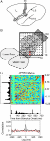
Fig. 1.
An example of correlated spike activity recorded from two adjacent electrodes in monkey 1. (A) A lateral schematic view of an owl monkey brain with area 3b shaded. Subdivisions representing the face and hand are outlined, including those that cannot be seen from a surface view. LS, lateral sulcus; STS, superior temporal sulcus. (B) The location of the array within the area 3b hand representation. The approximate representations of the digits (D1–5) and the digital (P1–3), thenar (PTh), hypothenar (PH) and insular (Pi) pads of the hand are identified and outlined. The red dots mark the electrodes where the two neurons were recorded for the analysis shown in C. Both neurons had RFs on the PTh pad. (C) An example of the spike timing synchrony of units recorded from adjacent electrodes. Two probes simultaneously indented the skin on the PTh and P1 pads. Spike synchrony between the two neurons is shown in the normalized joint peristimulus time histogram, the JPSTH matrix. The two PSTHs of the responses to 100 repetitions of 0.5-s skin indentations are shown to the left and below the matrix, with the cross-correlation histogram derived from the JPSTH analysis directly below. The colored pixels in the JPSTH matrix represent the magnitude of the normalized correlation at different lag times over a poststimulus time of 700 ms. Strong spike synchrony occurred around a 0-ms lag time throughout the period. The cross-correlation histogram (black) revealed a peak correlation of 0.16 that exceeded the mean correlation from the shuffled trials (red).
Fig. 2.
An example of widespread spike timing correlations and firing activity. Significant peak correlations across the sampled neuron-units in the 100-electrode array are displayed in a grid. A color map representing the peak firing rates of the units is overlaid on a schematic of the area 3b hand representation to indicate the approximate spatial locations of the electrodes. Shown is one example from monkey 1 when a single site on the thenar (PTh) palm was stimulated repeatedly (100 trials). Dots indicate electrode sites and significant correlations between units are represented by the lines connecting the dots. The size of the dots and the thickness of the connecting lines are visual representations of the peak magnitude of the correlation. Each colored box represents the peak firing rate of a unit at one electrode site. Peak firing rates are shown for those units included in the synchrony analysis. Dark blue squares indicate electrodes not analyzed for spike synchrony because units did not show sustained responses to stimulation.
Fig. 3.
Relationship of spike timing peak correlation magnitude to the distance between electrodes. The normalized, significant peak correlation magnitudes are plotted as a function of distance between the correlated unit pairs for monkeys 1 and 2 when nonadjacent sites were simultaneously stimulated (Dual-Site) or when single sites were stimulated as controls (Single-Site).
Similar articles
- Modular processing in the hand representation of primate primary somatosensory cortex coexists with widespread activation.
Reed JL, Qi HX, Pouget P, Burish MJ, Bonds AB, Kaas JH. Reed JL, et al. J Neurophysiol. 2010 Dec;104(6):3136-45. doi: 10.1152/jn.00566.2010. Epub 2010 Oct 6. J Neurophysiol. 2010. PMID: 20926605 Free PMC article. - Effects of spatiotemporal stimulus properties on spike timing correlations in owl monkey primary somatosensory cortex.
Reed JL, Pouget P, Qi HX, Zhou Z, Bernard MR, Burish MJ, Kaas JH. Reed JL, et al. J Neurophysiol. 2012 Dec;108(12):3353-69. doi: 10.1152/jn.00414.2011. Epub 2012 Sep 26. J Neurophysiol. 2012. PMID: 23019003 Free PMC article. - Response properties of neurons in primary somatosensory cortex of owl monkeys reflect widespread spatiotemporal integration.
Reed JL, Qi HX, Zhou Z, Bernard MR, Burish MJ, Bonds AB, Kaas JH. Reed JL, et al. J Neurophysiol. 2010 Apr;103(4):2139-57. doi: 10.1152/jn.00709.2009. Epub 2010 Feb 17. J Neurophysiol. 2010. PMID: 20164400 Free PMC article. - Multielectrode Recordings in the Somatosensory System.
Wiest M, Thomson E, Meloy J. Wiest M, et al. In: Nicolelis MAL, editor. Methods for Neural Ensemble Recordings. 2nd edition. Boca Raton (FL): CRC Press/Taylor & Francis; 2008. Chapter 6. In: Nicolelis MAL, editor. Methods for Neural Ensemble Recordings. 2nd edition. Boca Raton (FL): CRC Press/Taylor & Francis; 2008. Chapter 6. PMID: 21204443 Free Books & Documents. Review. - Sensory neuroscience: from skin to object in the somatosensory cortex.
Haggard P. Haggard P. Curr Biol. 2006 Oct 24;16(20):R884-6. doi: 10.1016/j.cub.2006.09.024. Curr Biol. 2006. PMID: 17055972 Review.
Cited by
- Intrinsic horizontal connections process global tactile features in the primary somatosensory cortex: neuroanatomical evidence.
Négyessy L, Pálfi E, Ashaber M, Palmer C, Jákli B, Friedman RM, Chen LM, Roe AW. Négyessy L, et al. J Comp Neurol. 2013 Aug 15;521(12):2798-817. doi: 10.1002/cne.23317. J Comp Neurol. 2013. PMID: 23436325 Free PMC article. - The relationship of anatomical and functional connectivity to resting-state connectivity in primate somatosensory cortex.
Wang Z, Chen LM, Négyessy L, Friedman RM, Mishra A, Gore JC, Roe AW. Wang Z, et al. Neuron. 2013 Jun 19;78(6):1116-26. doi: 10.1016/j.neuron.2013.04.023. Neuron. 2013. PMID: 23791200 Free PMC article. - Cortical topography of intracortical inhibition influences the speed of decision making.
Wilimzig C, Ragert P, Dinse HR. Wilimzig C, et al. Proc Natl Acad Sci U S A. 2012 Feb 21;109(8):3107-12. doi: 10.1073/pnas.1114250109. Epub 2012 Feb 6. Proc Natl Acad Sci U S A. 2012. PMID: 22315409 Free PMC article. - Differential Recovery of Submodality Touch Neurons and Interareal Communication in Sensory Input-Deprived Area 3b and S2 Cortices.
Wu R, Yang PF, Wang F, Liu Q, Gore JC, Chen LM. Wu R, et al. J Neurosci. 2022 Dec 14;42(50):9330-9342. doi: 10.1523/JNEUROSCI.0034-22.2022. Epub 2022 Nov 15. J Neurosci. 2022. PMID: 36379707 Free PMC article. - Peripheral and central changes combine to induce motor behavioral deficits in a moderate repetition task.
Coq JO, Barr AE, Strata F, Russier M, Kietrys DM, Merzenich MM, Byl NN, Barbe MF. Coq JO, et al. Exp Neurol. 2009 Dec;220(2):234-45. doi: 10.1016/j.expneurol.2009.08.008. Epub 2009 Aug 15. Exp Neurol. 2009. PMID: 19686738 Free PMC article.
References
- Niebur E, Hsiao SS, Johnson KO. Synchrony: A neuronal mechanism for attentional selection? Curr Opin Neurobiol. 2002;12:190–194. - PubMed
- Roy A, Steinmetz PN, Hsiao SS, Johnson KO, Niebur E. Synchrony: A neural correlate of somatosensory attention. J Neurophysiol. 2007;98:1645–1661. - PubMed
- Hsiao SS, O'Shaughnessy DM, Johnson KO. Effects of selective attention on spatial form processing in monkey primary and secondary somatosensory cortex. J Neurophysiol. 1993;70:444–447. - PubMed
Publication types
MeSH terms
Grants and funding
- R01 EY014680/EY/NEI NIH HHS/United States
- P30-EY08126/EY/NEI NIH HHS/United States
- EY014680-03/EY/NEI NIH HHS/United States
- F31-NS053231/NS/NINDS NIH HHS/United States
- F31 NS053231/NS/NINDS NIH HHS/United States
- NS16446/NS/NINDS NIH HHS/United States
- T32-GM07347/GM/NIGMS NIH HHS/United States
- P30 HD015052/HD/NICHD NIH HHS/United States
- R01 NS016446/NS/NINDS NIH HHS/United States
- P30 EY008126/EY/NEI NIH HHS/United States
- T32 GM007347/GM/NIGMS NIH HHS/United States
- P30-HD015052/HD/NICHD NIH HHS/United States
LinkOut - more resources
Full Text Sources
Other Literature Sources


