Differential expression of IRF8 in subsets of macrophages and dendritic cells and effects of IRF8 deficiency on splenic B cell and macrophage compartments - PubMed (original) (raw)
Differential expression of IRF8 in subsets of macrophages and dendritic cells and effects of IRF8 deficiency on splenic B cell and macrophage compartments
Chen-Feng Qi et al. Immunol Res. 2009.
Abstract
IRF8, a transcription factor restricted primarily to hematopoietic cells, is known to influence the differentiation and function of dendritic cells (DC), macrophages, granulocytes and B cells. In human tonsil, IRF8 is expressed at high levels by intrafollicular macrophages and DC, but at much lower levels by tingible body macrophages in germinal centers (GCs) and little, if at all, by follicular DC. Spleens of IRF8-deficient mice had reduced numbers of white pulp follicles and GCs that were irregular in shape. The frequency of follicular B cells was significantly reduced while the population of marginal zone (MZ) B cells was increased. In addition, MZ macrophages were reduced in number and abnormally distributed, while metallophilic macrophages were normal. These findings demonstrate differential requirements for IRF8 among distinct subsets of B cells, DC, and macrophages.
Figures
Fig. 1
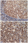
Anti-IRF8 IHC staining of Human tonsil samples. Large arrows show tangible body macrophages, small arrows show FDCs
Fig. 2
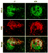
Confocal microscopic analyses of GCs from IRF8+/+(+/+) and IRF8−/− (−/−) spleens for expression of BCL6 and FDC-M1. The merged images are shown at the bottom
Fig. 3
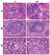
H&E staining of spleens from IRF8+/+(+/+) and IRF8−/− (−/−) mice. Original magnifications are indicated to the left. Germinal centers are circled in the lower four panels and arrows point to apoptotic bodies localized outside germinal centers
Fig. 4
Confocal microscopic analyses of normal spleen co-stained with antibodies to anti-IRF8 and FDC-M1. The merged images are shown at the bottom
Fig. 5
FASC analyses of spleen cells from IRF8+/+(+/+) and IRF8−/− (−/−) mice. The left panels, gated to show lymphocytes, show results obtained with antibodies to IgM and B220 and the gates used for characterization of B cells for expression of CD21 and CD23 shown in the right panels. Numbers in the right panel indicate the percentages of total B cells. In the right panels, the gates for follicular (FO) B cells (CD23hiCD21lo) and marginal zone (MZ) B cells (CD23lo/−CD21hi) are indicated together with the percentages of cells falling in the gates. Data are representative of five or more independent experiments
Fig. 6
Confocal microscopic analyses of GCs from IRF8+/+(+/+) and IRF8−/− (−/−) spleens for expression of the indicated pairs of cell surface markers
Fig. 7
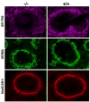
Confocal microscopic studies of GCs from IRF8+/+(+/+) and IRF8−/− (−/−) mice for expression of ERTR-9 on MZ macrophages, MOMA-1 on metallophilic macrophages, and MadCAM-1 on marginal sinus endothelial cells and activated FDCs
Similar articles
- IFN regulatory factor 8 restricts the size of the marginal zone and follicular B cell pools.
Feng J, Wang H, Shin DM, Masiuk M, Qi CF, Morse HC 3rd. Feng J, et al. J Immunol. 2011 Feb 1;186(3):1458-66. doi: 10.4049/jimmunol.1001950. Epub 2010 Dec 22. J Immunol. 2011. PMID: 21178004 Free PMC article. - Essential role of RelB in germinal center and marginal zone formation and proper expression of homing chemokines.
Weih DS, Yilmaz ZB, Weih F. Weih DS, et al. J Immunol. 2001 Aug 15;167(4):1909-19. doi: 10.4049/jimmunol.167.4.1909. J Immunol. 2001. PMID: 11489970 - Regulation of the germinal center gene program by interferon (IFN) regulatory factor 8/IFN consensus sequence-binding protein.
Lee CH, Melchers M, Wang H, Torrey TA, Slota R, Qi CF, Kim JY, Lugar P, Kong HJ, Farrington L, van der Zouwen B, Zhou JX, Lougaris V, Lipsky PE, Grammer AC, Morse HC 3rd. Lee CH, et al. J Exp Med. 2006 Jan 23;203(1):63-72. doi: 10.1084/jem.20051450. Epub 2005 Dec 27. J Exp Med. 2006. PMID: 16380510 Free PMC article. - IRF8 regulates myeloid and B lymphoid lineage diversification.
Wang H, Morse HC 3rd. Wang H, et al. Immunol Res. 2009;43(1-3):109-17. doi: 10.1007/s12026-008-8055-8. Immunol Res. 2009. PMID: 18806934 Free PMC article. Review. - Immune regulation by Fcα/μ receptor (CD351) on marginal zone B cells and follicular dendritic cells.
Shibuya A, Honda S. Shibuya A, et al. Immunol Rev. 2015 Nov;268(1):288-95. doi: 10.1111/imr.12345. Immunol Rev. 2015. PMID: 26497528 Review.
Cited by
- Molecular mapping of a core transcriptional signature of microglia-specific genes in schizophrenia.
Fiorito AM, Fakra E, Sescousse G, Ibrahim EC, Rey R. Fiorito AM, et al. Transl Psychiatry. 2023 Dec 13;13(1):386. doi: 10.1038/s41398-023-02677-y. Transl Psychiatry. 2023. PMID: 38092734 Free PMC article. - IRF8: Mechanism of Action and Health Implications.
Moorman HR, Reategui Y, Poschel DB, Liu K. Moorman HR, et al. Cells. 2022 Aug 24;11(17):2630. doi: 10.3390/cells11172630. Cells. 2022. PMID: 36078039 Free PMC article. Review. - Tagging single nucleotide polymorphisms in the IRF1 and IRF8 genes and tuberculosis susceptibility.
Ding S, Jiang T, He J, Qin B, Lin S, Li L. Ding S, et al. PLoS One. 2012;7(8):e42104. doi: 10.1371/journal.pone.0042104. Epub 2012 Aug 6. PLoS One. 2012. PMID: 22879909 Free PMC article. - IFN regulatory factor 8 restricts the size of the marginal zone and follicular B cell pools.
Feng J, Wang H, Shin DM, Masiuk M, Qi CF, Morse HC 3rd. Feng J, et al. J Immunol. 2011 Feb 1;186(3):1458-66. doi: 10.4049/jimmunol.1001950. Epub 2010 Dec 22. J Immunol. 2011. PMID: 21178004 Free PMC article. - Innate immune regulation by STAT-mediated transcriptional mechanisms.
Li HS, Watowich SS. Li HS, et al. Immunol Rev. 2014 Sep;261(1):84-101. doi: 10.1111/imr.12198. Immunol Rev. 2014. PMID: 25123278 Free PMC article. Review.
References
- Cozine CL, Wolniak KL, Waldschmidt TJ. The primary germinal center response in mice. Curr Opin Immunol. 2005;17:298–302. - PubMed
- Martin F, Kearney JF. Marginal-zone B cells. Nat Rev Immunol. 2002;2:323–35. - PubMed
- Mebius RE, Nolte MA, Kraal G. Development and function of the splenic marginal zone. Crit Rev Immunol. 2004;24:449–64. - PubMed
- Schwickert TA, Lindquist RL, Shakhar G, Livshits G, Skokos D, Kosco-Vilbois MH, et al. In vivo imaging of germinal centres reveals a dynamic open structure. Nature. 2007;446:83–7. - PubMed
- Allen CD, Okada T, Tang HL, Cyster JG. Imaging of germinal center selection events during affinity maturation. Science. 2007;315:528–31. - PubMed
Publication types
MeSH terms
Substances
LinkOut - more resources
Full Text Sources


