Frontal cortex mediates unconsciously triggered inhibitory control - PubMed (original) (raw)
Frontal cortex mediates unconsciously triggered inhibitory control
Simon van Gaal et al. J Neurosci. 2008.
Abstract
To further our understanding of the function of conscious experience we need to know which cognitive processes require awareness and which do not. Here, we show that an unconscious stimulus can trigger inhibitory control processes, commonly ascribed to conscious control mechanisms. We combined the metacontrast masking paradigm and the Go/No-Go paradigm to study whether unconscious No-Go signals can actively trigger high-level inhibitory control processes, strongly associated with the prefrontal cortex (PFC). Behaviorally, unconscious No-Go signals sometimes triggered response inhibition to the level of complete response termination and yielded a slow down in the speed of responses that were not inhibited. Electroencephalographic recordings showed that unconscious No-Go signals elicit two neural events: (1) an early occipital event and (2) a frontocentral event somewhat later in time. The first neural event represents the visual encoding of the unconscious No-Go stimulus, and is also present in a control experiment where the masked stimulus has no behavioral relevance. The second event is unique to the Go/No-Go experiment, and shows the subsequent implementation of inhibitory control in the PFC. The size of the frontal activity pattern correlated highly with the impact of unconscious No-Go signals on subsequent behavior. We conclude that unconscious stimuli can influence whether a task will be performed or interrupted, and thus exert a form of cognitive control. These findings challenge traditional views concerning the proposed relationship between awareness and cognitive control and stretch the alleged limits and depth of unconscious information processing.
Figures
Figure 1.
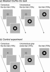
Stimuli and trial timing of the masked Go/No-Go task and the control experiment. The gray circle and black cross duration was 16.7 ms. Go signal duration was 100 ms. In conscious No-Go trials, the SOA between the No-Go signal and the Go signal was 83 ms. Participants had to respond to the Go signal (black metacontrast mask) but were instructed to withhold their response when a No-Go signal preceded the Go signal. In the masked Go/No-Go task, a gray circle served as a No-Go signal, whereas in the control experiment, the No-Go signal was a black cross. Therefore, the masked gray circle was associated with inhibition in the masked Go/No-Go task and thus served as an unconscious No-Go signal. In the control experiment, the unconscious gray circle was not associated with inhibition (and was task irrelevant) because participants were instructed to inhibit their responses on a black cross. Comparing processing of unconscious gray circles between both experiments enabled us to test whether (1) high-level inhibitory control processes can be triggered unconsciously, (2) unconscious No-Go signals reach prefrontal areas, and (3) task relevance influences the depth of processing of unconscious stimuli.
Figure 2.
Behavioral measures of unconsciously triggered response inhibition. In the masked Go/No-Go task, participants inhibited their responses more often on unconscious No-Go trials than on Go trials across all sessions (left; effect sizes: percentage of inhibited unconscious No-Go trials minus the percentage of inhibited Go trials). In the control experiment, participants did not inhibit their responses more often on unconscious gray circle trials than on Go trials. Additionally, the Fehrer–Raab effect (right) was significantly smaller in the masked Go/No-Go task (mean RT on unconscious No-Go trials minus mean RT on Go trials) than in the control experiment (mean RT on unconscious gray circle trials minus mean RT on Go trials). This finding supports the notion that unconscious No-Go signals triggered inhibitory control processes in the masked Go/No-Go task, whereas in the control experiment, unconscious gray circles did not (or less so).
Figure 3.
Typical ERP reported in the standard version of the Go/No-Go task. The average ERP at electrode FCz (for the masked Go/No-Go task) is depicted for responded Go trials as well as for conscious No-Go trials that were successfully inhibited (time locked to the onset of the Go signal). Scalp voltage maps show a characteristic frontocentral distribution of the N2 component and a more centroparietal distribution of the P3 component for successfully inhibited (conscious) No-Go trials. The vertical gray bars are an indication of the area that was selected for the computation of the voltage maps.
Figure 4.
The neural processing of unconscious gray circles. Scalp voltage maps show activations evoked by the unconscious stimulus as a difference between Go trials and unconscious No-Go trials (masked Go/No-Go task) and the difference between Go trials and unconscious gray circle trials (control experiment). The topography of the difference waveform between 0 and 496 ms is shown in 12 steps (t = 0 is the onset of the Go signal). In the masked Go/No-Go task, two neural events can be distinguished: (1) an early occipital difference at ∼125–164 ms and (2) a frontocentral difference at ∼332–414 ms. The first, early occipital event probably represents the visual encoding of the unconscious stimulus and is also present in the control experiment where the masked gray circle has no behavioral relevance. The second, frontal event is unique to the masked Go/No-Go experiment and probably represents the subsequent implementation of inhibitory control in the PFC.
Figure 5.
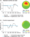
Frontal event-related potentials. a, ERP waveforms for unconscious No-Go trials and Go trials for the frontocentral ROI (pooling of electrodes FCz, FC1, FC2, Fz, F3, F4, AF3, and AF4, time locked to the onset of the Go signal). In the masked Go/No-Go task, unconscious No-Go trials differed significantly from Go trials between 309 and 418 ms. b, In the control experiment, unconscious gray circle trials did not differ from Go trials at any point in time between 0 and 500 ms after Go signal onset. Scalp voltage maps (right, pooled electrodes are shown in black) show the scalp distributions of the differential EEG activity between Go trials and unconscious No-Go trials (masked Go/No-Go task) and Go trials and unconscious gray circle trials (control experiment) between 309 and 418 ms.
Figure 6.
Frontal effects and correlations. a, Left, Correlation between the mean amplitude difference between unconscious No-Go trials and Go trials in the significant time window (309–418 ms) and increase in RT (electrode FCz). The scatter plot shows a strong positive correlation between the size of this frontocentral ERP effect and the increase in RT to subsequent Go signals in the masked Go/No-Go task (each dot is one subject). The map in the middle shows the scalp distribution of rho values for all 48 electrode sites (red, positive correlation; blue, negative correlation). The distribution of the frontal ERP effect (Fig. 5) strongly corresponds to the distribution of correlations in the masked Go/No-Go task. Right, Correlation between a moving average of EEG activity and the increase in RT across time at electrode FCz (the shown rho values are absolute). At the moment in time that frontocentral electrodes differentiate between unconscious No-Go trials and Go trials (309–418 ms), a strong positive correlation appears (p < 0.05, between 289 and 445 ms), which is absent at other times, as well as in the control experiment. b, Control experiment.
Figure 7.
Cortical activity evoked by unconscious No-Go signals. The reconstructed cortical sources at the peak of the differential ERP activity between unconscious No-Go trials and Go trials (352 ms) at the frontocentral ROI (in the masked Go/No-Go task). The source imaging revealed that the (especially right) lateral prefrontal cortex was active at this moment in time. Cortical current maps are represented on smoothed standardized cortex and shown in four different views (left view, right view, anterior view, and superior view). Activity of the reconstructed cortical sources is indicated by color (in current density units, Am), thresholded at 50% of the maximum value (yellow, 6.5 × 10⁻⁵ Am).
Figure 8.
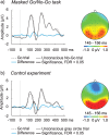
Occipital event-related potentials. a, ERP waveforms for unconscious No-Go trials and Go trials for the occipitoparietal ROI (pooling of electrodes Iz, I1, I2, Oz, O1, O2, PO7, P5, P7, PO8, P6, and P8, time locked to the onset of the Go signal). Unconscious No-Go trials differed significantly from Go trials between 145 and 156 ms in the masked Go/No-Go task. b, In the control experiment, unconscious gray circle trials differed significantly from Go trials between 141 and 172 ms and between 191 and 207 ms. Scalp voltage maps (right) show scalp distributions of the differential activity between 145 and 156 ms for both experiments (pooled electrodes are shown in black). The pattern of activity shows that unconscious gray circles were visually encoded alike in both conditions, but only triggered inhibitory control when they were associated with response inhibition (in the masked Go/No-Go task only).
Similar articles
- Unconscious activation of the prefrontal no-go network.
van Gaal S, Ridderinkhof KR, Scholte HS, Lamme VA. van Gaal S, et al. J Neurosci. 2010 Mar 17;30(11):4143-50. doi: 10.1523/JNEUROSCI.2992-09.2010. J Neurosci. 2010. PMID: 20237284 Free PMC article. - Conflict awareness dissociates theta-band neural dynamics of the medial frontal and lateral frontal cortex during trial-by-trial cognitive control.
Jiang J, Zhang Q, van Gaal S. Jiang J, et al. Neuroimage. 2015 Aug 1;116:102-11. doi: 10.1016/j.neuroimage.2015.04.062. Epub 2015 May 6. Neuroimage. 2015. PMID: 25957992 - Complete unconscious control: using (in)action primes to demonstrate completely unconscious activation of inhibitory control mechanisms.
Hepler J, Albarracin D. Hepler J, et al. Cognition. 2013 Sep;128(3):271-9. doi: 10.1016/j.cognition.2013.04.012. Epub 2013 Jun 4. Cognition. 2013. PMID: 23747649 Free PMC article. - To see or not to see--thalamo-cortical networks during blindsight and perceptual suppression.
Schmid MC, Maier A. Schmid MC, et al. Prog Neurobiol. 2015 Mar;126:36-48. doi: 10.1016/j.pneurobio.2015.01.001. Epub 2015 Feb 7. Prog Neurobiol. 2015. PMID: 25661166 Review. - Visual consciousness in health and disease.
Whatham AR, Vuilleumier P, Landis T, Safran AB. Whatham AR, et al. Neurol Clin. 2003 Aug;21(3):647-86, vi. doi: 10.1016/s0733-8619(02)00122-6. Neurol Clin. 2003. PMID: 13677817 Review.
Cited by
- Unconsciously triggered response inhibition requires an executive setting.
Chiu YC, Aron AR. Chiu YC, et al. J Exp Psychol Gen. 2014 Feb;143(1):56-61. doi: 10.1037/a0031497. Epub 2013 Jan 14. J Exp Psychol Gen. 2014. PMID: 23317085 Free PMC article. - Detecting analogies unconsciously.
Reber TP, Luechinger R, Boesiger P, Henke K. Reber TP, et al. Front Behav Neurosci. 2014 Jan 22;8:9. doi: 10.3389/fnbeh.2014.00009. eCollection 2014. Front Behav Neurosci. 2014. PMID: 24478656 Free PMC article. - Effect of pattern awareness on the behavioral and neurophysiological correlates of visual statistical learning.
Singh S, Daltrozzo J, Conway CM. Singh S, et al. Neurosci Conscious. 2017 Oct 7;2017(1):nix020. doi: 10.1093/nc/nix020. eCollection 2017. Neurosci Conscious. 2017. PMID: 29877520 Free PMC article. - Perceptual and attentional impairments of conscious access involve distinct neural mechanisms despite equal task performance.
Noorman S, Stein T, Fahrenfort JJ, van Gaal S. Noorman S, et al. Elife. 2025 May 1;13:RP97900. doi: 10.7554/eLife.97900. Elife. 2025. PMID: 40310881 Free PMC article. - Distinct brain mechanisms for conscious versus subliminal error detection.
Charles L, Van Opstal F, Marti S, Dehaene S. Charles L, et al. Neuroimage. 2013 Jun;73:80-94. doi: 10.1016/j.neuroimage.2013.01.054. Epub 2013 Feb 4. Neuroimage. 2013. PMID: 23380166 Free PMC article.
References
- Baars BJ. The conscious access hypothesis: origins and recent evidence. Trends Cogn Sci. 2002;6:47–52. - PubMed
- Baillet S, Mosher JC, Leahy RM. Electromagnetic brain mapping. IEEE Signal Process Mag. 2001;18:14–30.
- Bokura H, Yamaguchi S, Kobayashi S. Electrophysiological correlates for response inhibition in a Go/NoGo task. Clin Neurophysiol. 2001;112:2224–2232. - PubMed
- Casey BJ, Trainor RJ, Orendi JL, Schubert AB, Nystrom LE, Giedd JN, Castellanos FX, Haxby JV, Noll DC, Cohen JD, Forman SD, Dahl RE, Rapoport AJL. A developmental functional MRI study of prefrontal activation during performance of a go-no-go task. J Cogn Neurosci. 1997;9:835–847. - PubMed
- Dehaene S. Conscious and nonconscious processes: distinct forms of evidence accumulation? In: Engel C, Singer W, editors. Decision making, the human mind, and implications for institutions. Strüngmann forum reports. Cambridge, MA: MIT; 2008. pp. 21–49.
MeSH terms
LinkOut - more resources
Full Text Sources
Miscellaneous


