Rifampicin reduces alpha-synuclein in a transgenic mouse model of multiple system atrophy - PubMed (original) (raw)
Rifampicin reduces alpha-synuclein in a transgenic mouse model of multiple system atrophy
Kiren Ubhi et al. Neuroreport. 2008.
Abstract
Multiple system atrophy (MSA) is a progressive neurodegenerative disorder characterized by oligodendrocytic cytoplasmic inclusions containing abnormally aggregated alpha-synuclein. This aggregation has been linked to the neurodegeneration observed in MSA. Current MSA treatments are aimed at controlling symptoms rather than tackling the underlying cause of neurodegeneration. This study investigates the ability of the antibiotic rifampicin to reduce alpha-synuclein aggregation and the associated neurodegeneration in a transgenic mouse model of MSA. We report a reduction in monomeric and oligomeric alpha-synuclein and a reduction in phosphorylated alpha-synuclein (S129) upon rifampicin treatment. This reduction in alpha-synuclein aggregation was accompanied by reduced neurodegeneration. On the basis of its anti-aggregenic properties, we conclude that rifampicin may have therapeutic potential for MSA.
Figures
Fig. 1
Rifampicin reduced α-syn accumulation in MBP-α-syn tg mice. Brightfield microscope images of α-syn immunoreactivity in vehicle-treated MBP-α-syn tg mice (a), rifampicin-treated MBP-α-syn tg mice (b), phosphorylated α-syn (S129) in vehicle-treated MBP-α-syn tg mice (d), rifampicin-treated MBP-α-syn tg mice (e), nitrosylated α-syn immunoreactivity in vehicle-treated MBP-α-syn tg mice (g), and rifampicin-treated MBP-α-syn tg mice (h). Confocal microscope images of α-syn immunoreactivity in vehicle-treated MBP-α-syn tg mice (j) and rifampicin-treated MBP-α-syn tg mice (k). Scale bars represent 50 μM (b, e, and h) and 100 μM (k). c, f, i, and l represent stereological analyses of the number of α-syn-immunoreactive neurons (polyclonal antibody), phosphorylated α-syn (S129), nitrosylated α-syn and monoclonal α-syn, respectively.*Significant difference between rifampicin-treated MBP-α-syn tg mice (_n_=10) in comparison with vehicle-treated MBP-α-syn mice (_n_=8) (P<0.05, one-way ANOVA and post-hoc Fisher). Alpha-syn, α-synuclein; ANOVA, analysis of variance; MBP, myelin basic protein.
Fig. 2
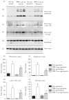
Rifampicin reduced α-syn accumulation in MBP-α-syn tg mice. Western blot analysis of α-syn, phosphorylated α-syn (S129), nitrosylated α-syn, and β-syn. Actin was used as a loading control (a). All blots were loaded with protein from the detergent-insoluble fraction. (b–e) Analyses of immunoreactivity for monomeric α-syn, oligomeric α-syn, phosphorylated α-syn (S129), and nitrosylated α-syn, respectively. Immunoreactivity signal was normalized over Actin. *Significant difference between vehicle-treated MBP-α-syn tg mice (_n_=8) in comparison with vehicle-treated non-tg controls (_n_=8) (P<0.05, one-way ANOVA and post-hoc Fisher). **Signifcant difference between rifampicin-treated MBP-α-syn tg mice (_n_=10) in comparison with vehicle-treated MBP-α-syn tg mice (_n_=8) (P<0.05, one-way ANOVA and post-hoc Fisher). Alpha-syn, α-synuclein; ANOVA, analysis of variance; MBP, myelin basic protein.
Fig. 3
Rifampicin reduced neurodegeneration in MBP-α-syn tg mice. Low-power confocal microscopic images of MAP2 immunoreactivity in vehicle-treated non-tg mice (a), vehicle-treated MBP-α-syn tg mice (b), and rifampicin-treated MBP-α-syn tg mice (c). Higher power confocal microscopic images of MAP2 immunoreactivity in vehicle-treated non-tg mice (d), vehicle-treated MBP-α-syn tg mice (e), and rifampicin-treated MBP-α-syn tg mice (f). Neocortical analysis of the area of the neuropil covered by MAP2 immunoreactivity was performed (g). Bright-field images of NeuN immunoreactivity in vehicle-treated non-tg mice (h), vehicle-treated MBP-α-syn tg mice (i) and rifampicin-treated MBP-α-syn tg mice (j) and GFAP immunoreactivity in vehicle-treated non-tg mice (l), vehicle-treated MBP-α-syn tg mice (m) and rifampicin-treated MBP-α-syn tg mice (n). Stereological analysis of NeuN immunoreactive neurons (k) and analysis of GFAP immunoreactivity (o). Scale bars represent 200 μM (a–c) and 50 μM (d–n). *Significant difference between vehicle-treated MBP-α-syn tg mice (_n_=8) in comparison with vehicle-treated non-tg controls (_n_=8) (P<0.05, one-way ANOVA and post-hoc Fisher). **Significant difference between rifampicin-treated MBP-α-syn tg mice (_n_=10) in comparison with vehicle-treated MBP-α-syn tg mice (_n_=8) (P<0.05, one-way ANOVA and post-hoc Fisher). ANOVA, analysis of variance; GFAP, glial fibrillary acidic protein; MBP, myelin basic protein.
Similar articles
- α-Synuclein-induced myelination deficit defines a novel interventional target for multiple system atrophy.
Ettle B, Kerman BE, Valera E, Gillmann C, Schlachetzki JC, Reiprich S, Büttner C, Ekici AB, Reis A, Wegner M, Bäuerle T, Riemenschneider MJ, Masliah E, Gage FH, Winkler J. Ettle B, et al. Acta Neuropathol. 2016 Jul;132(1):59-75. doi: 10.1007/s00401-016-1572-y. Epub 2016 Apr 8. Acta Neuropathol. 2016. PMID: 27059609 Free PMC article. - Anle138b modulates α-synuclein oligomerization and prevents motor decline and neurodegeneration in a mouse model of multiple system atrophy.
Heras-Garvin A, Weckbecker D, Ryazanov S, Leonov A, Griesinger C, Giese A, Wenning GK, Stefanova N. Heras-Garvin A, et al. Mov Disord. 2019 Feb;34(2):255-263. doi: 10.1002/mds.27562. Epub 2018 Nov 19. Mov Disord. 2019. PMID: 30452793 Free PMC article. - Novel therapeutic approaches in multiple system atrophy.
Palma JA, Kaufmann H. Palma JA, et al. Clin Auton Res. 2015 Feb;25(1):37-45. doi: 10.1007/s10286-014-0249-7. Epub 2014 Jun 14. Clin Auton Res. 2015. PMID: 24928797 Free PMC article. Review. - Reducing C-terminal truncation mitigates synucleinopathy and neurodegeneration in a transgenic model of multiple system atrophy.
Bassil F, Fernagut PO, Bezard E, Pruvost A, Leste-Lasserre T, Hoang QQ, Ringe D, Petsko GA, Meissner WG. Bassil F, et al. Proc Natl Acad Sci U S A. 2016 Aug 23;113(34):9593-8. doi: 10.1073/pnas.1609291113. Epub 2016 Aug 1. Proc Natl Acad Sci U S A. 2016. PMID: 27482103 Free PMC article. - Cellular pathology in multiple system atrophy.
Wakabayashi K, Takahashi H. Wakabayashi K, et al. Neuropathology. 2006 Aug;26(4):338-45. doi: 10.1111/j.1440-1789.2006.00713.x. Neuropathology. 2006. PMID: 16961071 Review.
Cited by
- Nasal Rifampicin Improves Cognition in a Mouse Model of Dementia with Lewy Bodies by Reducing α-Synuclein Oligomers.
Umeda T, Hatanaka Y, Sakai A, Tomiyama T. Umeda T, et al. Int J Mol Sci. 2021 Aug 6;22(16):8453. doi: 10.3390/ijms22168453. Int J Mol Sci. 2021. PMID: 34445158 Free PMC article. - Current Management and Emerging Therapies in Multiple System Atrophy.
Burns MR, McFarland NR. Burns MR, et al. Neurotherapeutics. 2020 Oct;17(4):1582-1602. doi: 10.1007/s13311-020-00890-x. Neurotherapeutics. 2020. PMID: 32767032 Free PMC article. Review. - Review: Multiple system atrophy: emerging targets for interventional therapies.
Stefanova N, Wenning GK. Stefanova N, et al. Neuropathol Appl Neurobiol. 2016 Feb;42(1):20-32. doi: 10.1111/nan.12304. Neuropathol Appl Neurobiol. 2016. PMID: 26785838 Free PMC article. Review. - The role of glia in α-synucleinopathies.
Fellner L, Stefanova N. Fellner L, et al. Mol Neurobiol. 2013 Apr;47(2):575-86. doi: 10.1007/s12035-012-8340-3. Epub 2012 Sep 2. Mol Neurobiol. 2013. PMID: 22941028 Free PMC article. Review. - Optimizing clinical trial design for multiple system atrophy: lessons from the rifampicin study.
Singer W, Low PA. Singer W, et al. Clin Auton Res. 2015 Feb;25(1):47-52. doi: 10.1007/s10286-015-0281-2. Epub 2015 Mar 13. Clin Auton Res. 2015. PMID: 25763826 Free PMC article. Review.
References
- Beyer K, Ariza A. Protein aggregation mechanisms in synucleinopathies: commonalities and differences. J Neuropathol Exp Neurol. 2007;66:965–974. - PubMed
- Wakabayashi K, Takahashi H. Cellular pathology in multiple system atrophy. Neuropathology. 2006;26:338–345. - PubMed
- Yoshida M. Multiple system atrophy: alpha-synuclein and neuronal degeneration. Neuropathology. 2007;27:484–493. - PubMed
- El-Agnaf OM, Jakes R, Curran MD, Middleton D, Ingenito R, Bianchi E, et al. Aggregates from mutant and wild-type alpha-synuclein proteins and NAC peptide induce apoptotic cell death in human neuroblastoma cells by formation of beta-sheet and amyloid-like filaments. FEBS Lett. 1998;440:71–75. - PubMed
Publication types
MeSH terms
Substances
Grants and funding
- R01 AG018440/AG/NIA NIH HHS/United States
- R37 AG018440/AG/NIA NIH HHS/United States
- AG022074/AG/NIA NIH HHS/United States
- P01 NS044233/NS/NINDS NIH HHS/United States
- NS044233/NS/NINDS NIH HHS/United States
- AG18440/AG/NIA NIH HHS/United States
- P01 AG022074/AG/NIA NIH HHS/United States
LinkOut - more resources
Full Text Sources
Molecular Biology Databases
Miscellaneous


