MicroRNA-221/222 confers tamoxifen resistance in breast cancer by targeting p27Kip1 - PubMed (original) (raw)
MicroRNA-221/222 confers tamoxifen resistance in breast cancer by targeting p27Kip1
Tyler E Miller et al. J Biol Chem. 2008.
Abstract
We explored the role of microRNAs (miRNAs) in acquiring resistance to tamoxifen, a drug successfully used to treat women with estrogen receptor-positive breast cancer. miRNA microarray analysis of MCF-7 cell lines that are either sensitive (parental) or resistant (4-hydroxytamoxifen-resistant (OHT(R))) to tamoxifen showed significant (>1.8-fold) up-regulation of eight miRNAs and marked down-regulation (>50%) of seven miRNAs in OHT(R) cells compared with parental MCF-7 cells. Increased expression of three of the most promising up-regulated (miR-221, miR-222, and miR-181) and down-regulated (miR-21, miR-342, and miR-489) miRNAs was validated by real-time reverse transcription-PCR. The expression of miR-221 and miR-222 was also significantly (2-fold) elevated in HER2/neu-positive primary human breast cancer tissues that are known to be resistant to endocrine therapy compared with HER2/neu-negative tissue samples. Ectopic expression of miR-221/222 rendered the parental MCF-7 cells resistant to tamoxifen. The protein level of the cell cycle inhibitor p27(Kip1), a known target of miR-221/222, was reduced by 50% in OHT(R) cells and by 28-50% in miR-221/222-overexpressing MCF-7 cells. Furthermore, overexpression of p27(Kip1) in the resistant OHT(R) cells caused enhanced cell death when exposed to tamoxifen. This is the first study demonstrating a relationship between miR-221/222 expression and HER2/neu overexpression in primary breast tumors that are generally resistant to tamoxifen therapy. This finding also provides the rationale for the application of altered expression of specific miRNAs as a predictive tamoxifen-resistant breast cancer marker.
Figures
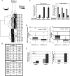
FIGURE 1.
miRNA expression profile of tamoxifen-sensitive and tamoxifen-resistant MCF-7 cells. A, shown are the results from cluster analysis. Total RNAs isolated from three biological replicates of tamoxifen-sensitive MCF-7 cells and tamoxifen-resistant MCF-7 cells (OHTR) were subjected to miRNA microarray analysis. miRNA expression data were normalized to the average median of all the genes present in the array. miRNAs expressed at least 1.5-fold higher or 50% lower in OHTR cells compared with MCF-7 cells were considered for cluster analysis. B, shown are the miRNAs that are up-regulated and down-regulated in OHTR cells.C, validation is shown. Total RNAs isolated from three biological replicates of MCF-7 and OHTR cells were subjected to real-time RT-PCR to validate differential expression of miR-221, miR-222, miR-181b, miR-21, miR-342, and miR-489. Each assay was done in triplicate, and expression of miRNA was normalized to snRNA RNU6B. D, the expression of miR-221 and miR-222 was analyzed in HER2/neu-positive and HER2/neu-negative primary human breast cancer tissues. Total RNAs isolated from formalin-fixed paraffin-embedded tissue sections were subjected to real-time RT-PCR with specific miRNA assay kits. Expression of snRNA RNU6B was used as normalizer. Normalized expression of the miRNAs was compared between the HER2/neu-positive (HER2/Neu +ve) and HER2/neu-negative (HER2/Neu -ve) samples using a Whisker plot. The asterisk indicate the outliers.E, total RNAs isolated from MCF-7 and ZR75.1 cells were subjected to real-time RT-PCR to analyze the expression levels of miR-221, miR-222, and HER2. miRNA levels were normalized to snRNA RNU6B, and HER2 levels were normalized to 18 S rRNA.
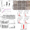
FIGURE 2.
Ectopic expression of miR-221/222 in MCF-7 cells results in increased tamoxifen resistance. A, expression of miR-221 and miR-222 was analyzed using total RNA isolated from miR-221/222-transfected MCF-7 cells by real-time RT-PCR and normalized to snRNA RNU6B. -Fold increase is shown below the bars. B, the G418-selected pool of miR-221/222-expressing MCF-7 cells, G418-selected clone 9 (MCF-7/221/222), and the empty vector-transfected MCF-7 cell pool were treated with 5 μ
m
tamoxifen for 72 h. Cell metabolic activity in the presence of the drug was measured every 24 h using the MTT assay. The metabolic activity of cells at 0 h was taken as 1. The results are the means ± S.D. of triplicate assays. C, vector- and miR-221/222-expressing MCF-7 cells were treated with 0, 15, and 20 μ
m
tamoxifen for 16 h. The cells were photographed using a phase-contrast microscope. O/E, overexpressing. D, whole cell extracts from the vector- and miR-221/222-transfected MCF-7 cells were separated by SDS-PAGE and probed with antibodies against PARP and caspase-7 that detect respective intact and cleaved products. The blot was reprobed with anti-Ku-70 antibody to ensure equal protein loading. The signal in each band was quantified using Kodak Imaging software. Quantification of both caspase-7 and PARP (uncleaved and cleaved) was plotted after normalization to Ku-70. The results are representative of two independent experiments. E, the vector- and miR-221/222-expressing MCF-7 cells were treated with 20 μ
m
tamoxifen (Tam) for 24 h. The cells were fixed and stained with propidium iodide, and cell cycle distribution was monitored by flow cytometry in a FACSCalibur. The percentage of apoptotic cells (sub-G1 peak) in vector (Vec)- and miR-221/222-expressing cells is represented in the bar diagram. Similar results were obtained from three independent experiments.
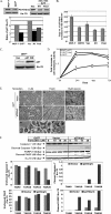
FIGURE 3.
p27Kip1, a target of miR-221/222, imparts tamoxifen sensitivity to MCF-7 cells. A, whole cell extracts from MCF-7 cells, OHTR cells, and miR-221/222-transfected and vector (Vec)-transfected MCF-7 cells were subjected to SDS-PAGE and probed with anti-p27Kip1 antibody. The membrane was reprobed with anti-Ku-70 antibody, and p27Kip1 expression was normalized to Ku-70 protein. O/E, overexpressing. B, total RNAs from MCF-7 cells, OHTR cells, and miR-221/222-transfected and vector-transfected MCF-7 cells were analyzed by real-time RT-PCR with primers specific for p27Kip1 and 18 S rRNA. C, whole cell extract from the p27Kip1-transfected OHTR cell pool was subjected to Western blot analysis with anti-p27Kip1 antibody and reprobed with anti-Ku-70 antibody. D, OHTR cells transfected with empty vector and p27 expression vector were treated with 5 μ
m
tamoxifen for 72 h. Cell metabolic activity was measured every 24 h using the MTT assay. The metabolic activity of cells at 0 h was taken as 1. The results are the means ± S.D. of triplicate assays.E, vector-overexpressing (panels A–C) and p27Kip1-overexpressing (D–F) OHTR cells were treated with 0 and 15 μ
m
tamoxifen for 16 h. The cells were photographed using a phase-contrast microscope. The small arrows in_panels G_ and H indicate autophagosome-like bodies.F, whole cell extracts from the vector-transfected and p27Kip1-transfected OHTR cells were subjected to SDS-PAGE and probed with antibodies against caspase-7 and PARP. The blot was reprobed with anti-Ku-70 antibody to normalize protein loading. The signal in each band was quantified using Kodak Imaging software. Quantification of uncleaved and cleaved caspase-7 and PARP is presented in the bar diagrams. The results are representative of three independent experiments. TAM, tamoxifen.
Similar articles
- Anti-microRNA-222 (anti-miR-222) and -181B suppress growth of tamoxifen-resistant xenografts in mouse by targeting TIMP3 protein and modulating mitogenic signal.
Lu Y, Roy S, Nuovo G, Ramaswamy B, Miller T, Shapiro C, Jacob ST, Majumder S. Lu Y, et al. J Biol Chem. 2011 Dec 9;286(49):42292-42302. doi: 10.1074/jbc.M111.270926. Epub 2011 Oct 18. J Biol Chem. 2011. PMID: 22009755 Free PMC article. Retracted. - Downregulation of miR-342 is associated with tamoxifen resistant breast tumors.
Cittelly DM, Das PM, Spoelstra NS, Edgerton SM, Richer JK, Thor AD, Jones FE. Cittelly DM, et al. Mol Cancer. 2010 Dec 20;9:317. doi: 10.1186/1476-4598-9-317. Mol Cancer. 2010. PMID: 21172025 Free PMC article. - miR-449a Suppresses Tamoxifen Resistance in Human Breast Cancer Cells by Targeting ADAM22.
Li J, Lu M, Jin J, Lu X, Xu T, Jin S. Li J, et al. Cell Physiol Biochem. 2018;50(1):136-149. doi: 10.1159/000493964. Epub 2018 Oct 2. Cell Physiol Biochem. 2018. PMID: 30278449 - MiR 221/222 as New Players in Tamoxifen Resistance.
Alamolhodaei NS, Behravan J, Mosaffa F, Karimi G. Alamolhodaei NS, et al. Curr Pharm Des. 2016;22(46):6946-6955. doi: 10.2174/1381612822666161102100211. Curr Pharm Des. 2016. PMID: 27809747 Review. - The Impact of miRNAs on the Efficacy of Tamoxifen in Breast Cancer Treatment: A Systematic Review.
Kavishahi NN, Rezaee A, Jalalian S. Kavishahi NN, et al. Clin Breast Cancer. 2024 Jun;24(4):341-350. doi: 10.1016/j.clbc.2024.01.015. Epub 2024 Jan 26. Clin Breast Cancer. 2024. PMID: 38413339 Review.
Cited by
- Cell-free Circulating miRNA Biomarkers in Cancer.
Mo MH, Chen L, Fu Y, Wang W, Fu SW. Mo MH, et al. J Cancer. 2012;3:432-48. doi: 10.7150/jca.4919. Epub 2012 Oct 13. J Cancer. 2012. PMID: 23074383 Free PMC article. - Recent trends in microRNA research into breast cancer with particular focus on the associations between microRNAs and intrinsic subtypes.
Kurozumi S, Yamaguchi Y, Kurosumi M, Ohira M, Matsumoto H, Horiguchi J. Kurozumi S, et al. J Hum Genet. 2017 Jan;62(1):15-24. doi: 10.1038/jhg.2016.89. Epub 2016 Jul 21. J Hum Genet. 2017. PMID: 27439682 Review. - The role of microRNAs in breast cancer migration, invasion and metastasis.
Tang J, Ahmad A, Sarkar FH. Tang J, et al. Int J Mol Sci. 2012 Oct 18;13(10):13414-37. doi: 10.3390/ijms131013414. Int J Mol Sci. 2012. PMID: 23202960 Free PMC article. Review. - Causes and consequences of microRNA dysregulation.
Iorio MV, Croce CM. Iorio MV, et al. Cancer J. 2012 May-Jun;18(3):215-22. doi: 10.1097/PPO.0b013e318250c001. Cancer J. 2012. PMID: 22647357 Free PMC article. Review. - Secreted uPAR isoform 2 (uPAR7b) is a novel direct target of miR-221.
Falkenberg N, Anastasov N, Schaub A, Radulovic V, Schmitt M, Magdolen V, Aubele M. Falkenberg N, et al. Oncotarget. 2015 Apr 10;6(10):8103-14. doi: 10.18632/oncotarget.3516. Oncotarget. 2015. PMID: 25797271 Free PMC article.
References
- Early Breast Cancer Trialists' Collaborative Group (2005) Lancet 365 1687-1717 - PubMed
- Shou, J., Massarweh, S., Osborne, C. K., Wakeling, A. E., Ali, S., Weiss, H., and Schiff, R. (2004) J. Natl. Cancer Inst. 96 926-935 - PubMed
- Carlomagno, C., Perrone, F., Gallo, C., De Laurentiis, M., Lauria, R., Morabito, A., Pettinato, G., Panico, L., D'Antonio, A., Bianco, A. R., and De Placido, S. (1996) J. Clin. Oncol. 14 2702-2708 - PubMed
- Lewis, J. S., and Jordan, V. C. (2005) Mutat. Res. 591 247-263 - PubMed
- Croce, C. M. (2008) N. Engl. J. Med. 358 502-511 - PubMed
Publication types
MeSH terms
Substances
Grants and funding
- R01 CA086978/CA/NCI NIH HHS/United States
- CA101956/CA/NCI NIH HHS/United States
- P01 CA101956/CA/NCI NIH HHS/United States
- CA086978/CA/NCI NIH HHS/United States
- R21 CA122523/CA/NCI NIH HHS/United States
- CA122523/CA/NCI NIH HHS/United States
LinkOut - more resources
Full Text Sources
Other Literature Sources
Medical
Research Materials
Miscellaneous


