The RNAseIII enzyme Drosha is critical in T cells for preventing lethal inflammatory disease - PubMed (original) (raw)
The RNAseIII enzyme Drosha is critical in T cells for preventing lethal inflammatory disease
Mark M W Chong et al. J Exp Med. 2008.
Erratum in
- J Exp Med. 2008 Sep 29;205(10). doi: 10.1084/jem.20071219090508c. Rundensky, Alexander Y [corrected to Rudensky, Alexander Y]
Abstract
MicroRNAs (miRNAs) are implicated in the differentiation and function of many cell types. We provide genetic and in vivo evidence that the two RNaseIII enzymes, Drosha and Dicer, do indeed function in the same pathway. These have previously been shown to mediate the stepwise maturation of miRNAs (Lee, Y., C. Ahn, J. Han, H. Choi, J. Kim, J. Yim, J. Lee, P. Provost, O. Radmark, S. Kim, and V.N. Kim. 2003. Nature. 425:415-419), and genetic ablation of either within the T cell compartment, or specifically within Foxp3(+) regulatory T (T reg) cells, results in identical phenotypes. We found that miRNA biogenesis is indispensable for the function of T reg cells. Specific deletion of either Drosha or Dicer phenocopies mice lacking a functional Foxp3 gene or Foxp3(+) cells, whereas deletion throughout the T cell compartment also results in spontaneous inflammatory disease, but later in life. Thus, miRNA-dependent regulation is critical for preventing spontaneous inflammation and autoimmunity.
Figures
Figure 1.
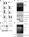
Drosha deficiency abrogates miRNA but not rRNA processing. (A) CD4-cre efficiently deletes exon 9 from the conditional Drosha transcript from the DP thymocyte stage onwards. cDNA from the indicated populations was analyzed for expression of the Drosha exons 5/6 or exon 9/10 junction by quantitative RT-PCR. DN4, CD90+TCRβloCD4/8/44/25−; DP, CD4+8+; CD4SP, TCRβhiCD24lo4+8−; CD8SP, TCRβhiCD24lo4−8+. (B) Drosha protein expression in DP thymocytes, and peripheral TCRβ+ T cells and B220+ B cells from DroshaF/Δ CD4-cre and control DroshaF/+ CD4-cre mice. Molecular weights are shown. (C) Total RNA from DP thymocytes and peripheral T and B cells was resolved on a polyacrylamide gel and Northern blotted for expression of mature miRNAs. Also shown is the ethidium bromide gel for expression of small rRNA and transfer RNA species. (D) Total RNA was resolved on an agarose-formaldehyde gel and Northern blotted for expression of pri-miR-150. Also shown is the ethidium bromide gel for expression of the large rRNA subunits.
Figure 2.
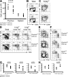
Drosha deficiency at the DP thymocyte stage partially perturbs thymocyte output. (A) Total thymocyte and splenocyte numbers in DroshaF/Δ CD4-cre and DroshaF/+ CD4-cre (control) mice. (B) TCRβ versus TCRγδ expression on total thymocytes. (C) CD24 and CD69 expression on TCRβhi gated thymocytes. (D) CD4 versus CD8 expression on total, postselected (TCRβhiCD24hi/69+) and mature (TCRβhiCD24lo/69−) thymocytes. (E and F) FACS analysis of populations in total splenocytes (E) and TCRβ+ splenocytes (F). Also shown is the effect of Dicer deficiency. Percentages of cells are shown in B–F. (G) Absolute CD4+8+ DP, mature thymocyte, and TCRβ+ splenocyte numbers. (H) CD4/CD8 ratio in the mature thymocyte and TCRβ+ splenocyte compartments.
Figure 3.
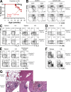
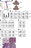
T cell–specific Drosha deficiency results in spontaneous inflammatory disease and premature mortality. (A) Kaplan-Meyer survival plot of DroshaF/Δ CD4-cre and control mice (DroshaF/F, DroshaΔ/+ , or DroshaF/+ CD4-cre). (B) Aberrant activation of CD4+ T cells in lymphoid organs of moribund DroshaF/Δ CD4-cre mice. (C and D) Cytokine expression by CD4+ (C) and CD8+ (D) T cells. (E) Frequent IFN-γ and IL-17A coexpression by CD4+ T cells. (F) Granulocyte (Gr-1+) and γδT cells populations in lymphoid organs. Percentages of cells are shown in B–F. (G) H&E-stained sections of the lung (i and iii) and liver (ii, iv, and v). Bars, 100 μm.
Figure 4.
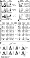
Drosha- and Dicer-dependent pathways are required for efficient induction of Foxp3 expression and function of T reg cells. (A) Reduced TCRβ+Foxp3+ T reg cell populations in the thymus and spleen of DroshaF/Δ CD4-cre and DicerF/F CD4-cre mice. (B) Foxp3 expression in peripheral T reg cell populations in the absence of Drosha or Dicer. (C and D) Drosha and Dicer are required for efficient differentiation of induced T reg cells. Naive CD62Lhi44lo25− CD4+ T cells from DroshaF/Δ CD4-cre, DicerF/F CD4-cre, and control DroshaF/+ CD4-cre mice were activated in vitro with anti-CD3/28 antibodies plus 10 U/ml IL-2, and TGF-β (C) or TGF-β plus 10 nM RA (D) for 3 d. The cells were restimulated with PMA/ionomycin plus GolgiStop for 3 h, and Foxp3 and IL-17A expression was analyzed by intracellular FACS. Percentages of cells are shown in A–D. (E) FACS-purified naive CD4+ cells from Cd45.1 mice were loaded with CFSE and mixed with purified CD4+25+ cells from DroshaF/Δ CD4-cre or DroshaF/+ CD4-cre (Cd45.2) mice at the indicated ratios. These were then incubated with inactivated splenocytes and anti-CD3 antibody. After 4 d, the CD45.1+ cells were analyzed for CFSE dilution.
Figure 5.
Drosha deficiency does not prevent polarization of naive CD4+ T cells into Th1 or Th2 cells. Naive CD62Lhi44lo25− CD4+ T cells from DroshaF/Δ CD4-cre and control DroshaF/+ CD4-cre mice were activated in vitro with anti-CD3/28 antibodies and 10 U/ml IL-2, plus (A) IL-12 and 1 μg/ml anti–IL-4 antibody or (B) IL-4 and 1 μg/ml anti-IFN-γ antibody for 4 d. The anti-CD3/28 antibodies were removed from the culture for the last 24 h, after which the cells were restimulated with PMA/ionomycin for 3 h in the presence of GolgiStop and analyzed for intracellular IFN-γ and IL-4 expression by FACS. Percentages of cells are shown.
Figure 6.
T reg cell–specific Drosha or Dicer deficiency recapitulates the scurfy phenotype. (A) Kaplan-Meyer survival plot of DroshaF/Δ Foxp3Cre/Cre(Y), DicerF/F Foxp3Cre/Cre(Y), and control mice (DroshaF/+ Foxp3Cre/Cre(Y), DroshaF/Δ Foxp3Cre/+, DroshaF/+ Foxp3Cre/+, DicerF/+ Foxp3Cre/Cre(Y), and DicerF/+ Foxp3Cre/+). (B) Massive lymphadenopathy in a female DroshaF/Δ Foxp3Cre/Cre mouse. Shown are the cervical lymph nodes. (C) Cell counts of lymphoid organs from moribund DroshaF/Δ Foxp3Cre/Cre or DroshaF/Δ Foxp3Cre/Y mice and littermate controls. Data represent the mean ± SD of four mice at 2.5–3 wk of age. (D) Only marginal changes in the proportions of B (CD19+), γδT (TCRγδ+), myeloid (CD11b+), and T cells (TCRβ+) in the spleen and lymph nodes of DroshaF/Δ Foxp3Cre/Cre mice. (E) Aberrant T cell activation in the spleen and lymph nodes of moribund DroshaF/Δ Foxp3Cre/Cre mice. (F and G) Cytokine expression by CD8+ (F) and CD4+ (G) T cells from DroshaF/Δ Foxp3Cre/Cre mice. (H and I) Foxp3 expression in total splenocytes (H) and CD4+ T cells (I). Percentages of cells are shown in D–I. (J) H&E-stained sections of the lung (i and iii) and liver (ii and iv). Designation of Foxp3Cre/Cre(Y) alleles indicates that both males and females were included in the experiments. Bars, 100 μm.
Similar articles
- Canonical and alternate functions of the microRNA biogenesis machinery.
Chong MM, Zhang G, Cheloufi S, Neubert TA, Hannon GJ, Littman DR. Chong MM, et al. Genes Dev. 2010 Sep 1;24(17):1951-60. doi: 10.1101/gad.1953310. Epub 2010 Aug 16. Genes Dev. 2010. PMID: 20713509 Free PMC article. - Re-evaluation of the roles of DROSHA, Export in 5, and DICER in microRNA biogenesis.
Kim YK, Kim B, Kim VN. Kim YK, et al. Proc Natl Acad Sci U S A. 2016 Mar 29;113(13):E1881-9. doi: 10.1073/pnas.1602532113. Epub 2016 Mar 14. Proc Natl Acad Sci U S A. 2016. PMID: 26976605 Free PMC article. - Dicer-dependent microRNA pathway safeguards regulatory T cell function.
Liston A, Lu LF, O'Carroll D, Tarakhovsky A, Rudensky AY. Liston A, et al. J Exp Med. 2008 Sep 1;205(9):1993-2004. doi: 10.1084/jem.20081062. Epub 2008 Aug 25. J Exp Med. 2008. PMID: 18725526 Free PMC article. - Selective miRNA disruption in T reg cells leads to uncontrolled autoimmunity.
Zhou X, Jeker LT, Fife BT, Zhu S, Anderson MS, McManus MT, Bluestone JA. Zhou X, et al. J Exp Med. 2008 Sep 1;205(9):1983-91. doi: 10.1084/jem.20080707. Epub 2008 Aug 25. J Exp Med. 2008. PMID: 18725525 Free PMC article. - Dicer cuts the kidney.
Ho JJ, Marsden PA. Ho JJ, et al. J Am Soc Nephrol. 2008 Nov;19(11):2043-6. doi: 10.1681/ASN.2008090986. Epub 2008 Oct 15. J Am Soc Nephrol. 2008. PMID: 18923053 Review. No abstract available.
Cited by
- Natural killer cell regulation by microRNAs in health and disease.
Leong JW, Sullivan RP, Fehniger TA. Leong JW, et al. J Biomed Biotechnol. 2012;2012:632329. doi: 10.1155/2012/632329. Epub 2012 Nov 19. J Biomed Biotechnol. 2012. PMID: 23226942 Free PMC article. Review. - miR-29ab1 deficiency identifies a negative feedback loop controlling Th1 bias that is dysregulated in multiple sclerosis.
Smith KM, Guerau-de-Arellano M, Costinean S, Williams JL, Bottoni A, Mavrikis Cox G, Satoskar AR, Croce CM, Racke MK, Lovett-Racke AE, Whitacre CC. Smith KM, et al. J Immunol. 2012 Aug 15;189(4):1567-76. doi: 10.4049/jimmunol.1103171. Epub 2012 Jul 6. J Immunol. 2012. PMID: 22772450 Free PMC article. - Canonical and alternate functions of the microRNA biogenesis machinery.
Chong MM, Zhang G, Cheloufi S, Neubert TA, Hannon GJ, Littman DR. Chong MM, et al. Genes Dev. 2010 Sep 1;24(17):1951-60. doi: 10.1101/gad.1953310. Epub 2010 Aug 16. Genes Dev. 2010. PMID: 20713509 Free PMC article. - Control of Immunoregulatory Molecules by miRNAs in T Cell Activation.
Rodríguez-Galán A, Fernández-Messina L, Sánchez-Madrid F. Rodríguez-Galán A, et al. Front Immunol. 2018 Sep 25;9:2148. doi: 10.3389/fimmu.2018.02148. eCollection 2018. Front Immunol. 2018. PMID: 30319616 Free PMC article. Review. - Micro (mi) RNA and Diabetic Retinopathy.
Sadashiv, Sharma P, Dwivedi S, Tiwari S, Singh PK, Pal A, Kumar S. Sadashiv, et al. Indian J Clin Biochem. 2022 Jul;37(3):267-274. doi: 10.1007/s12291-021-01018-4. Epub 2022 Jan 14. Indian J Clin Biochem. 2022. PMID: 35873619 Free PMC article. Review.
References
- Bennett, C.L., M.E. Brunkow, F. Ramsdell, K.C. O'Briant, Q. Zhu, R.L. Fuleihan, A.O. Shigeoka, H.D. Ochs, and P.F. Chance. 2001. A rare polyadenylation signal mutation of the FOXP3 gene (AAUAAA→AAUGAA) leads to the IPEX syndrome. Immunogenetics. 53:435–439. - PubMed
- Brunkow, M.E., E.W. Jeffery, K.A. Hjerrild, B. Paeper, L.B. Clark, S.A. Yasayko, J.E. Wilkinson, D. Galas, S.F. Ziegler, and F. Ramsdell. 2001. Disruption of a new forkhead/winged-helix protein, scurfin, results in the fatal lymphoproliferative disorder of the scurfy mouse. Nat. Genet. 27:68–73. - PubMed
- Kim, J.M., J.P. Rasmussen, and A.Y. Rudensky. 2007. Regulatory T cells prevent catastrophic autoimmunity throughout the lifespan of mice. Nat. Immunol. 8:191–197. - PubMed
- Wildin, R.S., F. Ramsdell, J. Peake, F. Faravelli, J.L. Casanova, N. Buist, E. Levy-Lahad, M. Mazzella, O. Goulet, L. Perroni, et al. 2001. X-linked neonatal diabetes mellitus, enteropathy and endocrinopathy syndrome is the human equivalent of mouse scurfy. Nat. Genet. 27:18–20. - PubMed
Publication types
MeSH terms
Substances
LinkOut - more resources
Full Text Sources
Other Literature Sources
Molecular Biology Databases
Research Materials
