A new approach yields high rates of radiographic progression in knee osteoarthritis - PubMed (original) (raw)
. 2008 Oct;35(10):2047-54.
Epub 2008 Sep 15.
Affiliations
- PMID: 18793000
- PMCID: PMC2758234
A new approach yields high rates of radiographic progression in knee osteoarthritis
David T Felson et al. J Rheumatol. 2008 Oct.
Abstract
Objective: Progression of knee osteoarthritis (OA) has typically been assessed in the medial tibiofemoral (TF) compartment on the anteroposterior (AP) or posteroanterior (PA) view. We propose a new approach using multiple views and compartments that is likely to be more sensitive to change and reveals progression throughout the knee.
Methods: We tested our approach in the Multicenter Osteoarthritis Study, a study of persons with OA or at high risk of disease. At baseline and 30 months, subjects provided PA (fixed flexion without fluoro) and lateral weight-bearing knee radiographs. Paired radiographs were read by 2 readers who scored joint space (JS) using a 0-3 atlas-based scale. When JS narrowed but narrowing did not reach a full grade on the scale, readers used half-grades. Change was scored in medial and lateral TF compartments on both PA and lateral views and in the patellofemoral (PF) joint on lateral view. A knee showed progression when there was at least a half-grade worsening in JS width in any compartment at followup. Disagreements were adjudicated by a panel of 3 readers. To validate progression, we tested definitions for TF progression to see if malalignment on long-limb radiographs at baseline (>or=3 degrees malaligned in any direction with nonmalaligned knees being reference) increased risk of progression. A valid definition of progression would show that malalignment strongly predicted progression.
Results: We studied 842 knees with either Kellgren-Lawrence grade>or=2 or PF OA at baseline in 606 subjects (age range 50-79 yrs, mean 63.9 yrs; 66.6% women). Mean body mass index was 31.9, and 32.8% of knees had frequent knee pain at baseline. Of these, 500 knees (59.4%) showed progression. Of the 500, 75 (15%) had progression only in the PF joint, while the remainder had progression in the TF joint. Malalignment increased the risk of overall progression in TF joint and increased the risk of half-grade progression, suggesting that half-grade progression had validity.
Conclusion: PA and lateral views obtained in persons at high risk of OA progression can produce a cumulative incidence of progression above 50% at 30 months. Keys to increasing the yield include imaging PF and lateral compartments, using semiquantitative scales designed to detect change, and examining more than one radiographic view.
Figures
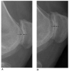
Figure 1
Examples of half-grade radiographic changes seen on posteroanterior lateral view. A: the baseline radiograph; B: the followup radiograph.
Figure 2
Examples of half-grade radiographic changes seen in the tibiofemoral compartment. A: the baseline radiograph; B: the followup radiograph.
Figure 3
Examples of half-grade radiographic changes seen on lateral view in the patellofemoral joint. A: the baseline radiograph; B: the followup radiograph.
Figure 4
Where is progression? Left: a depiction of tibiofemoral (TF) compared to patellofemoral (PF) compartment as a source of progression. Top panel: TF progression in posteroanterior (PA) view versus lateral view versus both. Middle panel shows which TF compartment showed progression. Bottom panel shows the percentage of half-grade versus full-grade TF progression.
Similar articles
- The lateral view radiograph for assessment of the tibiofemoral joint space in knee osteoarthritis: its reliability, sensitivity to change, and longitudinal validity.
LaValley MP, McLaughlin S, Goggins J, Gale D, Nevitt MC, Felson DT. LaValley MP, et al. Arthritis Rheum. 2005 Nov;52(11):3542-7. doi: 10.1002/art.21374. Arthritis Rheum. 2005. PMID: 16255043 - Valgus malalignment is a risk factor for lateral knee osteoarthritis incidence and progression: findings from the Multicenter Osteoarthritis Study and the Osteoarthritis Initiative.
Felson DT, Niu J, Gross KD, Englund M, Sharma L, Cooke TD, Guermazi A, Roemer FW, Segal N, Goggins JM, Lewis CE, Eaton C, Nevitt MC. Felson DT, et al. Arthritis Rheum. 2013 Feb;65(2):355-62. doi: 10.1002/art.37726. Arthritis Rheum. 2013. PMID: 23203672 Free PMC article. - Body mass index and alignment and their interaction as risk factors for progression of knees with radiographic signs of osteoarthritis.
Yusuf E, Bijsterbosch J, Slagboom PE, Rosendaal FR, Huizinga TW, Kloppenburg M. Yusuf E, et al. Osteoarthritis Cartilage. 2011 Sep;19(9):1117-22. doi: 10.1016/j.joca.2011.06.001. Epub 2011 Jun 16. Osteoarthritis Cartilage. 2011. PMID: 21722745 - Patterns of Compartment Involvement in End-stage Knee Osteoarthritis in a Chinese Orthopedic Center: Implications for Implant Choice.
Wang WJ, Sun MH, Palmer J, Liu F, Bottomley N, Jackson W, Qiu Y, Weng WJ, Price A. Wang WJ, et al. Orthop Surg. 2018 Aug;10(3):227-234. doi: 10.1111/os.12395. Orthop Surg. 2018. PMID: 30152607 Free PMC article. Review. - Sensitivity of standing radiographs to detect knee arthritis: a systematic review of Level I studies.
Duncan ST, Khazzam MS, Burnham JM, Spindler KP, Dunn WR, Wright RW. Duncan ST, et al. Arthroscopy. 2015 Feb;31(2):321-8. doi: 10.1016/j.arthro.2014.08.023. Epub 2014 Oct 11. Arthroscopy. 2015. PMID: 25312767 Review.
Cited by
- Investigating the relationship between radiographic joint space width loss and deep learning-derived magnetic resonance imaging-based cartilage thickness loss in the medial weight-bearing region of the tibiofemoral joint.
Minnig MCC, Arbeeva L, Niethammer M, Nissman D, Lund JL, Marron JS, Golightly YM, Nelson AE. Minnig MCC, et al. Osteoarthr Cartil Open. 2024 Aug 3;6(3):100508. doi: 10.1016/j.ocarto.2024.100508. eCollection 2024 Sep. Osteoarthr Cartil Open. 2024. PMID: 39238657 Free PMC article. - Symptoms of Knee Instability as Risk Factors for Recurrent Falls.
Nevitt MC, Tolstykh I, Shakoor N, Nguyen US, Segal NA, Lewis C, Felson DT; Multicenter Osteoarthritis Study Investigators. Nevitt MC, et al. Arthritis Care Res (Hoboken). 2016 Aug;68(8):1089-97. doi: 10.1002/acr.22811. Arthritis Care Res (Hoboken). 2016. PMID: 26853236 Free PMC article. - Structural predictors of response to intra-articular steroid injection in symptomatic knee osteoarthritis.
Maricar N, Parkes MJ, Callaghan MJ, Hutchinson CE, Gait AD, Hodgson R, Felson DT, O'Neill TW. Maricar N, et al. Arthritis Res Ther. 2017 May 8;19(1):88. doi: 10.1186/s13075-017-1292-2. Arthritis Res Ther. 2017. PMID: 28482926 Free PMC article. - Predictive validity of within-grade scoring of longitudinal changes of MRI-based cartilage morphology and bone marrow lesion assessment in the tibio-femoral joint--the MOST study.
Roemer FW, Nevitt MC, Felson DT, Niu J, Lynch JA, Crema MD, Lewis CE, Torner J, Guermazi A. Roemer FW, et al. Osteoarthritis Cartilage. 2012 Nov;20(11):1391-8. doi: 10.1016/j.joca.2012.07.012. Epub 2012 Jul 27. Osteoarthritis Cartilage. 2012. PMID: 22846715 Free PMC article. - Association of leg-length inequality with knee osteoarthritis: a cohort study.
Harvey WF, Yang M, Cooke TD, Segal NA, Lane N, Lewis CE, Felson DT. Harvey WF, et al. Ann Intern Med. 2010 Mar 2;152(5):287-95. doi: 10.7326/0003-4819-152-5-201003020-00006. Ann Intern Med. 2010. PMID: 20194234 Free PMC article.
References
- Mazzuca SA, Brandt KD. Plain radiography as an outcome measure in clinical trials involving patients with knee osteoarthritis. Rheum Dis Clin North Am. 1999;25:467–80. ix. - PubMed
- Mazzuca SA, Brandt KD, Katz BP. Is conventional radiography suitable for evaluation of a disease-modifying drug in patients with knee osteoarthritis? Osteoarthritis Cartilage. 1997;5:217–26. - PubMed
- Bingham CO, Buckland-Wright JC, Garnero P, et al. Risedronate decreases biochemical markers of cartilage degradation but does not decrease symptoms or slow radiographic progression in patients with medial compartment osteoarthritis of the knee: Results of the two-year multinational knee osteoarthritis structural arthritis study. Arthritis Rheum. 2006;54:3494–507. - PubMed
- LaValley MP, McLaughlin S, Goggins J, Gale D, Nevitt MC, Felson DT. The lateral view radiograph for assessment of the tibiofemoral joint space in knee osteoarthritis: its reliability, sensitivity to change, and longitudinal validity. Arthritis Rheum. 2005;52:3542–7. - PubMed
- Felson DT, Niu JB, Clancy M, et al. Low levels of vitamin D and worsening of knee osteoarthritis: results from two longitudinal studies. Arthritis Rheum. 2007;56:129–36. - PubMed
Publication types
MeSH terms
Grants and funding
- U01 AG18820/AG/NIA NIH HHS/United States
- AR47785/AR/NIAMS NIH HHS/United States
- U01 AG18947/AG/NIA NIH HHS/United States
- U01 AG018947/AG/NIA NIH HHS/United States
- P60 AR047785/AR/NIAMS NIH HHS/United States
- U01 AG18832/AG/NIA NIH HHS/United States
- U01 AG018832/AG/NIA NIH HHS/United States
- U01 AG19069/AG/NIA NIH HHS/United States
- P60 AR047785-07/AR/NIAMS NIH HHS/United States
- U01 AG018820-08S1/AG/NIA NIH HHS/United States
- U01 AG019069/AG/NIA NIH HHS/United States
- U01 AG018820/AG/NIA NIH HHS/United States
LinkOut - more resources
Full Text Sources
Miscellaneous


