Generation and regulation of human CD4+ IL-17-producing T cells in ovarian cancer - PubMed (original) (raw)
Generation and regulation of human CD4+ IL-17-producing T cells in ovarian cancer
Yoshihiro Miyahara et al. Proc Natl Acad Sci U S A. 2008.
Abstract
Despite the important role of Th17 cells in the pathogenesis of many autoimmune diseases, their prevalence and the mechanisms by which they are generated and regulated in cancer remain unclear. Here, we report the presence of a high percentage of CD4(+) Th17 cells at sites of ovarian cancer, compared with a low percentage of Th17 cells in peripheral blood mononuclear cells from healthy donors and cancer patients. Analysis of cytokine production profiles revealed that ovarian tumor cells, tumor-derived fibroblasts, and antigen-presenting cells (APCs) secreted several key cytokines including IL-1beta, IL-6, TNF-alpha and TGF-beta, which formed a cytokine milieu that regulated and expanded human IL-17-producing T-helper (Th17) cells. We further show that IL-1beta was critically required for the differentiation and expansion of human Th17 cells, whereas IL-6 and IL-23 may also play a role in the expansion of memory Th17 cells, even though IL-23 levels are low or undetectable in ovarian cancer. Further experiments demonstrated that coculture of naïve or memory CD4(+) T cells with tumor cells, APCs, or both could generate high percentages of Th17 cells. Treatment with anti-IL-1 alone or a combination of anti-IL-1 and anti-IL-6 reduced the ability of tumor cells to expand memory Th17 cells. Thus, we have identified a set of key cytokines secreted by ovarian tumor cells and tumor-associated APCs that favor the generation and expansion of human Th17 cells. These findings should accelerate efforts to define the function of this important subset of CD4(+) T cells in the human immune response to cancer.
Conflict of interest statement
The authors declare no conflict of interest.
Figures
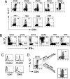
Fig. 1.
Identification of Th17 cells in ovarian tumor-derived T cell population. (A) Prevalence of Th17 cells among ovarian tumor-derived T cells. Bulk T cells isolated from ovarian tumor tissues were stimulated with PMA and ionomycin and stained with anti-CD4 and anti-IL-17 to determine the percentage of IL-17-producing T cells. OVA1–5 denotes T cells isolated from different ovarian tumor specimens. T cells from healthy donors (HD) served as controls. (B) Evaluation of IL-17- and IFN-γ-producing T cells in the total tumor-derived T cell population. (C) Phenotypic analysis of ovarian tumor-derived T cells. Bulk T cells generated from OVA1 were stained with antibodies against CD45RA, CD45RO, CCR7, and CD62L molecules and analyzed by FACS. (D) Th17-containing T cells were cultured in the presence of anti-CD3 antibody (OKT3) and IL-2 (300 units/ml) or cultured with tumor cells plus the same concentration of IL-2. After 2 weeks, IL-17-producing T cells in the total T cell population were determined after stimulation with PMA and ionomycin.
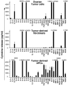
Fig. 2.
Proinflammatory cytokines produced by ovarian tumor cells, tumor-derived fibroblasts, and APCs. Supernatants of tumor cell lines and fibroblasts established from ovarian tumor samples were harvested and used for a cytokine assay with a Bio-Plex cytokine assay system. Tumor APCs were first stimulated with OKT3 and LPS, respectively, and the corresponding supernatants were collected for cytokine assays. 〉 indicates that the readout value for a particular cytokine exceeds the highest concentration of a particular cytokine standard. Cytokine concentrations (pg/ml) are reported as mean ± SD value from three experiments.
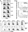
Fig. 3.
Critical roles of key cytokines in the generation and expansion of Th17 cells from naïve and memory CD4+ T cells. (A) IL-17-producing T cells detected in naïve and memory T cells after culturing with different cytokines. The purified naïve and memory CD4+ T cells were stimulated with plate-bound OKT3 in the presence of IL-1α, IL-β, IL-6, and IL-23. Eight days after activation, the cells were restimulated with PMA and ionomycin and analyzed for IL-17 and IFN-γ production after intracellular staining. (B) IL-1α and IL-1β play a dominant role in the differentiation and expansion of IL-17-producing T cells. Naïve and memory CD4+ T cells were purified from different PBMCs from healthy donors and used in experiments similar to that in B. Bars represent the average fold increases of IL-17-producing T cells. (C) Naïve and memory CD4+ T cells were purified from healthy donors and then stained with CFSE (final 4.5 μM) for 15 min at room temperature. After intensive washes, CFSE-labeled cells were stimulated with plate-bound OKT3 in the presence of IL-1β (20 ng/ml). The frequency of Th17 cells was monitored at the indicated time points.
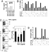
Fig. 4.
Tumor-secreted TGF-β inhibited Th17 cell expansion but promoted Treg cells. (A) Ovarian tumor cells secrete latent (inactive) and active TGF-β in culture. Cell supernatants were harvested and used to determine the concentration of TGF-β by ELISA according to the manufacturer's instructions. (B) TGF-β inhibits the expansion of Th17 cells by other cytokines. The purified human CD4+ T cells were stimulated with plate-bound OKT3 in the presence of the indicated cytokines, either alone or in various combinations, in T cell medium. Ten days after activation, the cells were restimulated with PMA/ionomycin and subjected to intracellular cytokine staining. The percentages of IL-17 expressing T cells in T cell medium served as a baseline. The data represent one of two independent experiments. (C) TGF-β inhibits the expansion of IL-17-producing T cells in a dose-dependent manner. Memory T cells were cultured in the presence of plate-bound OKT3 in the presence of the indicated concentrations of TGF-β. Results for the purified memory T cells of three healthy donors are plotted as mean ± SD value. *, P <0.05. (D) TGF-β promoted the generation of Foxp3+ T cells, but this effect was blocked by IL-6. The purified human CD4+ T cells were stimulated with plate-bound OKT3 in the presence of TGF-β either alone or in various combinations with other cytokines. Ten days after activation, the cells were analyzed for Foxp3 expression after intracellular cytokine staining with anti-Foxp3.
Fig. 5.
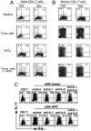
Generation and expansion of human Th17 cells by tumor cells and APCs. (A) Differentiation of naïve T cells to Th17 cells after stimulation by tumor cells plus APCs. The purified naïve CD4+ T cells failed to differentiate to Th17 cells except when they were cultured with tumor cells plus APCs. Such differentiation was minimal when the naïve CD4+ T cells were cultured with tumor cells alone or APCs alone. (B) Efficient expansion of Th17 cells from memory CD4+ T cells by tumor cells, APCs, or both. The purified CD4+ memory T cells were cultured with tumor cells, APCs, or both. CD4+ T cells with medium served as a control. (C) Anti-IL-1 reduces the expansion of memory Th17 cells by tumor cells or LPS-treated APCs. Purified memory CD4+ T cells were cultured with tumor cells or LPS-treated APCs in the presence of recombinant human IL-1Rα (final 5 μg/ml) and/or anti-human IL-6R mAb (final 10 μg/ml). Isotype antibody served as a control.
Similar articles
- Interleukin 2-mediated conversion of ovarian cancer-associated CD4+ regulatory T cells into proinflammatory interleukin 17-producing helper T cells.
Leveque L, Deknuydt F, Bioley G, Old LJ, Matsuzaki J, Odunsi K, Ayyoub M, Valmori D. Leveque L, et al. J Immunother. 2009 Feb-Mar;32(2):101-8. doi: 10.1097/CJI.0b013e318195b59e. J Immunother. 2009. PMID: 19238008 - Fetal BM-derived mesenchymal stem cells promote the expansion of human Th17 cells, but inhibit the production of Th1 cells.
Guo Z, Zheng C, Chen Z, Gu D, Du W, Ge J, Han Z, Yang R. Guo Z, et al. Eur J Immunol. 2009 Oct;39(10):2840-9. doi: 10.1002/eji.200839070. Eur J Immunol. 2009. PMID: 19637224 - Phenotypical characterization of human Th17 cells unambiguously identified by surface IL-17A expression.
Brucklacher-Waldert V, Steinbach K, Lioznov M, Kolster M, Hölscher C, Tolosa E. Brucklacher-Waldert V, et al. J Immunol. 2009 Nov 1;183(9):5494-501. doi: 10.4049/jimmunol.0901000. J Immunol. 2009. PMID: 19843935 - Induction, function and regulation of IL-17-producing T cells.
Mills KH. Mills KH. Eur J Immunol. 2008 Oct;38(10):2636-49. doi: 10.1002/eji.200838535. Eur J Immunol. 2008. PMID: 18958872 Review. - Properties and origin of human Th17 cells.
Romagnani S, Maggi E, Liotta F, Cosmi L, Annunziato F. Romagnani S, et al. Mol Immunol. 2009 Nov;47(1):3-7. doi: 10.1016/j.molimm.2008.12.019. Epub 2009 Feb 3. Mol Immunol. 2009. PMID: 19193443 Review.
Cited by
- Skewed immunological balance between Th17 (CD4(+)IL17A (+)) and Treg (CD4 (+)CD25 (+)FOXP3 (+)) cells in human oral squamous cell carcinoma.
Gaur P, Qadir GA, Upadhyay S, Singh AK, Shukla NK, Das SN. Gaur P, et al. Cell Oncol (Dordr). 2012 Oct;35(5):335-43. doi: 10.1007/s13402-012-0093-5. Epub 2012 Sep 7. Cell Oncol (Dordr). 2012. PMID: 22956260 - Human CCR4+ CCR6+ Th17 cells suppress autologous CD8+ T cell responses.
Zhao F, Hoechst B, Gamrekelashvili J, Ormandy LA, Voigtländer T, Wedemeyer H, Ylaya K, Wang XW, Hewitt SM, Manns MP, Korangy F, Greten TF. Zhao F, et al. J Immunol. 2012 Jun 15;188(12):6055-62. doi: 10.4049/jimmunol.1102918. Epub 2012 May 21. J Immunol. 2012. PMID: 22615204 Free PMC article. - The changes of Th17 cells and the related cytokines in the progression of human colorectal cancers.
Wang J, Xu K, Wu J, Luo C, Li Y, Wu X, Gao H, Feng G, Yuan BZ. Wang J, et al. BMC Cancer. 2012 Sep 21;12:418. doi: 10.1186/1471-2407-12-418. BMC Cancer. 2012. PMID: 22994684 Free PMC article. - The Th17/Treg balance and the expression of related cytokines in Uygur cervical cancer patients.
Chen Z, Ding J, Pang N, Du R, Meng W, Zhu Y, Zhang Y, Ma C, Ding Y. Chen Z, et al. Diagn Pathol. 2013 Apr 15;8:61. doi: 10.1186/1746-1596-8-61. Diagn Pathol. 2013. PMID: 23587428 Free PMC article.
References
- Wang R-F. The role of MHC class II-restricted tumor antigens and CD4+ T cells in antitumor immunity. Trends Immunol. 2001;22:269–276. - PubMed
- Wang HY, Wang RF. Regulatory T cells and cancer. Curr Opin Immunol. 2007;19:217–223. - PubMed
- Weaver CT, Harrington LE, Mangan PR, Gavrieli M, Murphy KM. Th17: An effector CD4 T cell lineage with regulatory T cell ties. Immunity. 2006;24:677–688. - PubMed
- Langowski JL, et al. IL-23 promotes tumour incidence and growth. Nature. 2006;442:461–465. - PubMed
Publication types
MeSH terms
Substances
LinkOut - more resources
Full Text Sources
Other Literature Sources
Medical
Research Materials
Miscellaneous
