Nonlinear optical imaging of cellular processes in breast cancer - PubMed (original) (raw)
Nonlinear optical imaging of cellular processes in breast cancer
Paolo P Provenzano et al. Microsc Microanal. 2008 Dec.
Abstract
Nonlinear optical imaging techniques such as multiphoton and second harmonic generation (SHG) microscopy used in conjunction with novel signal analysis techniques such as spectroscopic and fluorescence excited state lifetime detection have begun to be used widely for biological studies. This is largely due to their promise to noninvasively monitor the intracellular processes of a cell together with the cell's interaction with its microenvironment. Compared to other optical methods these modalities provide superior depth penetration and viability and have the additional advantage in that they are compatible technologies that can be applied simultaneously. Therefore, application of these nonlinear optical approaches to the study of breast cancer holds particular promise as these techniques can be used to image exogeneous fluorophores such as green fluorescent protein as well as intrinsic signals such as SHG from collagen and endogenous fluorescence from nicotinamide adenine dinucleotide or flavin adenine dinucleotide. In this article the application of multiphoton excitation, SHG, and fluorescence lifetime imaging microscopy to relevant issues regarding the tumor-stromal interaction, cellular metabolism, and cell signaling in breast cancer is described. Furthermore, the ability to record and monitor the intrinsic fluorescence and SHG signals provides a unique tool for researchers to understand key events in cancer progression in its natural context.
Figures
Figure 1
Live mammary tumor. Combined MPE and SHG at λex = 890 nm facilitates imaging of intact live mammary tumor tissue. a: Six planes of a 30-plane z-stack acquired every 10 μm (i.e., 300 μm total stack) into the tumor that was rendered and oriented with VisBio. b–g: The flat images for each corresponding imaging plane. Combined, a–g clearly show variations in endogenous cellular fluorescence as well as collagen surrounding and within the tumor validating the ability to image deep into live tissue and obtain meaningful information. Bar = 50 _μ_m.
Figure 2
MPE and SHG signal separation in live tumor. Since MPE excitation follows classical energy loss behavior while SHG signals are conserved, filtering techniques can be employed to separate the two signals following excitation with the same wavelength. Following excitation of live mammary tumor tissue with a wavelength (λex = 890 nm) that elicits both endogenous cellular fluorescence (of FAD) and SHG (a), the resultant emissions were separated using a 445 nm narrow band pass filter for SHG (b: pseudocolored green) and a 464 nm (cut-on) long pass filter for MPE (c: pseudocolored red). This approach allows clear visualization of the collagenous stroma (b) as well as stromal cells, localized within the collagen matrix, and tumor cells (c), while merging the two pseudocolored signals (d) helps reveal cell matrix interactions associated with the tumor-stromal interface. As such, the use of combined MPE/SHG has the potential to help identify and differentiate additional features that are not readily obtained with more traditional fluorescent microscopy techniques. Bar = 25 _μ_m.
Figure 3
Multiphoton FLIM of live mammary tumor. Following excitation at 890 nm, lifetime data were collected for 60 s using the Optical Work Station (described in the Instrumentation section) and displayed as (a) intensity or (b) color-mapped lifetimes (color bar 0 to 1.2 ns; red to blue) for a two-term exponential model (see equation (3)). The color mapping represents the weighted average of the short and long components: τm = (_a_1_τ_1 + _a_2_τ_2)/(_a_1 + _a_2), although the relative contribution of each component for cells in the tumor is shown in c. Additionally, due to the fact that signals from collagen principally arise from the conserved SHG polarization, the resulting signal has a theoretical lifetime of zero, which is supported by the fact that the majority of the collagen signal closely follows the instrumental response function.
Figure 4
Invasive breast carcinoma cells in 3D matrices. Highly invasive and migratory MDA-MB-231 breast carcinoma cells were cultured within reconstituted 3D collagen matrices to further examine the utility of combined MPE/SHG to probe the cell-matrix interaction. After 3 h in collagen gels (a–c), MDA-MB-231 cells expressing GFP-Vinculin form 3D matrix adhesions by presenting filopodia that interact with collagen fibers (arrows in a). As is shown in b, this interaction can be highlighted by separating the SHG signal (445 nm narrow band pass filter; green pseudocolor) and the GFP signal (480–550 nm band pass filter; red pseudocolor), while c is a magnified region (dashed box) of b clearly demonstrating vinculin positive filopodia interacting with collagen fibers (colocalization = yellow). After 24 h in 3D collage matrices (d: transmitted light), MDA-MB-231(GFP-vinculin) cells have aligned collagen fibers with vinculin localization at the cell-matrix interface (e, f: GFP = green pseudocolor; SHG blue pseudocolor). Hence, simultaneous imaging of cellular processes and matrix organization and structure with combined MPE/SHG not only facilitates imaging of nonnative and endogenous signals in vivo, but also provides a robust tool to study tumor-stromal interactions in live unfixed, unstained, nonsectioned) cells in vitro, which can further our understanding of the in vivo condition. Bar (a–c) = 10 _μ_m; bar (d–f) = 25 _μ_m.
Figure 5
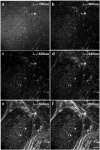
Changes in breast tumor cell and matrix signals as a function of excitation wavelength. By increasing the excitation wavelength from 780 to 880 nm in 20 nm increments, changes in the intensity and localization of endogenous fluorescence as well the emergence of collagen SHG is detectable. At λex = 780 nm (a), a two-photon wavelength that excites NADH, the cytoplasm of the tumor cells display a high degree of autofluorescence, which decreases as the excitation wavelength is increased (b–d). Additionally, minimal SHG signal from collagen is detectable below λex = 860 nm. At λex = 860 nm (e) and 880 nm (f), two-photon wavelengths that excite FAD and produce collagen SHG, strong autofluorescent signal and clear collagen SHG signals are present. The strong signal at λex = 780 nm, which decreases until λex = 860 nm likely indicates emission from the endogenous fluorophore NADH at 780 nm that decreases and transitions to endogenous fluorescence from FAD as the excitation wavelength approaches 900 nm. Hence, MPLSM facilitates the ability to image two important endogenous fluorophores, NADH and FAD, as well as fibrillar collagen, allowing visualization of breast tumor cell behavior and for detecting and studying changes in tumor cell metabolism. Bar = 25 _μ_m.
Figure 6
Fluorescent lifetime of NADH in T47D breast carcinoma cells. TPE at 740 nm elicits endogenous fluorescence of NADH, with a fluorescence lifetime that is independent of NADH concentration. As such, imaging fluorescence intensity with MPE and fluorescence lifetime with FLIM facilitate direct monitoring of normal and tumor cell metabolic enzyme amount and state in key processes such as tumorigenesis and metastasis, as well as response to antitumor therapeutics. Color mapping represents the weighted average of the short and long components: τm = (_a_1_τ_1 + _a_2_τ_2)/(_a_1 + _a_2); color bar = 0.4 to 3 ns, red to blue.
Figure 7
Fluorescent lifetime of MDA-MB-231 breast carcinoma cells in 3D. 890 nm excitation of MDA-MB-231 breast carcinoma cells in 3D collagen gels. The color map represents the weighted average of the two-term model components [τm = (_a_1_τ_1 + _a_2_τ_2)/(_a_1 + _a_2)] and validates the ability to detect changes in cellular lifetime of highly invasive breast carcinoma cells within a 3D culture environment. Color bar = 100 to 800 ps, red to blue.
Figure 8
Combined MPE/SHG of GFP-cdc42 in MDA-MB-231 cells within 3D collagen matrices. Live cells within type I collagen gels were imaged with combined MPE/SHG at λex = 890 nm. After 6 h within 3D collagen gels, differences in cell morphology and the cell matrix interaction could be detected as a function of cdc42 state. Cells expressing constitutively active cdc42 (b) were more spread and presented more cell protrusions than both control GFP cells (a) and dominant negative expressing cells (c). Additionally overexpression of wild type cdc42 resulted in cell protrusion (d), but was not as spread as cells expressing constitutively active cdc42 (e). Moreover, separation of the GFP signal (480–550 nm band pass filter; green pseudocolor) from SHG (445 narrow band pass filter, blue pseudocolor) reveals increased cell matrix attachments in cdc42(61L) cells with cdc42 localized to cell protrusions that are interacting with the collagen matrix.
Figure 9
Fluorescent lifetime imaging of a GFP-tagged molecule elucidates novel subdomains within the plasma membrane. GFP fused to constitutively active R-Ras (GFP-R-Ras(38V)) was transfected into Cos 7 cells and imaged by MPLSM: (a) intensity image and (b) color map of the FLIM of GFP-R-ras(38V) mapped from 1500 to 2500 ns; blue to red. (c) Blow up of plasma membrane region shown in b. Note that GFP-R-Ras(38V) localizes to active regions of the plasma membrane, which appear uniform in intensity via MPE in a, but map to distinct lifetime domains in b and c. FLIM was collected at 900 nm utilizing a 60× plan apo (Nikon) lens utilizing a lab-built MPLSM as described in Bird et al. (2004, .
Figure 10
FLIM of a lipophilic fluorescent molecule, filipin, reveals subdomains within the plasma membrane. Cos-7 cells were incubated in 50 _μ_M of the cholesterol-binding fluorophore filipin for 30 min in order to detect microdomains of high cholesterol within the plasma membrane. Multiphoton excitation images of two exemplar cells are shown on the left. Color mapped (7 to 17 ns; red to blue) images representing the fluorescence lifetime of filipin were generated (right panels). The lifetime of this fluorophore was uncharacteristically long (>16 ns) throughout the entire membrane; however, there are subregions with distinguishable lifetimes, which map as distinct colors in discrete plasma membrane domains that are not detectable with standard multiphoton microscopy. FLIM was collected at 900 nm utilizing a 60× plan apo (Nikon) lens utilizing a lab-built MPLSM as described in Bird et al. (2004, .
Figure 11
SLIM analysis of tumor sections. Utilizing a recently developed MPLSM system (Yan et al., 2006) in the Laboratory for Optical and Computational Instrumentation, spectral lifetime data were obtained from hematoxylin and eosin stained polyoma middle-T mammary tumor sections. The emission spectra were separated into 10 nm components over 16 channels: (a) intensity image, (b) total FLIM signaling, and (c–f) the 510, 520, 530, and 540 nm emission components of the emission spectrum, respectively. Color mapping represents the weighted average of the two-term model components [τm = (_a_1_τ_1 + _a_2_τ_2)/(_a_1 + _a_2)] with the color bar extending from 0 to 5 ns; red to blue. Of particular note is the observation that the total signal appears largely as background (b), yet the 530 nm emission component (e) reveals a low lifetime for the stroma (yellow) and a longer lifetime for the tumor cells (mostly green and blue) indicating that narrowing the detection spectra range while removing adjacent spectral components, that are most likely contributing significant noise, allows imaging and analysis of otherwise masked information.
References
- Abramoff MD, Magelhaes PJ, Ram SJ. Image processing with ImageJ. Biophoton Int. 2004;11(7):36–42.
- Ada-Nguema AS, Xenias H, Sheetz MP, Keely PJ. The small GTPase R-Ras regulates organization of actin and drives membrane protrusions through the activity of PLC{epsilon} J Cell Sci. 2006;119(Pt 7):1307–1319. - PubMed
- Becker W, Bergmann A, Hink MA, Konig K, Benndorf K, Biskup C. Fluorescence lifetime imaging by time-correlated single-photon counting. Microsc Res Tech. 2004;63(1):58–66. - PubMed
- Bird D, Gu M. Fibre-optic two-photon scanning fluorescence microscopy. J Microsc. 2002a;208(Pt 1):35–48. - PubMed
- Bird D, Gu M. Resolution improvement in two-photon fluorescence microscopy with a single-mode fiber. Appl Opt. 2002b;41(10):1852–1857. - PubMed
Publication types
MeSH terms
LinkOut - more resources
Full Text Sources
Other Literature Sources
Medical


