Protective effect of delayed treatment with low-dose glibenclamide in three models of ischemic stroke - PubMed (original) (raw)
Protective effect of delayed treatment with low-dose glibenclamide in three models of ischemic stroke
J Marc Simard et al. Stroke. 2009 Feb.
Abstract
Background and purpose: Ischemia/hypoxia induces de novo expression of the sulfonylurea receptor 1-regulated NC(Ca-ATP) channel. In rodent models of ischemic stroke, early postevent administration of the sulfonylurea, glibenclamide, is highly effective in reducing edema, mortality, and lesion volume, and in patients with diabetes presenting with ischemic stroke, pre-event plus postevent use of sulfonylureas is associated with better neurological outcome. However, the therapeutic window for treatment with glibenclamide has not been studied.
Methods: We examined the effect of low-dose (nonhypoglycemogenic) glibenclamide in 3 rat models of ischemic stroke, all involving proximal middle cerebral artery occlusion (MCAo): a thromboembolic model, a permanent suture occlusion model, and a temporary suture occlusion model with reperfusion (105 minutes occlusion, 2-day reperfusion). Treatment was started at various times up to 6 hours post-MCAo. Lesion volumes were measured 48 hours post-MCAo using 2,3,5-triphenyltetrazolium chloride.
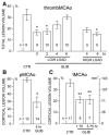
Results: Glibenclamide reduced total lesion volume by 53% in the thromboembolic MCAo model at 6 hours, reduced corrected cortical lesion volume by 51% in the permanent MCAo model at 4 hours, and reduced corrected cortical lesion volume by 41% in the temporary MCAo model at 5.75 hours (P<0.05 for all 3). Analysis of pooled data from the permanent MCAo and temporary MCAo series indicated a sigmoidal relationship between hemispheric swelling and corrected cortical lesion volume with the half-maximum cortical lesion volume being observed with 10% hemispheric swelling.
Conclusions: Low-dose glibenclamide has a strong beneficial effect on lesion volume and has a highly favorable therapeutic window in several models of ischemic stroke.
Figures
Figure 1
Glibenclamide reduces lesion volume. A–C, Lesion volumes are given for the thromboembolic MCAo model (A), the permanent MCAo model (B), and the temporary MCAo model (C) with vehicle versus glibenclamide administered at the indicated times; the number of rats in each group is indicated; values in B and C are for the “corrected” cortical lesion volume; the relationship between “percent hemisphere” and actual volume (mm3) is shown in C. *P<0.05; **P<0.01.
Figure 2
Hemispheric swelling predicts cortical lesion volume. Scatterplot of the corrected cortical lesion volume versus hemispheric swelling for treated and untreated rats in the permanent MCAo and temporary MCAo series (n=52). Fit to a sigmoid function indicated half-maximum cortical lesion (35%) occurring with 9.8% hemispheric swelling (maximum cortical lesion volume, 70%).
Figure 3
The doses of glibenclamide used are not hypoglycemogenic. A–B, Serum glucose levels at various times for vehicle-treated rats and rats treated with glibenclamide 5¾ hours post-MCAo (temporary MCAo series; loading dose, 10 _μ_g/mL; infusion dose, 200 ng/h; A) and for sham rats (no MCAo) administered a higher dose (loading dose, 80 _μ_g/mL; infusion dose, 600 ng/h; B).
Figure 4
Glibenclamide reduces the POI in the lateral and inferior/lateral cortex. A–C, Maps of the spatial density of POI were constructed using images from the control group (A), the group treated 1¾ hours post-MCAo (B), and the group treated 5¾ hours post-MCAo (C); temporary MCAo series; POI=1 is shown in white pseudocolor and POI=0 is shown in black pseudocolor; numbers indicate cortical Regions 1 to 4 (see text). D, The POI in cortical Regions 1 to 4 for the 3 groups, as indicated.
Figure 5
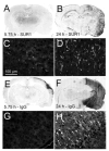
SUR1 expression, protein extravasation, and cellular damage continue beyond 5¾ hours after temporary MCAo. A–D, Immunolabeling for SUR1 at 5¾ hours (A, C) and at 24 hours (B, D) post-MCAo (temporary MCAo model with no treatment); C and D are from Region 4, as defined in Figure 4. E–H, Immunolabeling for IgG at 5¾ hours (E, G) and at 24 hours (F, H) post-MCAo; G and H are from Region 4. The data shown are representative of findings in 3 rats at each time.
Similar articles
- Novel approaches to the primary prevention of edema after ischemia.
Sheth KN. Sheth KN. Stroke. 2013 Jun;44(6 Suppl 1):S136. doi: 10.1161/STROKEAHA.113.001821. Stroke. 2013. PMID: 23709712 No abstract available. - Glibenclamide improves neurological function in neonatal hypoxia-ischemia in rats.
Zhou Y, Fathali N, Lekic T, Tang J, Zhang JH. Zhou Y, et al. Brain Res. 2009 May 13;1270:131-9. doi: 10.1016/j.brainres.2009.03.010. Epub 2009 Mar 21. Brain Res. 2009. PMID: 19306849 Free PMC article. - Glibenclamide enhances neurogenesis and improves long-term functional recovery after transient focal cerebral ischemia.
Ortega FJ, Jolkkonen J, Mahy N, Rodríguez MJ. Ortega FJ, et al. J Cereb Blood Flow Metab. 2013 Mar;33(3):356-64. doi: 10.1038/jcbfm.2012.166. Epub 2012 Nov 14. J Cereb Blood Flow Metab. 2013. PMID: 23149556 Free PMC article. - Glibenclamide in cerebral ischemia and stroke.
Simard JM, Sheth KN, Kimberly WT, Stern BJ, del Zoppo GJ, Jacobson S, Gerzanich V. Simard JM, et al. Neurocrit Care. 2014 Apr;20(2):319-33. doi: 10.1007/s12028-013-9923-1. Neurocrit Care. 2014. PMID: 24132564 Free PMC article. Review. - SUR1-TRPM4 channels, not KATP, mediate brain swelling following cerebral ischemia.
Woo SK, Tsymbalyuk N, Tsymbalyuk O, Ivanova S, Gerzanich V, Simard JM. Woo SK, et al. Neurosci Lett. 2020 Jan 23;718:134729. doi: 10.1016/j.neulet.2019.134729. Epub 2019 Dec 31. Neurosci Lett. 2020. PMID: 31899311 Free PMC article. Review.
Cited by
- Drug development in targeting ion channels for brain edema.
Luo ZW, Ovcjak A, Wong R, Yang BX, Feng ZP, Sun HS. Luo ZW, et al. Acta Pharmacol Sin. 2020 Oct;41(10):1272-1288. doi: 10.1038/s41401-020-00503-5. Epub 2020 Aug 27. Acta Pharmacol Sin. 2020. PMID: 32855530 Free PMC article. - Does inhibiting Sur1 complement rt-PA in cerebral ischemia?
Simard JM, Geng Z, Silver FL, Sheth KN, Kimberly WT, Stern BJ, Colucci M, Gerzanich V. Simard JM, et al. Ann N Y Acad Sci. 2012 Sep;1268:95-107. doi: 10.1111/j.1749-6632.2012.06705.x. Ann N Y Acad Sci. 2012. PMID: 22994227 Free PMC article. Review. - ATP-binding cassette transporters in immortalised human brain microvascular endothelial cells in normal and hypoxic conditions.
Lindner C, Sigrüner A, Walther F, Bogdahn U, Couraud PO, Schmitz G, Schlachetzki F. Lindner C, et al. Exp Transl Stroke Med. 2012 May 3;4(1):9. doi: 10.1186/2040-7378-4-9. Exp Transl Stroke Med. 2012. PMID: 22553972 Free PMC article. - Antidiabetic treatment, stroke severity and outcome.
Magkou D, Tziomalos K. Magkou D, et al. World J Diabetes. 2014 Apr 15;5(2):84-8. doi: 10.4239/wjd.v5.i2.84. World J Diabetes. 2014. PMID: 24748923 Free PMC article. Review. - Safety and efficacy of glibenclamide combined with rtPA in acute cerebral ischemia with occlusion/stenosis of anterior circulation (SE-GRACE): a randomized, double-blind, placebo-controlled trial.
Huang K, Zhao X, Zhao Y, Yang G, Zhou S, Yang Z, Huang W, Weng G, Chen P, Duan C, Lin Z, Wang S, Liu X, Huang Y, Zhang J, Zhang X, Li H, Ye S, Gu Y, Zhu M, Chen W, Quan W, Liu N, Chen Q, Chang Y, He J, Ji Z, Wu Y, Pan S; SE-GRACE Collaborators. Huang K, et al. EClinicalMedicine. 2023 Nov 1;65:102305. doi: 10.1016/j.eclinm.2023.102305. eCollection 2023 Nov. EClinicalMedicine. 2023. PMID: 37965431 Free PMC article.
References
Publication types
MeSH terms
Substances
Grants and funding
- HL051932/HL/NHLBI NIH HHS/United States
- R01 HL051932/HL/NHLBI NIH HHS/United States
- NS048260/NS/NINDS NIH HHS/United States
- R01 HL082517/HL/NHLBI NIH HHS/United States
- HL082517/HL/NHLBI NIH HHS/United States
- R01 NS048260/NS/NINDS NIH HHS/United States
- R01 HL082517-02/HL/NHLBI NIH HHS/United States
LinkOut - more resources
Full Text Sources
Other Literature Sources
Medical


