Nodal signalling is involved in left-right asymmetry in snails - PubMed (original) (raw)
. 2009 Feb 19;457(7232):1007-11.
doi: 10.1038/nature07603. Epub 2008 Dec 21.
Affiliations
- PMID: 19098895
- PMCID: PMC2661027
- DOI: 10.1038/nature07603
Nodal signalling is involved in left-right asymmetry in snails
Cristina Grande et al. Nature. 2009.
Abstract
Many animals display specific internal or external features with left-right asymmetry. In vertebrates, the molecular pathway that leads to this asymmetry uses the signalling molecule Nodal, a member of the transforming growth factor-beta superfamily, which is expressed in the left lateral plate mesoderm, and loss of nodal function produces a randomization of the left-right asymmetry of visceral organs. Orthologues of nodal have also been described in other deuterostomes, including ascidians and sea urchins, but no nodal orthologue has been reported in the other two main clades of Bilateria: Ecdysozoa (including flies and nematodes) and Lophotrochozoa (including snails and annelids). Here we report the first evidence for a nodal orthologue in a non-deuterostome group. We isolated nodal and Pitx (one of the targets of Nodal signalling) in two species of snails and found that the side of the embryo that expresses nodal and Pitx is related to body chirality: both genes are expressed on the right side of the embryo in the dextral (right-handed) species Lottia gigantea and on the left side in the sinistral (left-handed) species Biomphalaria glabrata. We pharmacologically inhibited the Nodal pathway and found that nodal acts upstream of Pitx, and that some treated animals developed with a loss of shell chirality. These results indicate that the involvement of the Nodal pathway in left-right asymmetry might have been an ancestral feature of the Bilateria.
Figures
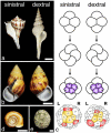
Figure 1. Chirality in snails
a, Species with different chirality: sinistral Busycon pulleyi (left) and dextral Fusinus salisbury (right). b, Sinistral (left) and dextral (right) shells of Amphidromus perversus, a species with chiral dimorphism. c, Early cleavage in dextral and sinistral species (based on Shibazaki _et al._27). In sinistral species, the third cleavage is in a counterclockwise direction, but clockwise in dextral species. In the next divisions the four quadrants (A, B, C, and D) are oriented as indicated. Cells colored in yellow have an endodermal fate and those in red have an endomesodermal fate in P. vulgata (dextral) and B. glabrata (sinistral) . d, B. glabrata possesses a sinistral shell and sinistral cleavage and internal organ organization. e, L. gigantea displays a dextral cleavage pattern and internal organ organization, and a relatively flat shell characteristic of limpets. Scale bars: 2.0 cm in a; 1.0 cm in b; 0.5 cm in d; 1.0 cm in e.
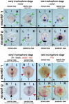
Figure 2. nodal and Pitx expression in snails
Anterior is up, L and R indicate left and right sides. Blue arrowhead in c-h indicates non-specific staining of the shell. a-b, nodal is expressed in the right cephalic region (upper green arrowhead) and right lateral ectoderm (lower green arrowhead) in L. gigantea as seen from dorsal (a) and right lateral views (b). c, Expression is maintained in the right lateral ectoderm (green arrowhead); right lateral view (d) shows that nodal expression (green arrowhead) is near the right side of the developing shell (blue arrowhead). e-h, nodal is expressed in the left lateral ectoderm (green arrowhead) in B. glabrata as seen from dorsal (e) and posterior views (f); g-h, Expression is maintained in the left lateral ectoderm (green arrowhead); posterior view (h) shows that nodal expression (green arrowhead) is near the left side of the developing shell (blue arrowhead). i-l, hedgehog (black arrowheads in i and k) is expressed along the ventral midline and nodal (red arrowheads in j and l) is expressed on the right side of L. gigantea (j) and on the left side of B. glabrata (l). m-n, Pitx is expressed in the visceral mass (orange arrow) and right lateral ectoderm (orange arrowhead) in L. gigantea as seen from dorsal (m) and right lateral views (n). o-p, Pitx is expressed in the stomodeum (orange arrow), visceral mass (orange triangle) and the left lateral ectoderm (orange arrowhead) in B. glabrata as seen from dorsal (o) and posterior views (p). Scale bars: 50 μm in all panels.
Figure 3. Early expression of nodal and Pitx in snails
a, 32-cell stage L. gigantea expressing nodal in a single cell. b, Group of cells expressing Pitx in L. gigantea. c, Onset of nodal expression in B. glabrata. d, Group of cells expressing Pitx in B. glabrata. e, 32-cell L. gigantea expressing nodal (red) in a single cell (2c) and brachyury (black) in two cells (3D and 3c). f-h, brachyury (black) is expressed in a symmetric fashion in progeny of 3c and 3d blastomeres (blue triangles in g), thus marking the bilateral axis and nodal (red) is expressed on the right side of L. gigantea in the progeny of 2c and 1c blastomeres, as seen from the lateral (f) and posterior (g, h) views of the same embryo. i, Group of cells expressing nodal (red) in the C quadrant and Pitx (black) in the D quadrant of the 120-cell stage embryo of L. gigantea. j, nodal (red) and Pitx (black) expression in adjacent areas of the right lateral ectoderm in L. gigantea. L and R indicate the left and right sides of the embryo, respectively. The black triangle in b and i, the green, yellow, and pink arrows in f and i, and the black and pink arrows in f and h point to the equivalent cells. Scale bars: 50 μm in all panels.
Figure 4. Wild type coiled and drug-treated non-coiled shells of B. glabrata and Pitx expression in drug-treated embryos
Control animals (a, e) display the normal sinistral shell morphology. Drug treated animals (b-d, f-g, exposed to SB-431542 from the 2 cell stage onwards) have straight shells. b-d are three different living individuals; f and g are a fourth individual, ethanol fixed, and shown from the side (f) and slightly rotated (g). h-k, Pitx expression in embryos exposed to SB-431542. Dorsal (h) and posterior views (i) of an embryo showing reduced levels of expression. Pitx expression is maintained in the stomodeum (orange arrow in h) and the visceral domain (orange triangle in i), but asymmetric expression in left ectoderm is greatly reduced (orange arrowhead). Dorsal (j) and posterior views (k) of an embryo in which the asymmetric ectodermal expression of Pitx is undetectable (orange arrowhead in j and k shows where expression would be expected), although the stomodeal (orange arrow in j) and visceral domain (orange triangle in k) expression of Pitx is normal. Pitx expression levels shown in h-k should be compared to levels in wildtype embryos in figure 2 o-p, which are the same levels seen in DMSO treated animals. L and R indicate the left and right sides of the embryo. Scale bars: 1.0 mm in a-d; 0.5 mm in e-g; 50 μm in h-k.
Similar articles
- Lophotrochozoa get into the game: the nodal pathway and left/right asymmetry in bilateria.
Grande C, Patel NH. Grande C, et al. Cold Spring Harb Symp Quant Biol. 2009;74:281-7. doi: 10.1101/sqb.2009.74.044. Epub 2010 Apr 22. Cold Spring Harb Symp Quant Biol. 2009. PMID: 20413706 Review. - Evolution, divergence and loss of the Nodal signalling pathway: new data and a synthesis across the Bilateria.
Grande C, Martín-Durán JM, Kenny NJ, Truchado-García M, Hejnol A. Grande C, et al. Int J Dev Biol. 2014;58(6-8):521-32. doi: 10.1387/ijdb.140133cg. Int J Dev Biol. 2014. PMID: 25690967 - Chiral blastomere arrangement dictates zygotic left-right asymmetry pathway in snails.
Kuroda R, Endo B, Abe M, Shimizu M. Kuroda R, et al. Nature. 2009 Dec 10;462(7274):790-4. doi: 10.1038/nature08597. Nature. 2009. PMID: 19940849 - Spiral cleavages determine the left-right body plan by regulating Nodal pathway in monomorphic gastropods, Physa acuta.
Abe M, Takahashi H, Kuroda R. Abe M, et al. Int J Dev Biol. 2014;58(6-8):513-20. doi: 10.1387/ijdb.140087rk. Int J Dev Biol. 2014. PMID: 25690966 - Shells and heart: are human laterality and chirality of snails controlled by the same maternal genes?
Oliverio M, Digilio MC, Versacci P, Dallapiccola B, Marino B. Oliverio M, et al. Am J Med Genet A. 2010 Oct;152A(10):2419-25. doi: 10.1002/ajmg.a.33655. Am J Med Genet A. 2010. PMID: 20830800 Review.
Cited by
- Molecular and cellular basis of left-right asymmetry in vertebrates.
Hamada H. Hamada H. Proc Jpn Acad Ser B Phys Biol Sci. 2020;96(7):273-296. doi: 10.2183/pjab.96.021. Proc Jpn Acad Ser B Phys Biol Sci. 2020. PMID: 32788551 Free PMC article. Review. - The physical basis of mollusk shell chiral coiling.
Chirat R, Goriely A, Moulton DE. Chirat R, et al. Proc Natl Acad Sci U S A. 2021 Nov 30;118(48):e2109210118. doi: 10.1073/pnas.2109210118. Proc Natl Acad Sci U S A. 2021. PMID: 34810260 Free PMC article. - Selective accumulation of germ-line associated gene products in early development of the sea star and distinct differences from germ-line development in the sea urchin.
Fresques T, Zazueta-Novoa V, Reich A, Wessel GM. Fresques T, et al. Dev Dyn. 2014 Apr;243(4):568-87. doi: 10.1002/dvdy.24038. Epub 2013 Dec 25. Dev Dyn. 2014. PMID: 24038550 Free PMC article. - Chiral Materials for Optics and Electronics: Ready to Rise?
Ham SH, Han MJ, Kim M. Ham SH, et al. Micromachines (Basel). 2024 Apr 15;15(4):528. doi: 10.3390/mi15040528. Micromachines (Basel). 2024. PMID: 38675339 Free PMC article. Review. - Investigating chiral morphogenesis of gold using generative cellular automata.
Im SW, Zhang D, Han JH, Kim RM, Choi C, Kim YM, Nam KT. Im SW, et al. Nat Mater. 2024 Jul;23(7):977-983. doi: 10.1038/s41563-024-01889-x. Epub 2024 May 1. Nat Mater. 2024. PMID: 38693448
References
- Massagué J, Gomis RR. The logic of TGFbeta signaling. FEBS Lett. 2006;580:2811–2820. - PubMed
- Hamada H, Meno C, Watanabe D, Saijoh Y. Establishment of vertebrate left-right asymmetry. Nat. Rev. Genet. 2002;2:103–113. - PubMed
- Okada Y, et al. Abnormal nodal flow precedes situs inversus in iv and inv mice. Mol. Cell. 1999;4:459–468. - PubMed
- Morokuma J, Ueno M, Kawanishi H, Saiga H, Nishida H. HrNodal, the ascidian nodal-related gene, is expressed in the left side of the epidermis, and lies upstream of HrPitx. Dev. Genes Evol. 2002;212:439–446. - PubMed
Publication types
MeSH terms
Substances
LinkOut - more resources
Full Text Sources
Other Literature Sources
Miscellaneous


