Neuroadaptations in the cellular and postsynaptic group 1 metabotropic glutamate receptor mGluR5 and Homer proteins following extinction of cocaine self-administration - PubMed (original) (raw)
Neuroadaptations in the cellular and postsynaptic group 1 metabotropic glutamate receptor mGluR5 and Homer proteins following extinction of cocaine self-administration
M Behnam Ghasemzadeh et al. Neurosci Lett. 2009.
Abstract
This study examined the role of group1 metabotropic glutamate receptor mGluR5 and associated postsynaptic scaffolding protein Homer1b/c in behavioral plasticity after three withdrawal treatments from cocaine self-administration. Rats self-administered cocaine or saline for 14 days followed by a withdrawal period during which rats underwent extinction training, remained in their home cages, or were placed in the self-administration chambers in the absence of extinction. Subsequently, the tissue level and distribution of proteins in the synaptosomal fraction associated with the postsynaptic density were examined. Cocaine self-administration followed by home cage exposure reduced the mGluR5 protein in nucleus accumbens (NA) shell and dorsolateral striatum. While extinction training reduced mGluR5 protein in NAshell, NAcore and dorsolateral striatum did not display any change. The scaffolding protein PSD95 increased in NAcore of the extinguished animals. Extinction of drug seeking was associated with a significant decrease in the synaptosomal mGluR5 protein in NAshell and an increase in dorsolateral striatum, while that of NAcore was not modified. Interestingly, both Homer1b/c and PSD95 scaffolding proteins were decreased in the synaptosomal fraction after extinction training in NAshell but not NAcore. Extinguished drug-seeking behavior was also associated with an increase in the synaptosomal actin proteins in dorsolateral striatum. Therefore, extinction of cocaine seeking is associated with neuroadaptations in mGluR5 expression and distribution that are region-specific and consist of extinction-induced reversal of cocaine-induced adaptations as well as emergent extinction-induced alterations. Concurrent plasticity in the scaffolding proteins further suggests that mGluR5 receptor neuroadaptations may have implications for synaptic function.
Figures
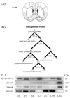
Figure 1. Subcellular fractionation analysis
Panel A shows the dissection of the nucleus accumbens core and shell and dorsolateral striatum. The numbers on the coronal brain section represent distance from Bregma. Panel B shows a schematic of the subcellular fractionation procedure (adopted from [9]), as described in Materials and Methods. The H and LP1 fractions were used for western blot analysis. Panel C shows representative immunoblots from nucleus accumbens tissue characterizing the content of each fraction.
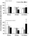
Figure 2. Tissue and synaptosomal membrane fraction levels of mGluR5 receptor protein after cocaine self-administration and Home, Box, and Extinction treatments
At the end of the withdrawal period, all animals were decapitated and brain tissue was dissected for the areas of interest. At the tissue level, mGluR5 protein was reduced significantly in the NAshell of Home and Extinction animals and showed near significant decrease in DL Striatum in Home animals but was not changed under other withdrawal conditions or in other brain regions. In the synaptosomal membrane fraction, mGluR5 receptor protein was increased in NAcore of Home animals and DL striatum of Extinction animals. However, the receptor protein was significantly decreased in the NAshell of extinction animals. N= 7–11 rats per treatment group. * p < 0.05, + p= 0.066 compared to respective saline control. @ p < 0.05 compared to Box treatment, # p < 0.05 compared to Home treatment.
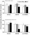
Figure 3. Tissue and synaptosomal membrane fraction levels of Homer 1b/c scaffolding protein after cocaine self-administration and Home, Box, and Extinction treatments
At the end of the withdrawal period, all animals were decapitated and brain tissue was dissected for the areas of interest. At the tissue level, the protein level was not modified in any withdrawal treatment group in any region examined. In the synaptosomal membrane fraction, Homer 1b/c protein was reduced in NAshell and exhibited a decreasing trend in the dorsolateral Striatum of Extinction animals. There were no changes in the protein level in the NAcore. N= 7–11 rats per treatment group. * p < 0.05, + p= 0.065 compared to respective saline control. @ p < 0.05 compared to Box treatment, # p < 0.05 compared to Home treatment.
Similar articles
- Behavioral sensitization to cocaine is associated with increased glutamate receptor trafficking to the postsynaptic density after extended withdrawal period.
Ghasemzadeh MB, Mueller C, Vasudevan P. Ghasemzadeh MB, et al. Neuroscience. 2009 Mar 3;159(1):414-26. doi: 10.1016/j.neuroscience.2008.10.027. Epub 2008 Nov 1. Neuroscience. 2009. PMID: 19105975 - Extinction training after cocaine self-administration induces glutamatergic plasticity to inhibit cocaine seeking.
Knackstedt LA, Moussawi K, Lalumiere R, Schwendt M, Klugmann M, Kalivas PW. Knackstedt LA, et al. J Neurosci. 2010 Jun 9;30(23):7984-92. doi: 10.1523/JNEUROSCI.1244-10.2010. J Neurosci. 2010. PMID: 20534846 Free PMC article. - Region-specific alterations in glutamate receptor expression and subcellular distribution following extinction of cocaine self-administration.
Ghasemzadeh MB, Vasudevan P, Mueller CR, Seubert C, Mantsch JR. Ghasemzadeh MB, et al. Brain Res. 2009 Apr 24;1267:89-102. doi: 10.1016/j.brainres.2009.01.047. Epub 2009 Feb 5. Brain Res. 2009. PMID: 19368820 - Extinction training regulates neuroadaptive responses to withdrawal from chronic cocaine self-administration.
Self DW, Choi KH, Simmons D, Walker JR, Smagula CS. Self DW, et al. Learn Mem. 2004 Sep-Oct;11(5):648-57. doi: 10.1101/lm.81404. Learn Mem. 2004. PMID: 15466321 Free PMC article. Review. - Brain-derived neurotrophic factor and cocaine addiction.
McGinty JF, Whitfield TW Jr, Berglind WJ. McGinty JF, et al. Brain Res. 2010 Feb 16;1314:183-93. doi: 10.1016/j.brainres.2009.08.078. Epub 2009 Sep 2. Brain Res. 2010. PMID: 19732758 Free PMC article. Review.
Cited by
- Marked global reduction in mGluR5 receptor binding in smokers and ex-smokers determined by [11C]ABP688 positron emission tomography.
Akkus F, Ametamey SM, Treyer V, Burger C, Johayem A, Umbricht D, Gomez Mancilla B, Sovago J, Buck A, Hasler G. Akkus F, et al. Proc Natl Acad Sci U S A. 2013 Jan 8;110(2):737-42. doi: 10.1073/pnas.1210984110. Epub 2012 Dec 17. Proc Natl Acad Sci U S A. 2013. PMID: 23248277 Free PMC article. - Glutamatergic plasticity in medial prefrontal cortex and ventral tegmental area following extended-access cocaine self-administration.
Ghasemzadeh MB, Vasudevan P, Giles C, Purgianto A, Seubert C, Mantsch JR. Ghasemzadeh MB, et al. Brain Res. 2011 Sep 21;1413:60-71. doi: 10.1016/j.brainres.2011.06.041. Epub 2011 Jun 23. Brain Res. 2011. PMID: 21855055 Free PMC article. - Homer2 within the nucleus accumbens core bidirectionally regulates alcohol intake by both P and Wistar rats.
Haider A, Woodward NC, Lominac KD, Sacramento AD, Klugmann M, Bell RL, Szumlinski KK. Haider A, et al. Alcohol. 2015 Sep;49(6):533-42. doi: 10.1016/j.alcohol.2015.03.009. Epub 2015 Jul 21. Alcohol. 2015. PMID: 26254965 Free PMC article. - Metabotropic glutamate receptor 5 (mGluR5) antagonists attenuate cocaine priming- and cue-induced reinstatement of cocaine seeking.
Kumaresan V, Yuan M, Yee J, Famous KR, Anderson SM, Schmidt HD, Pierce RC. Kumaresan V, et al. Behav Brain Res. 2009 Sep 14;202(2):238-44. doi: 10.1016/j.bbr.2009.03.039. Epub 2009 Apr 5. Behav Brain Res. 2009. PMID: 19463707 Free PMC article. - Calcium-permeable AMPA receptors in the VTA and nucleus accumbens after cocaine exposure: when, how, and why?
Wolf ME, Tseng KY. Wolf ME, et al. Front Mol Neurosci. 2012 Jun 27;5:72. doi: 10.3389/fnmol.2012.00072. eCollection 2012. Front Mol Neurosci. 2012. PMID: 22754497 Free PMC article.
References
- Anderson SM, Famous KR, Sadri-Vakili G, Kumaresan V, Schmidt HD, Bass CE, Terwilliger EF, Cha JJ, Pierce RC. CaMKII: a biochemical bridge linking accumbens dopamine and glutamate systems in cocaine seeking. Nature Neurosci. 2008;11:344–353. - PubMed
- Ango F, Robbe D, Tu JC, Xiao B, Worley PF, Pin JP, Bockaert J, Fagni L. Homer-dependent cell surface expression of metabotropic glutamate receptor type 5 in neurons. Mol. Cell. Neurosci. 2002;20:323–329. - PubMed
- Ary AW, Szumlinski KK. Regional differences in the effects of withdrawal from repeated cocaine upon Homer and glutamate receptor expression: A two-species comparison. Brain Res. 2007;1184:295–305. - PubMed
- Bäckström P, Bachteler D, Koch S, Hyytiä P, Spanagel R. mGluR5 antagonist MPEP reduces ethanol-seeking and relapse behavior. Neuropsychopharmacology. 2004;29:921–928. - PubMed
- Bouton ME. Context, ambiguity, and unlearning: sources of relapse after behavioral extinction. Biol. Psychiatry. 2002;52:976–986. - PubMed
Publication types
MeSH terms
Substances
Grants and funding
- R01 DA015758/DA/NIDA NIH HHS/United States
- R01 DA015758-03/DA/NIDA NIH HHS/United States
- R01 DA015758-02/DA/NIDA NIH HHS/United States
- R01 DA015758-05S1/DA/NIDA NIH HHS/United States
- DA15758/DA/NIDA NIH HHS/United States
- R01 DA015758-04/DA/NIDA NIH HHS/United States
- R01 DA014328/DA/NIDA NIH HHS/United States
- R01 DA015758-05/DA/NIDA NIH HHS/United States
- R01 DA015758-01/DA/NIDA NIH HHS/United States
- DA14328/DA/NIDA NIH HHS/United States
LinkOut - more resources
Full Text Sources
Miscellaneous


