Dicer is regulated by cellular stresses and interferons - PubMed (original) (raw)
Dicer is regulated by cellular stresses and interferons
Jennifer L Wiesen et al. Mol Immunol. 2009 Mar.
Abstract
The generation of microRNAs is dependent on the RNase III enzyme Dicer, the levels of which vary in different normal cells and in disease states. We demonstrate that Dicer protein expression in JAR trophoblast cells, and several other cell types, was inhibited by multiple stresses including reactive oxygen species, phorbol esters and the Ras oncogene. Additionally, double-stranded RNA and Type I interferons repress Dicer protein in contrast to IFN-gamma which induces Dicer. The effects of stresses and interferons are primarily post-transcriptional. The findings suggest that Dicer is a stress response component and identifies interferons as potentially important regulators of Dicer expression.
Figures
Figure 1. Dicer protein and activity are differentially regulated between cell types
A. Untreated JAR, JEG-3, HeLa, Raji, and Daudi cells were analyzed by western blotting for Dicer and βactin levels. B. Dicer and GAPDH mRNA levels were assessed by quantitative real time RT-PCR for each of the above cell lines and are expressed as CT values. C. Dicer activity was assessed for JAR, HeLa, and Raji cells. Cytoplasmic extract from each cell line or recombinant Dicer enzyme was assayed with a synthetic pre-miR-122a and buffer at 37°C for 2 hours. Activity was determined by quantitative RT-PCR of mature miR-122a levels relative to the recombinant dicer and is presented as Δ CT values. Constitutively expressed miRs were determined by miRNA microarray analyses and are presented as the percentage of miRs constitutively expressed in each cell line.
Figure 2. The HDACi, TSA, downregulates Dicer protein but not message levels in human JAR and JEG-3 trophoblasts and in a mouse B16 melanoma cell line
JAR (50nM), JEG-3 (250nM), and B16 (100nM) cell lines were treated in vitro with TSA for 24hrs; total RNA was recovered and whole cell lysates were prepared. Western blots for Dicer protein expression and real time RT-PCR analyses (reported as CT values) of Dicer mRNA levels are shown here for treated and untreated samples.
Figure 3. HDACi effects on Dicer activity and expression
A. Cytoplasmic extract from the JAR cell line or recombinant Dicer enzyme was assayed with 50nM TSA, a synthetic pre-miR-122a, and buffer at 37°C for 2 hours. Activity was determined by quantitative RT-PCR of mature miR-122a levels relative to the untreated samples and is presented as Δ CT values. B. JAR cells were treated with valproic acid (250μM for 24hrs) and TSA (50nM for 24hrs). Proteins were collected and analyzed by western blotting for Dicer and βactin levels.
Figure 4. Multiple stress pathways downregulate Dicer protein
A. JAR trophoblast cells were treated with 500μM H2O2 for 4hrs, washed, and then cultured for an additional 20 hours. Proteins were collected and analyzed by western blotting for Dicer and βactin levels. B. IMR-90 human fibroblast cells were transduced with either an empty retroviral vector or an activated Ras construct (18). Proteins were collected and analyzed by western blotting for Dicer and βactin levels. C. JAR cells were treated with PMA/IM (500ng/mL PMA for 4hrs followed by the addition of 10mM IM for 24hrs). Proteins were collected and analyzed by western blotting for Dicer and βactin levels.
Figure 5. Toll-like receptor and interferon mediated effects on Dicer expression
A. JAR cells were treated with poly I:C (2mg/mL for 24hrs), a TLR3 ligand, and IFN-α (1000U/mL for 72hrs), produced by multiple TLR stimuli. Proteins were collected and analyzed by western blotting for Dicer and βactin levels. B. JAR cells were also treated with 500U/mL IFN-γ for 24hrs to compare with IFN-α results. Proteins were collected and analyzed by western blotting for Dicer and βactin levels. Similar results to those shown above were seen in HeLa cells (data not shown).
Figure 6. Dicer protein levels are selectively regulated by cellular stresses in JAR trophoblast cells
Individual JAR whole cell extracts from the designated treatments (1,000U/mL IFN-α for 72hrs, 50nM TSA for 24hrs, and 250μM H2O2 for 15min) were analyzed by western blotting for Dicer, Argonaute 1, TTP, Hsp90, Calpain, Mad 1, GAPDH, and βactin. The results are representative of three independent experiments.
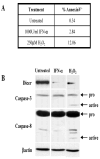
Figure 7. Apoptosis is not responsible for downregulation of Dicer with IFN-α treatment
JAR cells were treated with 1000U/mL IFN-αfor 72hrs. For a positive control, JAR cells were treated with 250μM H2O2 for 15minutes. A. The presence of apoptotic cells was analyzed by Annexin V surface staining of treated cells. Treated JAR cultures were stained with Annexin V-FITC (see Materials and Methods). After single cell gating, a marker was set based on the untreated cells and the percentage of cells stained with Annexin V was determined. B. Activation of the apoptotic signaling cascade was analyzed by SDS-PAGE and western blotting for Dicer, Caspase-3, Caspase-8, and βactin.
Figure 8. Dicer protein levels in fresh tissues of mice and humans
A. Splenocytes from C57BL/6 mice were treated in culture with 100U/mL IFN-γ for 24hrs or 1000U/mL IFN-αfor 72hrs. Protein was analyzed by SDS-PAGE and western blotting for Dicer and βactin. B. Fresh C57BL/6 kidneys were dissociated and cultured for 72hrs. Cultures were then treated with 1000U/mL IFN-α for 72hrs prior to whole cell lysate preparation and western analysis. C. Normal human kidney cells (obtained with IRB approval as excess tissue post-Pathology examination) were cultured and treated with 500U/mL IFN-γ for 24hrs or 10000U/mL IFN-α for 72hrs. Protein was analyzed by SDS-PAGE and western blotting.
Similar articles
- The epigenetic regulation of Dicer and microRNA biogenesis by Panobinostat.
Hoffend NC, Magner WJ, Tomasi TB. Hoffend NC, et al. Epigenetics. 2017 Feb;12(2):105-112. doi: 10.1080/15592294.2016.1267886. Epub 2016 Dec 9. Epigenetics. 2017. PMID: 27935420 Free PMC article. - MDA-7/IL-24 regulates the miRNA processing enzyme DICER through downregulation of MITF.
Pradhan AK, Bhoopathi P, Talukdar S, Scheunemann D, Sarkar D, Cavenee WK, Das SK, Emdad L, Fisher PB. Pradhan AK, et al. Proc Natl Acad Sci U S A. 2019 Mar 19;116(12):5687-5692. doi: 10.1073/pnas.1819869116. Epub 2019 Mar 6. Proc Natl Acad Sci U S A. 2019. PMID: 30842276 Free PMC article. - Overexpression of dicer as a result of reduced let-7 MicroRNA levels contributes to increased cell proliferation of oral cancer cells.
Jakymiw A, Patel RS, Deming N, Bhattacharyya I, Shah P, Lamont RJ, Stewart CM, Cohen DM, Chan EK. Jakymiw A, et al. Genes Chromosomes Cancer. 2010 Jun;49(6):549-59. doi: 10.1002/gcc.20765. Genes Chromosomes Cancer. 2010. PMID: 20232482 Free PMC article. - Crosstalk between NRF2 and Dicer through metastasis regulating MicroRNAs; mir-34a, mir-200 family and mir-103/107 family.
Nabih HK. Nabih HK. Arch Biochem Biophys. 2020 Jun 15;686:108326. doi: 10.1016/j.abb.2020.108326. Epub 2020 Mar 3. Arch Biochem Biophys. 2020. PMID: 32142889 Review. - The many faces of Dicer: the complexity of the mechanisms regulating Dicer gene expression and enzyme activities.
Kurzynska-Kokorniak A, Koralewska N, Pokornowska M, Urbanowicz A, Tworak A, Mickiewicz A, Figlerowicz M. Kurzynska-Kokorniak A, et al. Nucleic Acids Res. 2015 May 19;43(9):4365-80. doi: 10.1093/nar/gkv328. Epub 2015 Apr 16. Nucleic Acids Res. 2015. PMID: 25883138 Free PMC article. Review.
Cited by
- miR-3928 activates ATR pathway by targeting Dicer.
Chang L, Hu W, Ye C, Yao B, Song L, Wu X, Ding N, Wang J, Zhou G. Chang L, et al. RNA Biol. 2012 Oct;9(10):1247-54. doi: 10.4161/rna.21821. Epub 2012 Aug 24. RNA Biol. 2012. PMID: 22922797 Free PMC article. - MicroRNAs as regulatory elements in immune system logic.
Mehta A, Baltimore D. Mehta A, et al. Nat Rev Immunol. 2016 Apr 28;16(5):279-94. doi: 10.1038/nri.2016.40. Nat Rev Immunol. 2016. PMID: 27121651 Review. - Constitutive Dicer1 phosphorylation accelerates metabolism and aging in vivo.
Aryal NK, Pant V, Wasylishen AR, Parker-Thornburg J, Baseler L, El-Naggar AK, Liu B, Kalia A, Lozano G, Arur S. Aryal NK, et al. Proc Natl Acad Sci U S A. 2019 Jan 15;116(3):960-969. doi: 10.1073/pnas.1814377116. Epub 2018 Dec 28. Proc Natl Acad Sci U S A. 2019. PMID: 30593561 Free PMC article. - Mutant p53 regulates Dicer through p63-dependent and -independent mechanisms to promote an invasive phenotype.
Muller PA, Trinidad AG, Caswell PT, Norman JC, Vousden KH. Muller PA, et al. J Biol Chem. 2014 Jan 3;289(1):122-32. doi: 10.1074/jbc.M113.502138. Epub 2013 Nov 12. J Biol Chem. 2014. PMID: 24220032 Free PMC article. - MicroRNA as Type I Interferon-Regulated Transcripts and Modulators of the Innate Immune Response.
Forster SC, Tate MD, Hertzog PJ. Forster SC, et al. Front Immunol. 2015 Jul 8;6:334. doi: 10.3389/fimmu.2015.00334. eCollection 2015. Front Immunol. 2015. PMID: 26217335 Free PMC article. Review.
References
- Akira S, Takeda K. Toll-like receptor signaling. Nature Rev Immunol. 2004;4:499–511. - PubMed
- Bartel DP. MicroRNAs: Genomics, Biogenesis, Mechanism, and Function. Cell. 2004;116:281–297. - PubMed
- Bhattacharyya SN. Relief of microRNA-mediated translational repression in human cells subjected to stress. Cell. 2006;125:1111–1124. - PubMed
- Bihani T, Chicas A, Lo CP-K, Lin AW. Dissecting the Senescence-like Program in Tumor Cells Activated by Ras Signaling. J Biol Chem. 2007;282:2666–2675. - PubMed
Publication types
MeSH terms
Substances
Grants and funding
- HD 17013/HD/NICHD NIH HHS/United States
- P30 CA016056/CA/NCI NIH HHS/United States
- CA16056/CA/NCI NIH HHS/United States
- R01 HD017013-19/HD/NICHD NIH HHS/United States
- R01 CA124971-01A2/CA/NCI NIH HHS/United States
- CA 124971/CA/NCI NIH HHS/United States
- R01 HD017013/HD/NICHD NIH HHS/United States
- R01 CA124971/CA/NCI NIH HHS/United States
LinkOut - more resources
Full Text Sources
Other Literature Sources


