Prefrontal and midline interactions mediating behavioural control - PubMed (original) (raw)
Prefrontal and midline interactions mediating behavioural control
Catherine Fassbender et al. Eur J Neurosci. 2009 Jan.
Abstract
Top-down control processes are thought to interact with bottom-up stimulus-driven task demands to facilitate the smooth execution of behaviour. Frontal and midline brain areas in humans are believed to subserve these control processes but their distinct roles and the interactions between them remain to be fully elucidated. In this functional magnetic resonance imaging (fMRI) study, we utilized a GO/NO-GO task with cued and uncued inhibitory events to investigate the effect of cue-induced levels of top-down control on NO-GO trial response conflict. We found that, on a within-subjects, trial-for-trial basis, high levels of top-down control, as indexed by left dorsolateral prefrontal activation prior to the NO-GO, resulted in lower levels of activation on the NO-GO trial in the pre-supplementary motor area. These results suggest that prefrontal and midline regions work together to implement cognitive control and reveal that intra-subject variability is reflected in these lateral and midline interactions.
Figures
Figure 1
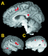
Areas activated during cue periods prior to a correct response. Activated areas include pre-SMA (A, Talairach: x = 2, y = 10, z = 46), right (B, 41, 14, 27) and left (C, -28, -7, 48) dorsolateral PFC as well as left parietal, occipital and right temporal areas.
Figure 2
Method of defining high and low control STOP events. A. represents the regressor for cued successful STOPs during the initial analysis. As shown in B., activations during the cue period in left dorsolateral PFC were divided into low activations, or low control cue periods (represented by the green curve) and high activations, or high control cue periods (represented by the blue curve). Successful STOPs from the original regressor (A) were then classified as high or low control STOPs based upon the cue period immediately preceding them, as shown in B. Therefore, the blue arrows represent STOPs that were preceded by high control cue periods and are high control STOPs, whereas the green arrows represent STOPs that were preceded by low control cue periods and are low control STOPs.
Figure 3
Correct inhibition activations from the left dorsolateral PFC split (A (i): pre-SMA (0, 1, 58), SMA (1, -11, 59) and (ii): rostral ACC (-2, 30, 28), caudal ACC (1, 13, 26)) and from the right dorsolateral PFC split (B (i): ACC (-2, 31, 26) and (ii): rostral SMA (1, -3, 60), caudal SMA (1, -18, 65)). Areas in which activation differed significantly between high and low control conditions are displayed in green.
Figure 4
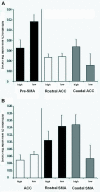
A: Mean activation in pre-SMA, rostral ACC and dorsal ACC for high and low control events as defined by the left dorsolateral PFC. B: Mean activation in ACC and rostral SMA and caudal SMA for high and low control events defined by the right dorsolateral PFC.
Figure 5
An area activated during errors in this task (from Hester et al, 2004b), including ACC extending into dorsal ACC, is represented in yellow. Pre-SMA, implicated in response-conflict monitoring in this study, is displayed in green. Orange represents the small area of overlap in pre-SMA between the two clusters. We found no evidence for any conflict-related activity in ACC, even at a liberal threshold.
Similar articles
- The prefrontal cortex and the executive control of attention.
Rossi AF, Pessoa L, Desimone R, Ungerleider LG. Rossi AF, et al. Exp Brain Res. 2009 Jan;192(3):489-97. doi: 10.1007/s00221-008-1642-z. Epub 2008 Nov 22. Exp Brain Res. 2009. PMID: 19030851 Free PMC article. - Changing plans: neural correlates of executive control in monkey and human frontal cortex.
Dias EC, McGinnis T, Smiley JF, Foxe JJ, Schroeder CE, Javitt DC. Dias EC, et al. Exp Brain Res. 2006 Sep;174(2):279-91. doi: 10.1007/s00221-006-0444-4. Epub 2006 Apr 25. Exp Brain Res. 2006. PMID: 16636795 - Interactions between decision making and performance monitoring within prefrontal cortex.
Walton ME, Devlin JT, Rushworth MF. Walton ME, et al. Nat Neurosci. 2004 Nov;7(11):1259-65. doi: 10.1038/nn1339. Epub 2004 Oct 24. Nat Neurosci. 2004. PMID: 15494729 - Limitations in information processing in the human brain: neuroimaging of dual task performance and working memory tasks.
Klingberg T. Klingberg T. Prog Brain Res. 2000;126:95-102. doi: 10.1016/S0079-6123(00)26009-3. Prog Brain Res. 2000. PMID: 11105642 Review. No abstract available. - Task set and prefrontal cortex.
Sakai K. Sakai K. Annu Rev Neurosci. 2008;31:219-45. doi: 10.1146/annurev.neuro.31.060407.125642. Annu Rev Neurosci. 2008. PMID: 18558854 Review.
Cited by
- Effect of foreknowledge on neural activity of primary "go" responses relates to response stopping and switching.
Xu B, Levy S, Butman J, Pham D, Cohen LG, Sandrini M. Xu B, et al. Front Hum Neurosci. 2015 Feb 4;9:34. doi: 10.3389/fnhum.2015.00034. eCollection 2015. Front Hum Neurosci. 2015. PMID: 25698959 Free PMC article. - Working memory in attention deficit/hyperactivity disorder is characterized by a lack of specialization of brain function.
Fassbender C, Schweitzer JB, Cortes CR, Tagamets MA, Windsor TA, Reeves GM, Gullapalli R. Fassbender C, et al. PLoS One. 2011;6(11):e27240. doi: 10.1371/journal.pone.0027240. Epub 2011 Nov 10. PLoS One. 2011. PMID: 22102882 Free PMC article. - Dissociating the role of prefrontal and premotor cortices in controlling inhibitory mechanisms during motor preparation.
Duque J, Labruna L, Verset S, Olivier E, Ivry RB. Duque J, et al. J Neurosci. 2012 Jan 18;32(3):806-16. doi: 10.1523/JNEUROSCI.4299-12.2012. J Neurosci. 2012. PMID: 22262879 Free PMC article. - Neural correlates of craving and impulsivity in abstinent former cocaine users: Towards biomarkers of relapse risk.
Bell RP, Garavan H, Foxe JJ. Bell RP, et al. Neuropharmacology. 2014 Oct;85:461-70. doi: 10.1016/j.neuropharm.2014.05.011. Epub 2014 Jun 18. Neuropharmacology. 2014. PMID: 24951856 Free PMC article. - Differences in time course activation of dorsolateral prefrontal cortex associated with low or high risk choices in a gambling task.
Bembich S, Clarici A, Vecchiet C, Baldassi G, Cont G, Demarini S. Bembich S, et al. Front Hum Neurosci. 2014 Jun 24;8:464. doi: 10.3389/fnhum.2014.00464. eCollection 2014. Front Hum Neurosci. 2014. PMID: 25009486 Free PMC article.
References
- Badre D, Wagner A. Selection, integration, and conflict monitoring: Assessing the nature and generality of prefrontal cognitive control mechanisms. Neuron. 2004;41:473–487. - PubMed
- Botvinick M, Nystrom LE, Fissell K, Carter CS, Cohen JD. Conflict monitoring versus selection-for-action in anterior cingulate cortex. Nature. 1999;402:179–81. - PubMed
- Botvinick MM, Braver TS, Barch DM, Carter CS, Cohen JD. Conflict monitoring and cognitive control. Psychol Rev. 2001;108:624–52. - PubMed
- Brass M, von Cramon DY. The role of the frontal cortex in task preparation. Cereb Cortex. 2002;12:908–14. - PubMed
- Brass M, von Cramon DY. Decomposing components of task preparation with functional magnetic resonance imaging. Journal of Cognitive Neuroscience. 2004;16:609–620. - PubMed
Publication types
MeSH terms
Grants and funding
- R01 DA014100/DA/NIDA NIH HHS/United States
- R01 MH065350/MH/NIMH NIH HHS/United States
- M01 RR000058/RR/NCRR NIH HHS/United States
- R03 MH063434/MH/NIMH NIH HHS/United States
- R03 MH063434-02/MH/NIMH NIH HHS/United States
- MH63434/MH/NIMH NIH HHS/United States
- M01 RR00058/RR/NCRR NIH HHS/United States
- MH65350/MH/NIMH NIH HHS/United States
- R01 MH065350-05/MH/NIMH NIH HHS/United States
- DA14100/DA/NIDA NIH HHS/United States
LinkOut - more resources
Full Text Sources


