Increased thymus- and decreased parathyroid-fated organ domains in Splotch mutant embryos - PubMed (original) (raw)
Increased thymus- and decreased parathyroid-fated organ domains in Splotch mutant embryos
Ann V Griffith et al. Dev Biol. 2009.
Abstract
Embryos that are homozygous for Splotch, a null allele of Pax3, have a severe neural crest cell (NCC) deficiency that generates a complex phenotype including spina bifida, exencephaly and cardiac outflow tract abnormalities. Contrary to the widely held perception that thymus aplasia or hypoplasia is a characteristic feature of Pax3(Sp/Sp) embryos, we find that thymic rudiments are larger and parathyroid rudiments are smaller in E11.5-12.5 Pax3(Sp/Sp) compared to Pax3(+/+) embryos. The thymus originates from bilateral third pharyngeal pouch primordia containing endodermal progenitors of both thymus and parathyroid glands. Analyses of Foxn1 and Gcm2 expression revealed a dorsal shift in the border between parathyroid- and thymus-fated domains at E11.5, with no change in the overall cellularity or volume of each shared primordium. The border shift increases the allocation of third pouch progenitors to the thymus domain and correspondingly decreases allocation to the parathyroid domain. Initial patterning in the E10.5 pouch was normal suggesting that the observed change in the location of the organ domain interface arises during border refinement between E10.5 and E11.5. Given the well-characterized NCC defects in Splotch mutants, these findings implicate NCCs in regulating patterning of third pouch endoderm into thymus- versus parathyroid-specified domains, and suggest that organ size is determined in part by the number of progenitor cells specified to a given fate.
Figures
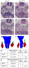
Figure 1. Thymus lobes in Pax3Sp/Sp embryos are large and ectopic
(A–D) Transverse H&E stained sections from E12.5 embryos. Dorsal is up. Pax3+/+ thymus lobes are located cranial to the heart and posterior to the laryngeo-tracheal groove (A). Thymus lobes are not evident at a comparable position in the _Pax3_Sp/Sp embryo (B). Hypoplastic dorsal root ganglia are apparent in _Pax3_Sp/Sp embryos. Cranial sections, reveal large, ectopic thymus lobes in _Pax3_Sp/Sp mutant (D), whereas only the anterior tip of one lobe is evident in the wildtype littermate (C). 3-D reconstructions of Pax3+/+ (E) and _Pax3_Sp/Sp (F) pharyngeal regions showing thymus (red), parathyroid (green), esophagus (blue) and trachea (yellow). Average volume of Pax3+/+ and _Pax3_Sp/Sp thymic (G) and parathyroid lobes (H). Abbreviations are drg, dorsal root ganglia; e, esophagus; tr, trachea ; th, thymus; lt, laryngeo-tracheal groove; ub, ultimobranchial bodies.
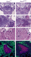
Figure 2. Delayed detachment phenotype is specific to the third pouch
Transverse H&E stained sections from (A,C,E) Pax3+/+ and (B,D,F) _Pax3_Sp/Sp E12.5 embryos. Panels C and D are higher magnification (20X) images of A and B (5X). The arrow in D shows attachment of _Pax3_Sp/Sp thymus rudiment to the pharynx. Normal ultimobranchial body detachment occurs in Pax3+/+ and _Pax3_Sp/Sp embryos (E,F). Abbreviations are thymus (th), parathyroid (pt), pharynx (ph) and ultimobranchial body (ub). Transverse frozen sections from (G) Pax3+/+ and (H) _Pax3_Sp/Sp E12.5 embryos were stained with anti-PDGFRα (green), anti-pancytokeratin (red) and DAPI (blue).
Figure 3. NCC localization is disrupted at E10.5 in Pax3Sp/Sp embryos
Sagittal sections from E10.5 Pax3+/+(A,C) and Pax3Sp/Sp (B,D) embryos were incubated with anti-sense riboprobe for CrabP1 to detect migrating NCC. (C and D) are higher magnification views of the third pouch region. Cranial is up and ventral is left. The clear space surrounding the pouches is an artifact of tissue processing. There is a paucity of CrabP1 expressing NCCs in pharyngeal arches and pouches of Pax3Sp/Sp embryos.
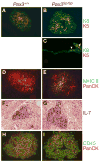
Figure 4. Epithelial differentiation is normal in E12.5 in Pax3Sp/Sp thymus lobes
Transverse frozen sections from wild type (A) and _Pax3_Sp/Sp embryos (B,C) stained with anti-K5 (red) or anti-K8 (green). Note the presence of K8+K5+ epithelial cells in the center of the _Pax3_Sp/Sp rudiment (arrow) even when it remains attached to the pharyngeal endoderm (arrowhead). Frozen thymus sections form Pax3+/+ (D) and _Pax3_Sp/Sp (E) embryos stained with anti-MHC class II (green) and anti-pan-cytokeratin (red). ISH analysis of IL-7 expression in sagittal sections of E12.5 thymus from Pax3+/+ (F) and _Pax3_Sp/Sp (G) embryos. Frozen transverse sections from Pax3+/+ (H) and _Pax3_Sp/Sp (I) E12.5 embryos stained with anti-CD45 (green) and anti-pan-cytokeratin (red).
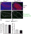
Figure 5. Proliferation and cellularity in Pax3Sp/Sp common primordia
Sagittal sections of E11.5 Pax3+/+(A, C) and Pax3Sp/Sp (B, D) embryos; cranial is up and dorsal is right. Staining for K8 (red) reveals epithelial cells in the common primordium; DAPI staining (blue) detects nuclei (A,B). Mitotic cells detected by costaining with anti-phosphohistone H3 antibody (green) (C,D). Average cell number (E) and percentage of mitotic cells (F) per section were determined by counting multiple sections from three Pax3+/+ and three Pax3Sp/Sp embryos.
Figure 6. Thymus/parathyroid fate boundary is shifted in Pax3Sp/Sp third pharyngeal pouch
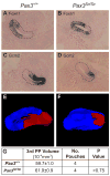
Sagittal sections of E11.5 Pax3+/+(A,C) and Pax3Sp/Sp (B,D) embryos analyzed by ISH for FoxN1 (A,B) and Gcm2 (C,D). The third pouch is indicated by a dotted line. Cranial is up and ventral is to the left. 3-D reconstructions of FoxN1 (blue) and Gcm2 (red) expression domains (E,F). Average volume of Pax3+/+ and _Pax3_Sp/Sp third pharyngeal pouch (G).
Figure 7. Third pharyngeal pouch patterning is normal at E10.5 in Pax3Sp/Sp embryos
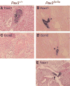
Sagittal sections from Pax3+/+ (A,C) and Pax3Sp/Sp (B,D) embryos at E10.5 were stained by ISH with anti-sense riboprobe for Bmp4 (A,B) or Gcm2 (C,D) and counterstained with nuclear fast red. In all panels, ventral is left and cranial is up. pp3, third pharyngeal pouch; pp4, fourth pharyngeal pouch. Neither Bmp4 nor Gcm2 expression is changed in the mutants.
Figure 8. Delayed separation and mixing of thymus and parathyroid fate domains in E12 Pax3Sp/Sp embryos
Transverse sections of E12.0 Pax3+/+ (A,C) and Pax3Sp/Sp (B,D,E) littermates analyzed for FoxN1 (A,B,E) and Gcm2 (C,D) expression by ISH. Ventral is down in (A–E). Arrowheads in B and D indicate mixing of FoxN1 and Gcm2 domains in Pax3Sp/Sp embryos. Note that the common primordium remains attached to the pharynx. (E) FoxN1 expression in Pax3Sp/Sp embryo extends into the pharyngeal endoderm exterior to the pouch.
Figure 9. Thymus lobes in E14.5 Pax3Cre/Cre embryos
Transverse H&E stained sections from E14.5 Pax3+/+ (A) and _Pax3_Cre/Cre (B) embryos. Abbreviations are; e, esophagus; tr, trachea ; th, thymus.
Similar articles
- Patterning of the third pharyngeal pouch into thymus/parathyroid by Six and Eya1.
Zou D, Silvius D, Davenport J, Grifone R, Maire P, Xu PX. Zou D, et al. Dev Biol. 2006 May 15;293(2):499-512. doi: 10.1016/j.ydbio.2005.12.015. Epub 2006 Mar 10. Dev Biol. 2006. PMID: 16530750 Free PMC article. - Hoxa3 and pax1 regulate epithelial cell death and proliferation during thymus and parathyroid organogenesis.
Su D, Ellis S, Napier A, Lee K, Manley NR. Su D, et al. Dev Biol. 2001 Aug 15;236(2):316-29. doi: 10.1006/dbio.2001.0342. Dev Biol. 2001. PMID: 11476574 - Ectopic TBX1 suppresses thymic epithelial cell differentiation and proliferation during thymus organogenesis.
Reeh KA, Cardenas KT, Bain VE, Liu Z, Laurent M, Manley NR, Richie ER. Reeh KA, et al. Development. 2014 Aug;141(15):2950-8. doi: 10.1242/dev.111641. Development. 2014. PMID: 25053428 Free PMC article. - Cellular and molecular mechanisms of the organogenesis and development, and function of the mammalian parathyroid gland.
Kameda Y. Kameda Y. Cell Tissue Res. 2023 Sep;393(3):425-442. doi: 10.1007/s00441-023-03785-3. Epub 2023 Jul 6. Cell Tissue Res. 2023. PMID: 37410127 Review. - Understanding the causes and prevention of neural tube defects: Insights from the splotch mouse model.
Greene ND, Massa V, Copp AJ. Greene ND, et al. Birth Defects Res A Clin Mol Teratol. 2009 Apr;85(4):322-30. doi: 10.1002/bdra.20539. Birth Defects Res A Clin Mol Teratol. 2009. PMID: 19180568 Review.
Cited by
- EphB-ephrin-B2 interactions are required for thymus migration during organogenesis.
Foster KE, Gordon J, Cardenas K, Veiga-Fernandes H, Makinen T, Grigorieva E, Wilkinson DG, Blackburn CC, Richie E, Manley NR, Adams RH, Kioussis D, Coles MC. Foster KE, et al. Proc Natl Acad Sci U S A. 2010 Jul 27;107(30):13414-9. doi: 10.1073/pnas.1003747107. Epub 2010 Jul 8. Proc Natl Acad Sci U S A. 2010. PMID: 20616004 Free PMC article. - Mechanisms of thymus organogenesis and morphogenesis.
Gordon J, Manley NR. Gordon J, et al. Development. 2011 Sep;138(18):3865-78. doi: 10.1242/dev.059998. Development. 2011. PMID: 21862553 Free PMC article. Review. - Gata3-deficient mice develop parathyroid abnormalities due to dysregulation of the parathyroid-specific transcription factor Gcm2.
Grigorieva IV, Mirczuk S, Gaynor KU, Nesbit MA, Grigorieva EF, Wei Q, Ali A, Fairclough RJ, Stacey JM, Stechman MJ, Mihai R, Kurek D, Fraser WD, Hough T, Condie BG, Manley N, Grosveld F, Thakker RV. Grigorieva IV, et al. J Clin Invest. 2010 Jun;120(6):2144-55. doi: 10.1172/JCI42021. Epub 2010 May 17. J Clin Invest. 2010. PMID: 20484821 Free PMC article. - Dynamics of thymus organogenesis and colonization in early human development.
Farley AM, Morris LX, Vroegindeweij E, Depreter ML, Vaidya H, Stenhouse FH, Tomlinson SR, Anderson RA, Cupedo T, Cornelissen JJ, Blackburn CC. Farley AM, et al. Development. 2013 May;140(9):2015-26. doi: 10.1242/dev.087320. Development. 2013. PMID: 23571219 Free PMC article. - It's Time to Unite: Diversity and Coordination of Thymic Stromal Cells for T Cell Selection and Organ Integrity.
Muro R, Nitta T. Muro R, et al. Immunol Rev. 2025 Jul;332(1):e70040. doi: 10.1111/imr.70040. Immunol Rev. 2025. PMID: 40464763 Free PMC article. Review.
References
- Anderson G, Jenkinson WE, Jones T, Parnell SM, Kinsella FA, White AJ, Pongrac’z JE, Rossi SW, Jenkinson EJ. Establishment and functioning of intrathymic microenvironments. Immunol Rev. 2006;209:10–27. - PubMed
- Auerbach R. Analysis of the developmental effects of a lethal mutation in the house mouse. J Exp Zool. 1954;127:305–329.
- Auerbach R. Morphogenetic interactions in the development of the mouse thymus gland. Dev Biol. 1960;2:271–384. - PubMed
- Bennett AR, Farley A, Blair NF, Gordon J, Sharp L, Blackburn CC. Identification and characterization of thymic epithelial progenitor cells. Immunity. 2002;16:803–814. - PubMed
Publication types
MeSH terms
Substances
Grants and funding
- R01 HD056315/HD/NICHD NIH HHS/United States
- P30 CA016672/CA/NCI NIH HHS/United States
- CA16672/CA/NCI NIH HHS/United States
- HD035920/HD/NICHD NIH HHS/United States
- P30 ES007784/ES/NIEHS NIH HHS/United States
- HD056315/HD/NICHD NIH HHS/United States
- ES07784/ES/NIEHS NIH HHS/United States
- R01 HD035920/HD/NICHD NIH HHS/United States
LinkOut - more resources
Full Text Sources
Molecular Biology Databases
