Transcription is required for establishment of germline methylation marks at imprinted genes - PubMed (original) (raw)
Transcription is required for establishment of germline methylation marks at imprinted genes
Mita Chotalia et al. Genes Dev. 2009.
Erratum in
- Genes Dev. 2009 Oct 1;23(19):2358
Abstract
Genomic imprinting requires the differential marking by DNA methylation of genes in male and female gametes. In the female germline, acquisition of methylation imprint marks depends upon the de novo methyltransferase Dnmt3a and its cofactor Dnmt3L, but the reasons why specific sequences are targets for Dnmt3a and Dnmt3L are still poorly understood. Here, we investigate the role of transcription in establishing maternal germline methylation marks. We show that at the Gnas locus, truncating transcripts from the furthest upstream Nesp promoter disrupts oocyte-derived methylation of the differentially methylated regions (DMRs). Transcription through DMRs in oocytes is not restricted to this locus but occurs across the prospective DMRs at many other maternally marked imprinted domains, suggesting a common requirement for transcription events. The transcripts implicated here in gametic methylation are protein-coding, in contrast to the noncoding antisense transcripts involved in the monoallelic silencing of imprinted genes in somatic tissues, although they often initiate from alternative promoters in oocytes. We propose that transcription is a third essential component of the de novo methylation system, which includes optimal CpG spacing and histone modifications, and may be required to create or maintain open chromatin domains to allow the methylation complex access to its preferred targets.
Figures
Figure 1.
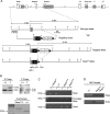
The Nesp transcript is detected in growing oocytes coincident with establishment of methylation of germline DMRs in the Gnas locus. (A) Scheme of the mouse Gnas locus (Plagge and Kelsey 2006), showing the organization of the overlapping, protein-coding transcripts Nesp, Gnasxl, and Gnas and the noncoding Nespas and 1A transcripts. Transcripts expressed from the maternal allele are indicated above the line, from the paternal allele below the line; Gnas exhibits tissue-specific imprinting, with repression of the paternal allele in a subset of tissues. The location of the DMRs is shown by the rows of filled circles on the methylated allele; the Gnas promoter resides in a biallelically unmethylated CGI (open circles). The positions of the PCR products for bisulphite analysis in B and Figures 3 and 4 are indicated by the hatched bars, and the primers for the RT–PCR assays in C and Figure 2 by labeled arrows. (B) Bisulphite sequences of the Nespas/Gnasxl and 1A DMRs in oocytes isolated at postnatal days 5, 10, and 15 and mature metaphase II (MII) oocytes from adult females. Each row represents the CpG sites of an individual sequenced clone, with filled circles depicting methylated CpG sites (missing circles represent CpGs for which sequence was ambiguous). Sequences obtained from two independent bisulphite treatments are indicated by bracketed sets of methylation profiles. Methylation of these DMRs in MII oocytes has been described previously (Liu et al. 2000; Coombes et al. 2003). (C) RT–PCRs for Nesp, Nespas, Gnasxl, 1A, and Gnas transcripts in day 5, 10, 15 oocytes. Co indicates amplification control with cDNA from an E13.5 embryo; H indicates no template control. The left lanes on each gel show a 100-bp marker ladder, with the 100- or 200-bp markers indicated.
Figure 2.
Truncation of Nesp transcription by insertion of a termination cassette. (A) Scheme of the targeting vector in relation to the Gnas locus, the targeted allele, and the Nesptrun allele after Cre-mediated excision of the selectable marker cassette. The rabbit β-globin cassette is represented by the black box, NeoR and URA3 selection cassettes as open boxes, and loxP sites as black arrowheads. The location of restriction sites AflII (A) and XhoI (X) for Southern blot detection of targeting events is given. (B) Southern blots of wild-type (+/+) and targeted (T) embryonic stem (ES) cell clones detecting correct recombination events by hybridization with 5′ and 3′ probes in AflII and XhoI digests, respectively. Below, PCRs using primers Lp-F and Lp-R to verify the Cre-mediated excision event after germline transmission (confirmed by sequencing of the PCR product) (data not shown) and N2-F2 and Lp-R to detect the wild-type allele. (C) RT–PCR analysis of neonatal brain RNA after paternal (+/Nesptrun) and maternal (Nesptrun/+) transmission of the Nesptrun allele. RT–PCRs for Nesp and Gnas were done primers using N2-F1 + E2-R1 and Gs1-F + E2-R2, respectively, as in Figure 1A. The RT–PCR for Nesp therefore assays the presence of wild-type, full-length Nesp transcripts, which are absent after maternal transmission. The panel labeled Truncated Nesp is a 3′RACE assay (primer N2-F1 and 3′RACE primer GR-R2) for the presence of prematurely terminated Nesp transcripts; sequencing these PCR products verified that the desired truncation event had occurred (Supplemental Fig. 2). (D) RT–PCR assay (primers as above) showing the absence of full-length Nesp transcripts in MII oocytes from a Nesptrun homozygous female.
Figure 3.
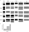
Maternal transmission of the Nesp truncation results in loss of methylation of maternal germline DMRs. (A) Bisulphite sequence profiles from neonatal brain DNA from Nesptrun/+ and wild-type (+/+) pups. A total of 34 Nesptrun/+ pups was analyzed by COBRA (see B), and sequences for one or more amplicon were obtained from seven mutants. Representative sequences from Nesptrun/+ mutants with complete LoM (#A25, #A62) or partial LoM (#A43, #A47) of the maternal germline DMRs are shown. Sequence polymorphisms allowed maternal and paternal alleles to be discriminated for products F1, F3, and F5. (B) Summary showing the frequency of LoM across the DMRs in neonatal brain in Nesptrun/+ mutants collected from 11 litters. The number of Nesptrun/+ pups typed by COBRA for amplicons F2, F3, and F6 was 34; for F4 was 22; and for F5 was 26; seven wild-type pups were similarly scored and found to have the normal pattern of DMRs. These data are a combination of COBRA results from crosses of Nesptrun/+ females with C57BL/6J males and with males carrying a _M. spretus_–derived Gnas locus; the variation in methylation pattern among Nesptrun/+ pups was similar in the two crosses. See Figure 1A for the location of the amplicons within the DMRs.
Figure 4.
Loss of methylation occurs in oocytes of Nesptrun/+ females. (A) Bisulphite sequences for the Nespas/Gnasxl (amplicons F2, F3, and F5) and 1A DMRs (F6) of brain and liver DNAs from the same pup (#1D) inheriting the Nesptrun allele maternally (Nesptrun/+). DNAs from four other pups from the same litter were assayed in both brain and liver by COBRA, revealing similar aberrant methylation profiles in the two tissues (Supplemental Fig. 4). (B) Bisulphite sequences for amplicons F2 and F6 obtained in GV oocytes from Nesptrun homozygous females in comparison with wild type (+/+). See Figure 1A for the location of the PCR products within the DMRs.
Figure 5.
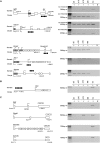
Transcription across maternal germline DMRs in oocytes is common among imprinted genes. (A) On the left, schemes representing the imprinted loci analyzed, Igf2r, Grb10, Kcnq1, Zac1, and Impact, showing the locations of the primers used for RT–PCRs in relation to the germline DMRs (filled circles). The schemes are not to scale, and for simplicity, not all exons of the genes are shown. Characterized start sites or start sites determined by 5′RACE analysis are indicated by arrows, with those above the line representing start sites detected in somatic cells and those below the line in oocytes. Novel exons identified by 5′RACE are shown as gray boxes. On the right, RT–PCRs for these loci for transcripts traversing the DMRs in day 5, day 10, day 15 and MII oocytes are shown. For both Igf2r and Grb10, RT–PCRs labeled with Ex1-F/Ex3-R1 assay transcripts from the canonical somatic promoter and RT–PCRs labeled with Un1-F/Ex3-R2 assay the novel start sites identified in oocytes by 5′RACE. The multiple bands for Zac1 reflect alternative splicing of the various 5′ untranslated exons, exon 8 being the first coding exon. (B) RT–PCR analysis of transcripts for the H19 paternal germline DMR. The RT–PCR represents amplicon 4 from Schoenfelder et al. (2007). (C) RT–PCR analysis of transcripts crossing the intragenic CGIs of four nonimprinted genes. Gene Sp6 represents locus PvuII 44, Chst8 is PstI 53, Sema6c is PstI 58, and Adra1b is PvuII 66 from Song et al. (2005). In the schemes to the left, the locations of the CGIs are represented by the open circles. In each panel, the lanes marked Co represent control amplification from E13.5 embryo RNA, and H indicates no template control. The left lanes on each gel show a 100-bp marker ladder, with size marker relevant to the RT–PCR product indicated by arrows.
Similar articles
- Transcription driven somatic DNA methylation within the imprinted Gnas cluster.
Mehta S, Williamson CM, Ball S, Tibbit C, Beechey C, Fray M, Peters J. Mehta S, et al. PLoS One. 2015 Feb 6;10(2):e0117378. doi: 10.1371/journal.pone.0117378. eCollection 2015. PLoS One. 2015. PMID: 25659103 Free PMC article. - Forced expression of DNA methyltransferases during oocyte growth accelerates the establishment of methylation imprints but not functional genomic imprinting.
Hara S, Takano T, Fujikawa T, Yamada M, Wakai T, Kono T, Obata Y. Hara S, et al. Hum Mol Genet. 2014 Jul 15;23(14):3853-64. doi: 10.1093/hmg/ddu100. Epub 2014 Mar 5. Hum Mol Genet. 2014. PMID: 24599402 Free PMC article. - The loss of imprinted DNA methylation in mouse blastocysts is inflicted to a similar extent by in vitro follicle culture and ovulation induction.
Saenz-de-Juano MD, Billooye K, Smitz J, Anckaert E. Saenz-de-Juano MD, et al. Mol Hum Reprod. 2016 Jun;22(6):427-41. doi: 10.1093/molehr/gaw013. Epub 2016 Feb 7. Mol Hum Reprod. 2016. PMID: 26908643 - Imprinting on chromosome 20: tissue-specific imprinting and imprinting mutations in the GNAS locus.
Kelsey G. Kelsey G. Am J Med Genet C Semin Med Genet. 2010 Aug 15;154C(3):377-86. doi: 10.1002/ajmg.c.30271. Am J Med Genet C Semin Med Genet. 2010. PMID: 20803660 Review. - Developmental regulation of somatic imprints.
John RM, Lefebvre L. John RM, et al. Differentiation. 2011 Jun;81(5):270-80. doi: 10.1016/j.diff.2011.01.007. Epub 2011 Feb 11. Differentiation. 2011. PMID: 21316143 Review.
Cited by
- PRKACB is a novel imprinted gene in marsupials.
Newman T, Bond DM, Ishihara T, Rizzoli P, Gouil Q, Hore TA, Shaw G, Renfree MB. Newman T, et al. Epigenetics Chromatin. 2024 Sep 28;17(1):29. doi: 10.1186/s13072-024-00552-8. Epigenetics Chromatin. 2024. PMID: 39342354 Free PMC article. - Maternal loss-of-function of Nlrp2 results in failure of epigenetic reprogramming in mouse oocytes.
Anvar Z, Jochum MD, Chakchouk I, Sharif M, Demond H, To AK, Kraushaar DC, Wan YW, Andrews S, Kelsey G, Veyver IB. Anvar Z, et al. Res Sq [Preprint]. 2024 Jun 4:rs.3.rs-4457414. doi: 10.21203/rs.3.rs-4457414/v1. Res Sq. 2024. PMID: 38883732 Free PMC article. Preprint. - Roles of endogenous retroviral elements in the establishment and maintenance of imprinted gene expression.
Fang S, Chang KW, Lefebvre L. Fang S, et al. Front Cell Dev Biol. 2024 Mar 5;12:1369751. doi: 10.3389/fcell.2024.1369751. eCollection 2024. Front Cell Dev Biol. 2024. PMID: 38505259 Free PMC article. Review. - GNAS AS2 methylation status enables mechanism-based categorization of pseudohypoparathyroidism type 1B.
Iwasaki Y, Reyes M, Jüppner H, Bastepe M. Iwasaki Y, et al. JCI Insight. 2024 Mar 8;9(5):e177190. doi: 10.1172/jci.insight.177190. JCI Insight. 2024. PMID: 38290008 Free PMC article. - Role of transcription in imprint establishment in the male and female germ lines.
Liao J, Szabó PE. Liao J, et al. Epigenomics. 2024 Jan;16(2):127-136. doi: 10.2217/epi-2023-0344. Epub 2023 Dec 21. Epigenomics. 2024. PMID: 38126127 Free PMC article. Review.
References
- Arnaud P., Monk D., Hitchins M., Gordon E., Dean W., Beechey C.V., Peters J., Craigen W., Preece M., Stanier P., et al. Conserved methylation imprints in the human and mouse GRB10 genes with divergent allelic expression suggests differential reading of the same mark. Hum. Mol. Genet. 2003;12:1005–1019. - PubMed
- Arnaud P., Hata K., Kaneda M., Li E., Sasaki H., Feil R., Kelsey G. Stochastic imprinting in the progeny of Dnmt3L−/− females. Hum. Mol. Genet. 2006;15:589–598. - PubMed
- Bastepe M., Frohlich L.F., Hendy G.N., Indridason O.S., Josse R.G., Koshiyama H., Korkko J., Nakamoto J.M., Rosenbloom A.L., Slyper A.H., et al. Autosomal dominant pseudohypoparathyroidism type Ib is associated with a heterozygous microdeletion that likely disrupts a putative imprinting control element of GNAS. J. Clin. Invest. 2003;112:1255–1263. - PMC - PubMed
- Bastepe M., Frohlich L.F., Linglart A., Abu-Zahra H.S., Tojo K., Ward L.M., Juppner H. Deletion of the NESP55 differentially methylated region causes loss of maternal GNAS imprints and pseudohypoparathyroidism type Ib. Nat. Genet. 2005;37:25–27. - PubMed
- Bernstein B.E., Kamal M., Lindblad-Toh K., Bekiranov S., Bailey D.K., Huebert D.J., McMahon S., Karlsson E.K., Kulbokas E.J., III, Gingeras T.R., et al. Genomic maps and comparative analysis of histone modifications in human and mouse. Cell. 2005;120:169–181. - PubMed
Publication types
MeSH terms
Substances
LinkOut - more resources
Full Text Sources
Other Literature Sources
Molecular Biology Databases


