Overexpression of myocilin in the Drosophila eye activates the unfolded protein response: implications for glaucoma - PubMed (original) (raw)
Overexpression of myocilin in the Drosophila eye activates the unfolded protein response: implications for glaucoma
Mary Anna Carbone et al. PLoS One. 2009.
Abstract
Background: Glaucoma is the world's second leading cause of bilateral blindness with progressive loss of vision due to retinal ganglion cell death. Myocilin has been associated with congenital glaucoma and 2-4% of primary open angle glaucoma (POAG) cases, but the pathogenic mechanisms remain largely unknown. Among several hypotheses, activation of the unfolded protein response (UPR) has emerged as a possible disease mechanism.
Methodology / principal findings: We used a transgenic Drosophila model to analyze whole-genome transcriptional profiles in flies that express human wild-type or mutant MYOC in their eyes. The transgenic flies display ocular fluid discharge, reflecting ocular hypertension, and a progressive decline in their behavioral responses to light. Transcriptional analysis shows that genes associated with the UPR, ubiquitination, and proteolysis, as well as metabolism of reactive oxygen species and photoreceptor activity undergo altered transcriptional regulation. Following up on the results from these transcriptional analyses, we used immunoblots to demonstrate the formation of MYOC aggregates and showed that the formation of such aggregates leads to induction of the UPR, as evident from activation of the fluorescent UPR marker, xbp1-EGFP. CONCLUSIONS / SIGNIFICANCE: Our results show that aggregation of MYOC in the endoplasmic reticulum activates the UPR, an evolutionarily conserved stress pathway that culminates in apoptosis. We infer from the Drosophila model that MYOC-associated ocular hypertension in the human eye may result from aggregation of MYOC and induction of the UPR in trabecular meshwork cells. This process could occur at a late age with wild-type MYOC, but might be accelerated by MYOC mutants to account for juvenile onset glaucoma.
Conflict of interest statement
Competing Interests: The authors have declared that no competing interests exist.
Figures
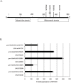
Figure 1. Expression of wt-MYOC and mutant MYOCs in transgenic flies.
A) Diagram of the MYOC polypeptide showing the position of the amino acid substitutions (R342K, Q368X, D380N and K423E) that were created by site-directed mutagenesis. The Q368X mutant is predicted to result in a premature stop codon (octagon). Also shown are the N-terminal myosin-like domain and the C-terminal olfactomedin domain. B) Gene expression by quantitative PCR of human wt- MYOC and mutant MYOCs in heads of transgenic flies. Expression of MYOC is evident where the Gal4 driver is combined with the UAS-transgenes, but not in the parental lines.
Figure 2. Eye phenotypes and phototactic responses of wild-type and mutant MYOC transgenic flies.
Fluid discharge from eyes of transgenic flies. The normal appearance of eyes from a fly of the gmr-Gal4/Sam w1118 control line is shown in panel A. A fly eye expressing the wt-MYOC with liquid discharge (white arrow) is shown in panel B. The flies expressing mutant MYOCs displaying a white salt residue (black arrows) after drying of the liquid residue (panels C–F).
Figure 3. Phototactic behavior of MYOC-expressing transgenic flies.
Progressive visual impairment of the MYOC-expressing flies compared to the gmr-Gal4/Sam w1118 control line is observed.
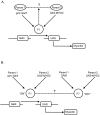
Figure 4. Diagrammatic representation of the expression microarray analysis.
A) One-way ANOVA was used to compare differences between expression levels for each probe set in the F1 hybrid and the expected mean value of the two parents according to the model Y = μ+G+e, where μ is the overall mean, G is the effect of genotype, and e is the error variance. B) Two-way ANOVA was used to compare differences between expression levels for each probe set between F1 hybrids in which the UAS-transgene was ‘on’ or ‘off’ according to the model Y = μ+E+C+E×C+e, where E represents the effect of MYOC expression (‘on’ or ‘off’), C is the effect of the MYOC construct (wt-MYOC, R342K, Q368X, D380N, K423E), E×C is the effect due to the interaction between expression and each construct, and e is the error variance.
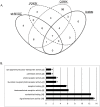
Figure 5. Probe sets with altered transcript abundance when expression in the F1 is compared to the predicted mean values of the parents.
A) Venn diagram depicting the overlap of probe sets among MYOC transgenes. Only one gene (1632731_at; CG8613) was differentially expressed in flies carrying the K423E construct and is not included in the diagram. The Q368X construct shows a uniquely high proportion of transcripts with altered expression (89%) that do not overlap with transcripts with altered expression in the other transgenic lines. B) Bar-chart showing the distribution of differentially expressed genes among Gene Ontology (GO) molecular function processes according to the analysis illustrated in Figure 4A. Parameters in DAVID were set to GO level “all”, count threshold of 2 and EASE threshold of 0.1. The output is sorted by percentage. Asterisks refer to modified Fisher-Exact P values (P<0.05, i.e. strongly enriched in the annotation categories) and the numbers in brackets refer to the number of genes in the category.
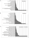
Figure 6. Gene ontology categories when transcript expression is analyzed across all genotypes comparing the ‘on’ and ‘off’ mode (Figure 4B).
Bar-chart showing the distribution of differentially expressed genes among Gene Ontology (GO) molecular function processes, for each of the 3 terms in the two-way ANOVA (expression, A; construct, B; expression×construct, C). Parameters in DAVID were set to GO level 3, count threshold of 2 and EASE threshold of 0.1. The output is sorted by percentage. Asterisks refer to modified Fisher-Exact _P-_values (P<0.05, i.e. strongly enriched in the annotation categories) and the numbers in brackets refer to the number of genes in the category.
Figure 7. Western blot of soluble (S) and insoluble MYOC proteins (I) isolated from heads of transgenic flies.
The wt-MYOC, R342K, Q368X and K423E represent the corresponding gmr-Gal4/UAS-MYOC genotypes; gmr-Gal4 designates the gmr-Gal4/Sam w1118 control. The solid-arrow indicates the MYOC proteins that are expressed in the gmr-GAL4/UAS-MYOC heads at ∼57 kDa. The dashed-arrow indicates the Q368X-MYOC protein at ∼41 kDa. MYOC proteins recovered in the insoluble fraction represent aggregated proteins prior to treatment with SDS. The high molecular weight band that is also observed in the gmr-Gal4/Sam w1118 control, likely represents cross-reactivity with the myosin heavy chain (arrow-head).
Figure 8. ER-stress activates xbp1-EGFP expression in MYOC-expressing transgenic flies.
All panels show eye-imaginal discs from third-instar F1 larvae after crossing gmr-Gal4/UAS-xbp1-EGFP flies to the following lines: A) gmr-Gal4/Sam w1118 (control), B) gmr-Gal4/UAS-NTE, C) Gal4/UAS-Q368X, D) gmr-Gal4/UAS-D380N. Panels C′ and D′ show 100× magnifications of the boxed areas in C and D, respectively. Punctate nuclear staining reflects activation of the UPR. Only background fluorescence is observed in flies that overexpress NTE (B) without the characteristic punctate nuclear staining.
Similar articles
- Histochemical Analysis of Glaucoma Caused by a Myocilin Mutation in a Human Donor Eye.
van der Heide CJ, Alward WLM, Flamme-Wiese M, Riker M, Syed NA, Anderson MG, Carter K, Kuehn MH, Stone EM, Mullins RF, Fingert JH. van der Heide CJ, et al. Ophthalmol Glaucoma. 2018 Sep-Oct;1(2):132-138. doi: 10.1016/j.ogla.2018.08.004. Epub 2018 Aug 17. Ophthalmol Glaucoma. 2018. PMID: 30906929 Free PMC article. - Transcription profiling in Drosophila eyes that overexpress the human glaucoma-associated trabecular meshwork-inducible glucocorticoid response protein/myocilin (TIGR/MYOC).
Borrás T, Morozova TV, Heinsohn SL, Lyman RF, Mackay TF, Anholt RR. Borrás T, et al. Genetics. 2003 Feb;163(2):637-45. doi: 10.1093/genetics/163.2.637. Genetics. 2003. PMID: 12618402 Free PMC article. - Reduction of ER stress via a chemical chaperone prevents disease phenotypes in a mouse model of primary open angle glaucoma.
Zode GS, Kuehn MH, Nishimura DY, Searby CC, Mohan K, Grozdanic SD, Bugge K, Anderson MG, Clark AF, Stone EM, Sheffield VC. Zode GS, et al. J Clin Invest. 2011 Sep;121(9):3542-53. doi: 10.1172/JCI58183. Epub 2011 Aug 8. J Clin Invest. 2011. PMID: 21821918 Free PMC article. - A review of genetic and structural understanding of the role of myocilin in primary open angle glaucoma.
Kanagavalli J, Pandaranayaka E, Krishnadas SR, Krishnaswamy S, Sundaresan P. Kanagavalli J, et al. Indian J Ophthalmol. 2004 Dec;52(4):271-80. Indian J Ophthalmol. 2004. PMID: 15693317 Review. - Myocilin-associated Glaucoma: A Historical Perspective and Recent Research Progress.
Sharma R, Grover A. Sharma R, et al. Mol Vis. 2021 Aug 20;27:480-493. eCollection 2021. Mol Vis. 2021. PMID: 34497454 Free PMC article. Review.
Cited by
- Applications of genome editing technology in the targeted therapy of human diseases: mechanisms, advances and prospects.
Li H, Yang Y, Hong W, Huang M, Wu M, Zhao X. Li H, et al. Signal Transduct Target Ther. 2020 Jan 3;5(1):1. doi: 10.1038/s41392-019-0089-y. Signal Transduct Target Ther. 2020. PMID: 32296011 Free PMC article. Review. - Comprehensive analysis of myocilin variants in east Indian POAG patients.
Banerjee D, Bhattacharjee A, Ponda A, Sen A, Ray K. Banerjee D, et al. Mol Vis. 2012;18:1548-57. Epub 2012 Jun 13. Mol Vis. 2012. PMID: 22736945 Free PMC article. - The effects of myocilin expression on functionally relevant trabecular meshwork genes: a mini-review.
Borrás T. Borrás T. J Ocul Pharmacol Ther. 2014 Mar-Apr;30(2-3):202-12. doi: 10.1089/jop.2013.0218. Epub 2014 Feb 24. J Ocul Pharmacol Ther. 2014. PMID: 24564495 Free PMC article. Review. - Screening of the Drug-Induced Effects of Prostaglandin EP2 and FP Agonists on 3D Cultures of Dexamethasone-Treated Human Trabecular Meshwork Cells.
Watanabe M, Ida Y, Furuhashi M, Tsugeno Y, Ohguro H, Hikage F. Watanabe M, et al. Biomedicines. 2021 Jul 31;9(8):930. doi: 10.3390/biomedicines9080930. Biomedicines. 2021. PMID: 34440134 Free PMC article. - Genetic architecture of natural variation in visual senescence in Drosophila.
Carbone MA, Yamamoto A, Huang W, Lyman RA, Meadors TB, Yamamoto R, Anholt RR, Mackay TF. Carbone MA, et al. Proc Natl Acad Sci U S A. 2016 Oct 25;113(43):E6620-E6629. doi: 10.1073/pnas.1613833113. Epub 2016 Oct 10. Proc Natl Acad Sci U S A. 2016. PMID: 27791033 Free PMC article.
References
- Tamm ER. Myocilin and glaucoma: facts and ideas. Prog Retin Eye Res. 2002;21:395–428. - PubMed
- Tan JC, Peters DM, Kaufman PL. Recent developments in understanding the pathophysiology of elevated intraocular pressure. Curr Opin Ophthalmol. 2006;17:168–174. - PubMed
- Bruttini M, Longo I, Frezzotti P, Ciappetta R, Randazzo A, et al. Mutations in the myocilin gene in families with primary open-angle glaucoma and juvenile open-angle glaucoma. Arch Ophthalmol. 2003;121:1034–1038. - PubMed
Publication types
MeSH terms
Substances
LinkOut - more resources
Full Text Sources
Other Literature Sources
Medical
Molecular Biology Databases


