Large-scale detection of ubiquitination substrates using cell extracts and protein microarrays - PubMed (original) (raw)
Large-scale detection of ubiquitination substrates using cell extracts and protein microarrays
Yifat Merbl et al. Proc Natl Acad Sci U S A. 2009.
Abstract
Identification of protein targets of post-translational modification is an important analytical problem in biology. Protein microarrays exposed to cellular extracts could offer a rapid and convenient means of identifying modified proteins, but this kind of biochemical assay, unlike DNA microarrays, depends on a faithful reconstruction of in vivo conditions. Over several years, concentrated cellular extracts have been developed, principally for cell cycle studies that reproduce very complex cellular states. We have used extracts that replicate the mitotic checkpoint and anaphase release to identify differentially regulated poyubiquitination. Protein microarrays were exposed to these complex extracts, and the polyubiquitinated products were detected by specific antibodies. We expected that among the substrates revealed by the microarray should be substrates of the anaphase promoting complex (APC). Among 8,000 proteins on the chip, 10% were polyubiquitinated. Among those, we found 11 known APC substrates (out of 16 present on the chip) to be polyubiquitinated. Interestingly, only 1.5% of the proteins were differentially ubiquitinated on exit from the checkpoint. When we arbitrarily chose 6 proteins thought to be involved in mitosis from the group of differentially modified proteins, all registered as putative substrates of the APC, and among 4 arbitrarily chosen non-mitotic proteins picked from the same list, 2 were ubiquitinated in an APC-dependent manner. The striking yield of potential APC substrates from a simple assay with concentrated cell extracts suggests that combining microarray analysis of the products of post-translational modifications with extracts that preserve the physiological state of the cell can yield information on protein modification under various in vivo conditions.
Conflict of interest statement
The authors declare no conflict of interest.
Figures
Fig. 1.
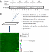
(A) Assaying the biological state and competence of the extracts. To evaluate the activity of the extracts, S35-labeled securin was added to extract samples to follow its degradation. The reactions were stopped at the indicted times by the addition of sample buffer and were then analyzed by on SDS/PAGE and autoradiography. The star (*) labeled lanes reflect the state of the extracts at the time when incubation on the protein microarrays were stopped. (B) Signal on microarrays for the detection of polyubiqutination extracts with an active E3 (e.g., checkpoint released) or with an inhibitor or lacking the activity (e.g., checkpoint extracts or extracts inhibited with emi1) were incubated on protein microarrays and detected (Human ProtoArray, Invitrogen) as described. An example of one block/subarray out of the 48 on each microarray is given (16 rows × 16 columns).
Fig. 2.
(A) The distribution of the signal intensity minus background of all of the spots on a microarray. Reactivates were divided into 100 equally sized bins and the number of spots (y axis) at different intensity levels (x axis) of CP-released (Left) and APC-inhibited (Right) extracts were plotted. The inset represents a 20× magnification of the positive signals where the y axis ranges between 0 and 250 and the x axis ranges between 0 and 45,000. (B) The reactivity level of 16 known APC substrates (red dots) compared with the reactivity level of the ‘buffer’ spots located in the same subarray (black stars). These reactivities were then compared using two-sample t test to determine their significance and the _P_-values were labeled below each substrate. (C) Scatter plots of the positive signal intensities on each microarray were plotted to compare the variability between 2 biological replicates (black dots; x axis, CP-released; y axis, CP-released) vs. the variability between signals from two different conditions (red spots; x axis, APC-inhibited; y axis, CP-released).
Fig. 3.
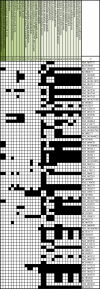
The Biological terms/functions specifically enriched in our gene list compared to the background list of proteins that were spotted on the microarray. We used functional annotation clustering in the Database for Annotation, Visualization, and Integrated Discovery. The proteins that were classified to each of the most enriched (P value <0.05) GO annotation terms are labeled in black. The percentage (%) of proteins that correspond to each GO category is also presented (see
Table S2
for a full report of the results).
Fig. 4.
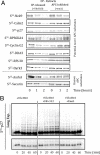
(A) Soluble degradation assays for identified substrates. S35-labeled putative substrates (Nek9, Calm2, RPS6KA4, cyclin G2, RPL30, and Dyrk3) were added to CP-synchronized HeLa S3 extracts with and without the addition of the APC-inhibitor emi1. Reactions were stopped at 0, 60, and 120 min, and analyzed by SDS/PAGE (4–15%) and autoradiography. (B) Ubiqutination and limited degradation of p27. S35-labeled p27 was added to CP-synchronized HeLa S3 extracts with the addition of UbcH10 (1 μL; 1 mg/mL), UbcH10 and MG-132 (200 μM), or UbcH10 and Emi1 (5 mg/mL). The bottom panel shows the change in stability of p27 under these conditions. The top panel is the same gel overexposed to detect p27-conjugated ubiquitin chains.
Similar articles
- Methods to measure ubiquitin-dependent proteolysis mediated by the anaphase-promoting complex.
Kraft C, Gmachl M, Peters JM. Kraft C, et al. Methods. 2006 Jan;38(1):39-51. doi: 10.1016/j.ymeth.2005.07.005. Methods. 2006. PMID: 16343932 Review. - Preparation of synchronized human cell extracts to study ubiquitination and degradation.
Williamson A, Jin L, Rape M. Williamson A, et al. Methods Mol Biol. 2009;545:301-12. doi: 10.1007/978-1-60327-993-2_19. Methods Mol Biol. 2009. PMID: 19475397 - Anaphase-promoting complex/cyclosome controls the stability of TPX2 during mitotic exit.
Stewart S, Fang G. Stewart S, et al. Mol Cell Biol. 2005 Dec;25(23):10516-27. doi: 10.1128/MCB.25.23.10516-10527.2005. Mol Cell Biol. 2005. PMID: 16287863 Free PMC article. - Inhibitory factors associated with anaphase-promoting complex/cylosome in mitotic checkpoint.
Braunstein I, Miniowitz S, Moshe Y, Hershko A. Braunstein I, et al. Proc Natl Acad Sci U S A. 2007 Mar 20;104(12):4870-5. doi: 10.1073/pnas.0700523104. Epub 2007 Mar 13. Proc Natl Acad Sci U S A. 2007. PMID: 17360335 Free PMC article. - The spindle checkpoint: how do cells delay anaphase onset?
Sczaniecka MM, Hardwick KG. Sczaniecka MM, et al. SEB Exp Biol Ser. 2008;59:243-56. SEB Exp Biol Ser. 2008. PMID: 18368927 Review.
Cited by
- Use of biotinylated ubiquitin for analysis of rat brain mitochondrial proteome and interactome.
Buneeva OA, Medvedeva MV, Kopylov AT, Zgoda VG, Medvedev AE. Buneeva OA, et al. Int J Mol Sci. 2012;13(9):11593-11609. doi: 10.3390/ijms130911593. Epub 2012 Sep 14. Int J Mol Sci. 2012. PMID: 23109873 Free PMC article. - Applications of proteomic technologies for understanding the premature proteolysis of CFTR.
Henderson MJ, Singh OV, Zeitlin PL. Henderson MJ, et al. Expert Rev Proteomics. 2010 Aug;7(4):473-86. doi: 10.1586/epr.10.42. Expert Rev Proteomics. 2010. PMID: 20653504 Free PMC article. Review. - Defining genome maintenance pathways using functional genomic approaches.
Bansbach CE, Cortez D. Bansbach CE, et al. Crit Rev Biochem Mol Biol. 2011 Aug;46(4):327-41. doi: 10.3109/10409238.2011.588938. Crit Rev Biochem Mol Biol. 2011. PMID: 21787120 Free PMC article. Review. - Enzyme-substrate relationships in the ubiquitin system: approaches for identifying substrates of ubiquitin ligases.
O'Connor HF, Huibregtse JM. O'Connor HF, et al. Cell Mol Life Sci. 2017 Sep;74(18):3363-3375. doi: 10.1007/s00018-017-2529-6. Epub 2017 Apr 28. Cell Mol Life Sci. 2017. PMID: 28455558 Free PMC article. Review. - O-GlcNAc Transferase Recognizes Protein Substrates Using an Asparagine Ladder in the Tetratricopeptide Repeat (TPR) Superhelix.
Levine ZG, Fan C, Melicher MS, Orman M, Benjamin T, Walker S. Levine ZG, et al. J Am Chem Soc. 2018 Mar 14;140(10):3510-3513. doi: 10.1021/jacs.7b13546. Epub 2018 Mar 5. J Am Chem Soc. 2018. PMID: 29485866 Free PMC article.
References
- Rape M, Reddy SK, Kirschner MW. The processivity of multiubiquitination by the APC determines the order of substrate degradation. Cell. 2006;124:89–103. - PubMed
- Ptacek J, et al. Global analysis of protein phosphorylation in yeast. Nature. 2005;438:679–684. - PubMed
- Schnack C, Danzer KM, Hengerer B, Gillardon F. Protein array analysis of oligomerization-induced changes in alpha-synuclein protein-protein interactions points to an interference with Cdc42 effector proteins. Neuroscience. 2008;154:1450–1457. - PubMed
- Emili AQ, Cagney G. Large-scale functional analysis using peptide or protein arrays. Nat Biotechnol. 2000;18:393–397. - PubMed
Publication types
MeSH terms
Substances
LinkOut - more resources
Full Text Sources


