Stress-induced dendritic remodeling in the prefrontal cortex is circuit specific - PubMed (original) (raw)
Stress-induced dendritic remodeling in the prefrontal cortex is circuit specific
Rebecca M Shansky et al. Cereb Cortex. 2009 Oct.
Abstract
Chronic stress exposure has been reported to induce dendritic remodeling in several brain regions, but it is not known whether individual neural circuits show distinct patterns of remodeling. The current study tested the hypothesis that the projections from the infralimbic (IL) area of the medial prefrontal cortex (mPFC) to the basolateral nucleus of the amygdala (BLA), a pathway relevant to stress-related mental illnesses like depression and post-traumatic stress disorder, would have a unique pattern of remodeling in response to chronic stress. The retrograde tracer FastBlue was injected into male rats' BLA or entorhinal cortex (EC) 1 week prior to 10 days of immobilization stress. After cessation of stress, FastBlue-labeled and unlabeled IL pyaramidal neurons were loaded with fluorescent dye Lucifer Yellow to visualize dendritic arborization and spine density. As has been previously reported, randomly selected (non-FastBlue-labeled) neurons showed stress-induced dendritic retraction in apical dendrites, an effect also seen in EC-projecting neurons. In contrast, BLA-projecting neurons showed no remodeling with stress, suggesting that this pathway may be particularly resilient against the effects of stress. No neurons showed stress-related changes in spine density, contrasting with reports that more dorsal areas of the mPFC show stress-induced decreases in spine density. Such region- and circuit-specificity in response to stress could contribute to the development of stress-related mental illnesses.
Figures
Figure 1.
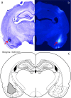
FastBlue injection. Representative syringe placement for retrograde tracer FastBlue injections in the BLA (a), representative Fastblue diffusion into the BLA (b), and schematic of maximal observed diffusion into the BLA (c). Adapted from Paxinos and Watson (2005).
Figure 2.
Ten days immobilization stress causes decreased weight gain. Stressed animals gained significantly less weight than control animals over the course of the study. *P < 0.04.
Figure 3.
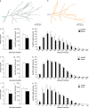
Randomly selected neurons and EC-projecting neurons, but not BLA-projecting neurons, show retraction with stress. Representative randomly selected neuron apical dendrite Neurolucida tracings from control (a) and stressed (b) animals. In rats exposed to stress, apical dendrites in randomly selected IL layer II/III pyramidal neurons show reduced number of branch points (c) and overall dendritic length (d) when compared with control rats. Sholl analysis suggests that these changes are accounted for by retraction in intermediate dendrites, approximately 120–180 μm from the soma (e). Apical dendrites in amygdala-projecting IL layer II/III pyramidal neurons show no change in branch points (f) or in overall dendritic length (g) when compared with control rats. Moreover, there were no group differences at any distance from the soma (h). Apical dendrites in EC-projecting layer II/III neurons showed significantly reduced number of branch points (i) and overall dendritic length (j), but no significant changes at individual distances from the soma (k). *P < 0.05; **P < 0.01.
Figure 4.
Stress does not alter spine density in the IL. Representative randomly selected neuron apical dendrite segments from control (a) and stressed (b) animals. Neither randomly selected neurons nor BLA-projecting nor EC-projecting neurons showed any significant change in spine density with stress.
Similar articles
- Chronic restraint stress induces depression-like behaviors and alterations in the afferent projections of medial prefrontal cortex from multiple brain regions in mice.
Ge MJ, Chen G, Zhang ZQ, Yu ZH, Shen JX, Pan C, Han F, Xu H, Zhu XL, Lu YP. Ge MJ, et al. Brain Res Bull. 2024 Jul;213:110981. doi: 10.1016/j.brainresbull.2024.110981. Epub 2024 May 21. Brain Res Bull. 2024. PMID: 38777132 - Estrogen promotes stress sensitivity in a prefrontal cortex-amygdala pathway.
Shansky RM, Hamo C, Hof PR, Lou W, McEwen BS, Morrison JH. Shansky RM, et al. Cereb Cortex. 2010 Nov;20(11):2560-7. doi: 10.1093/cercor/bhq003. Epub 2010 Feb 5. Cereb Cortex. 2010. PMID: 20139149 Free PMC article. - Chronic stress alters inhibitory networks in the medial prefrontal cortex of adult mice.
Gilabert-Juan J, Castillo-Gomez E, Guirado R, Moltó MD, Nacher J. Gilabert-Juan J, et al. Brain Struct Funct. 2013 Nov;218(6):1591-605. doi: 10.1007/s00429-012-0479-1. Epub 2012 Nov 21. Brain Struct Funct. 2013. PMID: 23179864 - Repeated social stress leads to contrasting patterns of structural plasticity in the amygdala and hippocampus.
Patel D, Anilkumar S, Chattarji S, Buwalda B. Patel D, et al. Behav Brain Res. 2018 Jul 16;347:314-324. doi: 10.1016/j.bbr.2018.03.034. Epub 2018 Mar 23. Behav Brain Res. 2018. PMID: 29580891 - Remodeling of axo-spinous synapses in the pathophysiology and treatment of depression.
Licznerski P, Duman RS. Licznerski P, et al. Neuroscience. 2013 Oct 22;251:33-50. doi: 10.1016/j.neuroscience.2012.09.057. Epub 2012 Oct 2. Neuroscience. 2013. PMID: 23036622 Free PMC article. Review.
Cited by
- The influence of stress and gonadal hormones on neuronal structure and function.
Farrell MR, Gruene TM, Shansky RM. Farrell MR, et al. Horm Behav. 2015 Nov;76:118-24. doi: 10.1016/j.yhbeh.2015.03.003. Epub 2015 Mar 25. Horm Behav. 2015. PMID: 25819727 Free PMC article. Review. - Sex differences and chronic stress effects on the neural circuitry underlying fear conditioning and extinction.
Farrell MR, Sengelaub DR, Wellman CL. Farrell MR, et al. Physiol Behav. 2013 Oct 2;122:208-15. doi: 10.1016/j.physbeh.2013.04.002. Epub 2013 Apr 23. Physiol Behav. 2013. PMID: 23624153 Free PMC article. Review. - Environmental and pharmacological modulations of cellular plasticity: role in the pathophysiology and treatment of depression.
Ota KT, Duman RS. Ota KT, et al. Neurobiol Dis. 2013 Sep;57:28-37. doi: 10.1016/j.nbd.2012.05.022. Epub 2012 Jun 9. Neurobiol Dis. 2013. PMID: 22691453 Free PMC article. Review. - Challenges and rewards of in vivo synaptic density imaging, and its application to the study of depression.
Asch RH, Abdallah CG, Carson RE, Esterlis I. Asch RH, et al. Neuropsychopharmacology. 2024 Nov;50(1):153-163. doi: 10.1038/s41386-024-01913-3. Epub 2024 Jul 22. Neuropsychopharmacology. 2024. PMID: 39039139 Review. - Chronic Stress Impairs the Structure and Function of Astrocyte Networks in an Animal Model of Depression.
Aten S, Du Y, Taylor O, Dye C, Collins K, Thomas M, Kiyoshi C, Zhou M. Aten S, et al. Neurochem Res. 2023 Apr;48(4):1191-1210. doi: 10.1007/s11064-022-03663-4. Epub 2022 Jul 7. Neurochem Res. 2023. PMID: 35796915 Free PMC article.
References
- Bouras C, Kovari E, Hof PR, Riederer BM, Giannakopoulos P. Anterior cingulate cortex pathology in schizophrenia and bipolar disorder. Acta Neuropathol. 2001;102:373–379. - PubMed
- Drevets WC. Neuroimaging abnormalities in the amygdala in mood disorders. Ann N Y Acad Sci. 2003;985:420–444. - PubMed
- Frodl T, Meisenzahl EM, Zetzsche T, Born C, Jager M, Groll C, Bottlender R, Leinsinger G, Moller HJ. Larger amygdala volumes in first depressive episode as compared to recurrent major depression and healthy control subjects. Biol Psychiatry. 2003;53:338–344. - PubMed
- Hendler T, Rotshtein P, Yeshurun Y, Weizmann T, Kahn I, Ben-Bashat D, Malach R, Bleich A. Sensing the invisible: differential sensitivity of visual cortex and amygdala to traumatic context. Neuroimage. 2003;19:587–600. - PubMed
Publication types
MeSH terms
Substances
LinkOut - more resources
Full Text Sources
Research Materials


