Expression of the multiple sclerosis-associated MHC class II Allele HLA-DRB1*1501 is regulated by vitamin D - PubMed (original) (raw)
. 2009 Feb;5(2):e1000369.
doi: 10.1371/journal.pgen.1000369. Epub 2009 Feb 6.
Narelle J Maugeri, Lahiru Handunnetthi, Matthew R Lincoln, Sarah-Michelle Orton, David A Dyment, Gabriele C Deluca, Blanca M Herrera, Michael J Chao, A Dessa Sadovnick, George C Ebers, Julian C Knight
Affiliations
- PMID: 19197344
- PMCID: PMC2627899
- DOI: 10.1371/journal.pgen.1000369
Expression of the multiple sclerosis-associated MHC class II Allele HLA-DRB1*1501 is regulated by vitamin D
Sreeram V Ramagopalan et al. PLoS Genet. 2009 Feb.
Abstract
Multiple sclerosis (MS) is a complex trait in which allelic variation in the MHC class II region exerts the single strongest effect on genetic risk. Epidemiological data in MS provide strong evidence that environmental factors act at a population level to influence the unusual geographical distribution of this disease. Growing evidence implicates sunlight or vitamin D as a key environmental factor in aetiology. We hypothesised that this environmental candidate might interact with inherited factors and sought responsive regulatory elements in the MHC class II region. Sequence analysis localised a single MHC vitamin D response element (VDRE) to the promoter region of HLA-DRB1. Sequencing of this promoter in greater than 1,000 chromosomes from HLA-DRB1 homozygotes showed absolute conservation of this putative VDRE on HLA-DRB1*15 haplotypes. In contrast, there was striking variation among non-MS-associated haplotypes. Electrophoretic mobility shift assays showed specific recruitment of vitamin D receptor to the VDRE in the HLA-DRB1*15 promoter, confirmed by chromatin immunoprecipitation experiments using lymphoblastoid cells homozygous for HLA-DRB1*15. Transient transfection using a luciferase reporter assay showed a functional role for this VDRE. B cells transiently transfected with the HLA-DRB1*15 gene promoter showed increased expression on stimulation with 1,25-dihydroxyvitamin D3 (P = 0.002) that was lost both on deletion of the VDRE or with the homologous "VDRE" sequence found in non-MS-associated HLA-DRB1 haplotypes. Flow cytometric analysis showed a specific increase in the cell surface expression of HLA-DRB1 upon addition of vitamin D only in HLA-DRB1*15 bearing lymphoblastoid cells. This study further implicates vitamin D as a strong environmental candidate in MS by demonstrating direct functional interaction with the major locus determining genetic susceptibility. These findings support a connection between the main epidemiological and genetic features of this disease with major practical implications for studies of disease mechanism and prevention.
Conflict of interest statement
The authors have declared that no competing interests exist.
Figures
Figure 1. HLA-DRB1 promoter.
Sequence shown is that for HLA-DRB1*15. Important regulatory elements (S, X and Y Boxes) are highlighted.
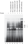
Figure 2. In vitro binding of VDR protein to the HLA-DRB1*15 VDRE.
Electrophoretic mobility shift assay showing binding of recombinant VDR and retinoic acid receptor beta (RXR) to radiolabelled oligoduplex probe corresponding to the VDRE in the proximal HLA-DRB1 promoter region for the HLA-DRB*15 haplotype. Two specific complexes are indicated, denoted I and II, together with a supershifted complex shown by an * symbol in the presence of antibody to VDR.
Figure 3. VDR is recruited to HLA-DRB1*15 VDRE in PGF cells.
Chromatin immunoprecipitation experiment using PGF cells either unstimulated (○) or after stimulation with 1,25-dihydroxyvitamin D3 (•). Input controls are shown (lanes 1 and 2), mock antibody immunoprecipitated controls (lanes 3 and 4) and VDR primary antibody immunoprecipitated DNA (lanes 5 and 6).
Figure 4. Reporter gene analysis of DRB1 promoter VDRE.
Raji B cells were transiently transfected with pGL3 luciferase constructs as indicated together with pRL_TK to normalise luciferase activity. Open bars indicate resting cells, grey shaded bars results following stimulation of transfected cells with 1,25-dihydroxyvitamin D3. Mean+/−SD of three independent transient transfection experiments are shown, each performed in quadruplicate.
Similar articles
- Contributions of vitamin D response elements and HLA promoters to multiple sclerosis risk.
Nolan D, Castley A, Tschochner M, James I, Qiu W, Sayer D, Christiansen FT, Witt C, Mastaglia F, Carroll W, Kermode A. Nolan D, et al. Neurology. 2012 Aug 7;79(6):538-46. doi: 10.1212/WNL.0b013e318263c407. Epub 2012 Jul 11. Neurology. 2012. PMID: 22786591 - Vitamin D responsive elements within the HLA-DRB1 promoter region in Sardinian multiple sclerosis associated alleles.
Cocco E, Meloni A, Murru MR, Corongiu D, Tranquilli S, Fadda E, Murru R, Schirru L, Secci MA, Costa G, Asunis I, Cuccu S, Fenu G, Lorefice L, Carboni N, Mura G, Rosatelli MC, Marrosu MG. Cocco E, et al. PLoS One. 2012;7(7):e41678. doi: 10.1371/journal.pone.0041678. Epub 2012 Jul 25. PLoS One. 2012. PMID: 22848563 Free PMC article. - Interaction of vitamin D receptor with HLA DRB1 0301 in type 1 diabetes patients from North India.
Israni N, Goswami R, Kumar A, Rani R. Israni N, et al. PLoS One. 2009 Dec 2;4(12):e8023. doi: 10.1371/journal.pone.0008023. PLoS One. 2009. PMID: 19956544 Free PMC article. - Multiple sclerosis, vitamin D, and HLA-DRB1*15.
Handunnetthi L, Ramagopalan SV, Ebers GC. Handunnetthi L, et al. Neurology. 2010 Jun 8;74(23):1905-10. doi: 10.1212/WNL.0b013e3181e24124. Neurology. 2010. PMID: 20530326 Free PMC article. Review. - HLA class II sequences and genetic susceptibility to insulin dependent diabetes mellitus.
Erlich HA. Erlich HA. Baillieres Clin Endocrinol Metab. 1991 Sep;5(3):395-411. doi: 10.1016/s0950-351x(05)80138-7. Baillieres Clin Endocrinol Metab. 1991. PMID: 1909860 Review.
Cited by
- Multiple sclerosis: major histocompatibility complexity and antigen presentation.
Ramagopalan SV, Ebers GC. Ramagopalan SV, et al. Genome Med. 2009 Nov 6;1(11):105. doi: 10.1186/gm105. Genome Med. 2009. PMID: 19895714 Free PMC article. - Radiological Association Between Multiple Sclerosis Lesions and Serum Vitamin D Levels.
Akhtar A, Neupane R, Singh A, Khan M. Akhtar A, et al. Cureus. 2022 Nov 23;14(11):e31824. doi: 10.7759/cureus.31824. eCollection 2022 Nov. Cureus. 2022. PMID: 36579263 Free PMC article. - Multiple sclerosis genetics--is the glass half full, or half empty?
Oksenberg JR, Baranzini SE. Oksenberg JR, et al. Nat Rev Neurol. 2010 Aug;6(8):429-37. doi: 10.1038/nrneurol.2010.91. Epub 2010 Jul 13. Nat Rev Neurol. 2010. PMID: 20625377 Review. - Extraskeletal effects and manifestations of Vitamin D deficiency.
Visweswaran RK, Lekha H. Visweswaran RK, et al. Indian J Endocrinol Metab. 2013 Jul;17(4):602-10. doi: 10.4103/2230-8210.113750. Indian J Endocrinol Metab. 2013. PMID: 23961475 Free PMC article. - Vitamin D and Its Synthetic Analogs.
Maestro MA, Molnár F, Carlberg C. Maestro MA, et al. J Med Chem. 2019 Aug 8;62(15):6854-6875. doi: 10.1021/acs.jmedchem.9b00208. Epub 2019 Apr 2. J Med Chem. 2019. PMID: 30916559 Free PMC article. Review.
References
- Noseworthy JH, Lucchinetti C, Rodriguez M, Weinshenker BG. Multiple sclerosis. N Engl J Med. 2000;343:938–952. - PubMed
- Ebers GC. Environmental factors and multiple sclerosis. Lancet Neurol. 2008;7:268–277. - PubMed
- Ramagopalan SV, Ebers GC. Genes for multiple sclerosis. Lancet. 2008;371:283–285. - PubMed
- Lundmark F, Duvefelt K, Iacobaeus E, Kockum I, Wallstrom E, et al. Variation in interleukin 7 receptor alpha chain (IL7R) influences risk of multiple sclerosis. Nat Genet 2007 - PubMed
- Hafler DA, Compston A, Sawcer S, Lander ES, Daly MJ, et al. Risk alleles for multiple sclerosis identified by a genomewide study. N Engl J Med. 2007;357:851–862. - PubMed
Publication types
MeSH terms
Substances
LinkOut - more resources
Full Text Sources
Other Literature Sources
Medical
Research Materials


