The mesopontine rostromedial tegmental nucleus: A structure targeted by the lateral habenula that projects to the ventral tegmental area of Tsai and substantia nigra compacta - PubMed (original) (raw)
The mesopontine rostromedial tegmental nucleus: A structure targeted by the lateral habenula that projects to the ventral tegmental area of Tsai and substantia nigra compacta
Thomas C Jhou et al. J Comp Neurol. 2009.
Abstract
Prior studies revealed that aversive stimuli and psychostimulant drugs elicit Fos expression in neurons clustered above and behind the interpeduncular nucleus that project strongly to the ventral tegmental area (VTA) and substantia nigra (SN) compacta (C). Other reports suggest that these neurons modulate responses to aversive stimuli. We now designate the region containing them as the "mesopontine rostromedial tegmental nucleus" (RMTg) and report herein on its neuroanatomy. Dense micro-opioid receptor and somatostatin immunoreactivity characterize the RMTg, as do neurons projecting to the VTA/SNC that are enriched in GAD67 mRNA. Strong inputs to the RMTg arise in the lateral habenula (LHb) and, to a lesser extent, the SN. Other inputs come from the frontal cortex, ventral striatopallidum, extended amygdala, septum, preoptic region, lateral, paraventricular and posterior hypothalamus, zona incerta, periaqueductal gray, intermediate layers of the contralateral superior colliculus, dorsal raphe, mesencephalic, pontine and medullary reticular formation, and the following nuclei: parafascicular, supramammillary, mammillary, ventral lateral geniculate, deep mesencephalic, red, pedunculopontine and laterodorsal tegmental, cuneiform, parabrachial, and deep cerebellar. The RMTg has meager outputs to the forebrain, mainly to the ventral pallidum, preoptic-lateral hypothalamic continuum, and midline-intralaminar thalamus, but much heavier outputs to the brainstem, including, most prominently, the VTA/SNC, as noted above, and to medial tegmentum, pedunculopontine and laterodorsal tegmental nuclei, dorsal raphe, and locus ceruleus and subceruleus. The RMTg may integrate multiple forebrain and brainstem inputs in relation to a dominant LHb input. Its outputs to neuromodulatory projection systems likely converge with direct LHb projections to those structures.
2009 Wiley-Liss, Inc.
Figures
Figure 1
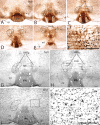
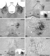
Photomicrographs illustrating the mesopontine rostromedial tegmental nucleus (RMTg) in preparations processed for μ-opioid receptor 1A (Mor) immunoreactivity (A–F), somatostatin (Som) immunoreactivity (G and H) and Nissl staining (I and J). The rat from which the sections shown in A–F, I and J were taken received an injection of methamphetamine (10 mg/kg) two hours prior to sacrifice in order to elicit the expression in the RMTg of Fos, which is shown as a second immunolabel in A–F (black dots [Fos-ir nuclei], in contrast to Mor-ir, which is brown). Note in panels B–E that strong immunoreactivity against Mor is coextensive with a dense cluster of Fos-immunoreactive nuclei. Note also that Mor-ir is denser in the medial (asterisk in C and F, an enlargement of the box in C) than lateral part of the RMTg. The RMTg is encircled on the left indicated by arrows on the right in A, B, D, E, G and H. Note the location of the RMTg in relation to the interpeduncular nucleus (IPN), medial lemniscus (ml) and the horizontal fascicles of the ventral tegmental (tgx) and superior cerebellar peduncle (xscp) decussations. In G and H, somatostatin immunoreactivity is shown at a rostral (G) and more caudal (H) level of the RMTg. Panel I shows a section adjacent to the one illustrated in C (note similar pattern of vessels in the IPN in C and I) and demonstrates that the RMTg is relatively undistinguished from surrounding regions in Nissl-stained preparations. Arrows in J, which is an enlargement of the boxed area in I, indicate the variety of small and small-medium sized neurons located in the RMTg. Scale bars: 1 mm in A–E, 200 μm in F (bar in F); 1 mm in G–I, 200 μm in J (bar in J).
Figure 2
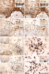
A–D. Photomicrographs of the rostromedial tegmental nucleus (RMTg, encircled in A–D) at sequentially more caudal levels, illustrating its relationship to tyrosine hydroxylase immunoreactive (TH-ir) elements in the ventral tegmental area (VTA) and retrorubral field (RRF). Note at rostral levels of the RMTg (A and B) that Fos immunoreactive nuclei (black dots) representing the RMTg (Fos expression was elicited by an i.p. methamphetamine injection) are embedded among TH-ir neurons and processes (the brown immunostained elements in the micrographs), which are fewer and more widely scattered at more caudal levels (C and D). The boxed areas in the insets are enlarged in the corresponding panels. E – H. Photomicrographs showing the rostromedial tegmental nucleus (RMTg, encircled in E and G) in sections processed with a cRNA probe against glutamate decarboxylase-67 mRNA (black label). Also shown is the distribution in the RMTg of Fluoro-Gold immunoreactive neurons (brown label) following an injection of retrogradely transported tracer into the ventral tegmental area (arrowed in the inset in H). The RMTg is shown at sequentially more caudal levels in panels E andG, which are enlargements of the boxed areas in the accompanying insets. Boxes in E and G are enlarged in panels F and H, respectively. Note, in panels F and H, that numerous neurons indicated by long arrows exhibit both labels, whereas other RMTg neurons exhibit only the black or brown (short arrows) label. Additional abbreviations: aq - cerebral aqueduct; cp - cerebral peduncle; IPN - interpeduncular nucleus; PAG - periaqueductal gray; SNR and SNC - substantia nigra reticulata and compacta; VTA - ventral tegmental area. Scale bars: 400 μm for A–D, E and G; 200 μm for F and H (bar in H); 1 mm for insets (bar in inset A).
Figure 3
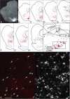
An injection of CtB into the ventral tegmental area (VTA), shown in a fluorescence micrograph in A, produces many labeled neurons in the midbrain, shown in frontal sections in rostrocaudal sequence (B–H). CtB labeled neurons expressing GAD 67 mRNA are represented by filled red circles, and are particularly enriched in the rostromedial tegmental nucleus (RMTg, boxed in black in C–G), while CtB-labeled neurons lacking GAD67 are represented as filled black dots, and predominate outside of the RMTg. The red spot in panel B represents a region just outside the CtB injection site, where labeled cells and fibers were too dense to identify reliably. A diagrammed sagittal section (I) illustrates that the region of CtB-labeled neurons expressing GAD67 extends caudodorsally from the VTA injection site (grey filled patch). The box in I is enlarged in J, within which the RMTg region is outlined. Panels K and L are photomicrographs of the green boxed area in panel G and are intended to serve as representative of tissue processed to reveal both CtB and GAD67. CtB is indicated by red fluorescent label (K), while GAD67 mRNA is indicated by silver grains (L). CtB neurons expressing GAD67 are indicated by cyan arrows, while those lacking GAD67 are indicated by blue arrowheads. Additional abbreviations: DRN - dorsal raphe nucleus; PPTg - pedunculopontine tegmental nucleus; SNC - substantia nigra compacta. Scale bar: 100μm.
Figure 4
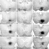
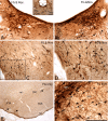
Photomicrographs illustrating, from top to bottom, four sequentially more caudal transverse sections through the mesencephalon and rostral pons from three cases. Corresponding levels are shown in each horizontal row. The rostromedial tegmental nucleus (RMTg) is encircled in panels E–H, which came from a rat (case 06130) in which Fos expression was elicited in the RMTg by cocaine administration. Sections from that brain were processed to exhibit Fos immunoreactive neurons, which are densely clustered within the RMTg (arrows on left and encircled on right in E–H). The sections shown in panels A–D came from a rat that received an injection of anterogradely transported PHAL (case 07027), while those shown in panels I–L came from a rat that received an injection of retrogradely transported Fluoro-Gold (case 08023), both of which can be seen to occupy the RMTg by reference to panels E–H. Maps illustrating the resulting transport of tracers are shown in Figures 5 (08023) and 10 (07027). Additional abbreviations: aq - cerebral aqueduct; cp - cerebral peduncle; IPN - interpeduncular nucleus; ml - medial lemniscus; PAG - periaqueductal gray; SNR - substantia nigra reticulata. Scale bar: 1 mm.
Figure 5
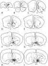
Map illustrating the distribution in the rat brain of retrogradely labeled neurons following injection of Fluoro-Gold into the medial tegmental nucleus (RMTg). The injection site is shown in Figure 4 I– L. Each dot represents one retrogradely labeled neuron. Selected groups of nitric oxide synthase immunoreactive neurons are indicated as gray dots in selected panels (K, O, and S–X). Abbreviations: 10 -dorsal motor nucleus of the vagus nerve; Acb - nucleus accumbens; Bar - Barrington's nucleus; vBST - bed nucleus of stria terminalis, ventral part; Cg - cingulate cortex; CN - deep cerebellar nuclei; CnF - cuneiform nucleus; DA - dorsal hypothalamic area; DP - dorsal peduncular cortex; DpMe - deep mesencephalic nucleus; DR - dorsal raphe; EPN - entopeduncular nucleus; FrA - frontal association cortex; GP - globus pallidus; IPN - interpeduncular nucleus; ISC - intermediate layers of the superior colliculus; LDTg - laterodorsal tegmental nucleus; LH - lateral hypothalamus; LHb - lateral habenula; LPO - lateral preoptic area; LS - lateral septum; M - mammillary nucleus; MPO - medial preoptic area; O - orbital cortex; Pa - hypothalamic paraventricular nucleus; PAG - periaqueductal gray; PB - parabrachial nucleus; PF - parafascicular nucleus; PH - posterior hypothalamic area; PPTg - pedunculopontine tegmental nucleus; PrL - prelimbic cortex; RN - red nucleus; Rt - reticular formation; SI - substantia innominata; sm - stria medullaris; SNC & R - substantia nigra pars compacta and reticulata; Sol - nucleus of the tractus solitarius; SuM - supramammillary nucleus; VDB - vertical limb of the diagonal band; VLG - ventral lateral geniculate nucleus; RMTg - medial tegmental nucleus; VP - ventral pallidum; VTA - ventral tegmental area; ZI - zona incerta.
Figure 6
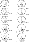
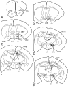
Maps illustrating Fluoro-Gold injection sites into the RMTg (08019, 08023, and 08025) and into control sites surrounding it (06160, 06164, 06166, 08022, 08031). Panels A–E and F–J are replicate sequences of idealized transverse sections through the mesencephalon and rostral pons at increasingly caudal levels from top to bottom. Four fill-coded injection sites are shown in each series. The RMTg is represented by clustered black dots, which reflect Fos immunoreactive neurons plotted in sections from a rat that received an injection of methamphetamine 2 hours prior to sacrifice. Brain structures in which retrogradely labeled neurons were observed in each indicated case are given in Table 1.
Figure 7
A sampling of sites where neurons that became retrogradely labeled (black reaction product) following Fluoro-Gold (FG) injections into the rostromedial tegmental nucleus (case 08023) are extensively co-distributed with nitric oxide synthase (Nos, panels A–D) or tyrosine hydroxylase (TH, panels E and F) immunoreactive (ir) neurons. Innumerable retrogradely labeled (black label) and Nos-ir (brown label) neurons occupy the laterodorsal tegmental nucleus (LDTg) ipsilateral to the injection site (B and inset in A) and many of these contain both labels. Fewer retrogradely labeled neurons are present in the contralateral LDTg (A). Likewise, retrogradely labeled (black) and Nos-ir (brown) neurons are extensively intermixed in the ipsilateral lateral hypothalamus (C) and many contain both labels (arrows in D, an enlargement of the box in C). Retrogradely labeled neurons in the ventral tegmental area and substantia nigra are shown in panel E (some examples are arrowed). In panel F, which illustrates a section adjacent to the one shown in E, retrogradely labeled neurons are shown in black (arrows) relative to TH-ir neurons (brown). Neurons exhibiting the retrograde label and TH-ir were not observed. Additional abbreviations: aq - cerebral aqueduct; CnF - cuneiform nucleus; cp - cerebral peduncle; DTg - dorsal tegmental nucleus; IC - inferior colliculus; ml - medial lemniscus; MR - median raphe; PPTg - pedunculopontine tegmental nucleus; SNC & R - substantia nigra pars compacta & reticulata. Scale bars: 400 μm in A–C, 200 μm in D and F, 1mm in E (bar in F); 1 mm in inset A.
Figure 8
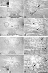
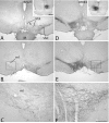
Photomicrographs illustrating a labeled projection to the ipsilateral and contralateral medial tegmental nucleus (RMTg) observed following injection of PHA-L into the substantia nigra pars compacta and adjacent reticulata (white x in inset in A). Panels A, C, E and G show sequentially more caudal sections through the RMTg (encircled on side, boxed on the other). Note that immunoperoxidase product is present in the boxes in A, C, E, and G, of which all, except the one in A, are ipsilateral to the injection site. B, D, F and H, respectively, are enlargements of the boxes in A, C, E and G and reveal that the labeling in the boxes reflects PHA-L labeled axonal projections. Arrows in A, C, E and G indicate labeling in the RMTg on the side not indicated by a box. Arrows in B, D, F and H indicate some of the labeled axons. Additional abbreviations: aq: cerebral aqueduct; cp - cerebral peduncle; IPN - interpeduncular nucleus; ml - medial leminscus; tgx - tegmental decussation; xscp - decussation of the superior cerebellar peduncle. Scale bars: 1 mm in A, C, E and G; 200 μm in B, D, F and H (bar in H); 1 mm in inset A.
Figure 9
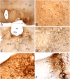
Photomicrographs illustrating a robust, selective projection from the lateral habenula (LHb) to the medial tegmental nucleus (RMTg). Panel A displays the prominent ipsilateral (right side) and less prominent contralateral distribution of retrogradely labeled neurons in the LHb following an injection of Fluoro-Gold into the RMTg (case 08023, injection site shown in Fig. 4 I–K and 6G–J). Panels B–D illustrate the prominent ipsilateral (right side) and less prominent contralateral distribution of anterogradely labeled fibers in the RMTg following a PHA-L injection into the LHb shown in the inset in B. Panels B and C (case 08004) show more rostral and caudal levels of the RMTg, respectively, and D shows the ipsilateral (right side) labeling in C, enlarged. Rat 08004 was administered 10 mg/kg of methamphetamine 2 hours prior to sacrifice to elicit Fos expression in the RMTg and this is reflected in the black dots [Fos-ir nuclei] shown in panels B–F. Note that the dense anterograde labeling is distributed coextensively with the dense clusters of Fos-ir neurons. Panels E and F (case 08011, also received methamphetamine) illustrate a more medial injection of PHA-L into the LHb (inset in E), which gives rise to anterograde labeling that involves only the medialmost part of the RMTg (see panel F, an enlargement of the ipsilateral anterograde labeling (right side) in panel E. Additional abbreviations: IPN - interpeduncular nucleus; MHb - medial habenula; ml - medial lemniscus; xscp - decussation of the superior cerebellar peduncle. Scale bars: 1 mm in A, B, C, and E, 400 μm in D and F (bar in F); 1mm in insets (bar in inset B).
Figure 10
Map illustrating the distribution in the rat brain of anterogradely labeled fibers following an injection of PHA-L into the rostromedial tegmental nucleus. The injection site is shown in Figure 4A–D. Symbols (pluses) indicate anterograde labeling in numbers that, based upon subjective impression, correspond approximately to the density of labeling observed in the sections. Selected groups of nitric oxide synthase immunoreactive neurons are indicated as gray dots in selected panels (D, H, M, N, O, R and S). The ventral tegmental area (VTA) and substantia nigra compacta (SNC) area outlined in panels I–K. The circumscribed injection site (inj) is indicated in panels M and N. Tyrosine hydroxylase immunoreactive neurons and fibers of the locus ceruleus (LC) and subceruleus (SC) are indicated as gray dots and pluses, respectively, in panel Q. Abbreviations: mBST - bed nucleus of stria terminalis, medial part; CnF - cuneiform nucleus; DA - dorsal hypothalamic area; d Hipp - dorsal hippocampus; DR - dorsal raphe; EPN - entopeduncular nucleus; FrA - frontal association cortex; IPAC - interstitial nucleus of the posterior limb of the anterior commissure; LC - locus ceruleus; LDTg - laterodorsal tegmental nucleus; LH - lateral hypothalamus; LHb - lateral habenula; LPO - lateral preoptic area; m-i Th - midline-intralaminar thalamic nuclei; PAG - periaqueductal gray; PB - parabrachial nucleus; PF - parafascicular nucleu; PPTg - pedunculopontine tegmental nucleus; Pr - nucleus prepositus; Rt - reticular formation; SC - subceruleus; Sol - nucleus of the tractus solitarius; VDB - vertical limb of the diagonal band; RMTg - medial tegmental nucleus; VP - ventral pallidum; VTA - ventral tegmental area; ZI - zona incerta.
Figure 11
Photomicrographs showing the projections from the rostromedial tegmental nucleus (RMTg) to the ventral tegmental area (VTA) - substantia nigra compacta (SNC) complex in two cases in which PHA-L was injected into the RMTg. Panels A–C illustrate case 07027, which is mapped in Figure 9 and has an injection site shown in Fig. 4 A–D. Panels A and B show more rostral and caudal levels of the VTA-SNC complex with ipsilateral anterograde labeling from the injection (shown in the inset in A) on the right. Contralateral labeling, within the box in B and enlarged in C, is less prominent. Panels D–E illustrate a similar injection (case 07033) that gives rise to denser ipsilateral (D and E, on the left) and contralateral anterograde labeling. The box in E is enlarged in F. Verticle lines and x in the insets approximate the midline and centers of the injections, respectively. The injection shown in panels A–C is centered slightly further from the midline and caudal relative to that shown in panels D–F. Additional abbreviation: fr - fasciculus retroflexus. Scale bars: 1mm in A–D, 200 μm in C and F (bar in F); 1 mm in insets (bar in inset A).
Figure 12
Diagram depicting idealized sections though the mesencephalon and rostral pons with centers of PHA-L injection sites coded to reflect the amount of anterograde labeling that they gave rise to in the ventral tegmental area (VTA) - substantia nigra compacta (SNC) complex. Gray injections produced little or no labeling in the VTA-SNC complex. Cross-hatched sites produced sparse to moderate labeling, whereas simple-hatched sites produced robust VTA-SNC anterograde labeling. The medial tegmental nucleus (RMTg) is indicated by the clusters of black dots. The cases shown in this diagram are also included in Table 2, which considers additional cases and provides a more detailed treatment of VTA-SNC labeling.
Figure 13
Photomicrographs of sections processed for PHA-L (black), nitric oxide synthase (Nos) immunoreactivity (brown in A–E) or tyrosine hydroxylase (TH, in F) immunoreactivity showing anterograde labeling of axons in the lateral hypothalamus (A and B), mesopontine tegmentum (C–E) and locus ceruleus (F) in case 07027, in which PHA-L was injected into the medial tegmental nucleus (RMTg). The injection site for 07027 is illustrated in Figure 4 and the case is mapped in Figure 10. Note the moderate numbers of labeled projections to the lateral hypothalamus (LH) are (black fibers in B) in comparison to the robust labeled projection within in the mesopontine reticular formation (Rt). The mesopontine labeling substantially invades the laterodorsal tegmental nucleus (LDTg in E, an enlargement of Box E in panel C) and the dissipated part of the pedunculopontine tegmental nucleus (dPPTg in D, an enlargement of box D in panel C), which extends between the LDTg and PPTg and contains scattered Nos-ir neurons. Panel F shows the locus ceruleus, processed for TH-ir (brown) and PHA-L-ir (black fibers), from the same case. Robust axonal labeling can be seen in the region containing the distal parts of TH-ir dendrites (long arrows) and with in the cell dense part of the locus ceruleus (short arrows). Additional abbreviations: III - 3rd ventricle; aq - cerebral aqueduct; CnF - cuneiform nucleus; f - fornix; GP - globus pallidus; ic - internal capsule; Me - medial amygdala; mfb - medial forebrain bundle; opt - optic tract; Pa - paraventricular nucleus; Th - thalamus. Scale bars: 1 mm in A and C, μ200 m in B and D, 400 μm in E and F (bar in F).
Figure 14
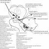
Summary diagram illustrating the input-output relationships of the rostromedial tegmental nucleus (RMTg). Inputs are given on the left, outputs on the right. Line widths and arrowhead sizes represent approximations of the relative robustness of the pathways. Anatomical data suggest that the major RMTg throughput involves strong inputs to the RMTg from the lateral habenula, supplemented by projections from neurons scattered along the interface between the substantia nigra compacta and reticulata, and strong outputs to the ventral tegmental area/substantia nigra compacta complex. Numerous other forebrain and brainstem structures (listed on lower left in approximate rostrocaudal order) also project varyingly strongly to the RMTg, which has about equivalently strong outputs to the dorsal raphe, pedunculopontine tegmental nucleus, laterodorsal tegmental nucleus and locus ceruleus, which give rise to ascending neuromodulatory (“state-setting”) pathways, and to the medial part of the mesencephalic and pontomedullary reticular formation, which gives rise to extensive, diffusely organized “level-setting” pathways to lower brainstem and spinal cord (see Discussion). Relatively meager outputs to the forebrain, by comparison, include the ventral pallidum, lateral preoptic area, lateral hypothalamus and midline-intralaminar thalamic groups. Abbreviations: contralat - contralateral, cx - cortex, div - division, intermed - intermediate, n - nucleus, nn - nuclei.
Comment in
- The mesopontine rostromedial tegmental nucleus and the emotional motor system: role in basic survival behavior.
Holstege G. Holstege G. J Comp Neurol. 2009 Apr 20;513(6):559-65. doi: 10.1002/cne.21990. J Comp Neurol. 2009. PMID: 19235226 No abstract available.
Similar articles
- Sources of input to the rostromedial tegmental nucleus, ventral tegmental area, and lateral habenula compared: A study in rat.
Yetnikoff L, Cheng AY, Lavezzi HN, Parsley KP, Zahm DS. Yetnikoff L, et al. J Comp Neurol. 2015 Nov 1;523(16):2426-56. doi: 10.1002/cne.23797. Epub 2015 Jun 10. J Comp Neurol. 2015. PMID: 25940654 Free PMC article. - Lateral habenula and the rostromedial tegmental nucleus innervate neurochemically distinct subdivisions of the dorsal raphe nucleus in the rat.
Sego C, Gonçalves L, Lima L, Furigo IC, Donato J Jr, Metzger M. Sego C, et al. J Comp Neurol. 2014 May 1;522(7):1454-84. doi: 10.1002/cne.23533. J Comp Neurol. 2014. PMID: 24374795 - Mesopontine rostromedial tegmental nucleus neurons projecting to the dorsal raphe and pedunculopontine tegmental nucleus: psychostimulant-elicited Fos expression and collateralization.
Lavezzi HN, Parsley KP, Zahm DS. Lavezzi HN, et al. Brain Struct Funct. 2012 Jul;217(3):719-34. doi: 10.1007/s00429-011-0368-z. Epub 2011 Dec 18. Brain Struct Funct. 2012. PMID: 22179106 Free PMC article. - Reward and aversion in a heterogeneous midbrain dopamine system.
Lammel S, Lim BK, Malenka RC. Lammel S, et al. Neuropharmacology. 2014 Jan;76 Pt B(0 0):351-9. doi: 10.1016/j.neuropharm.2013.03.019. Epub 2013 Apr 8. Neuropharmacology. 2014. PMID: 23578393 Free PMC article. Review. - The human substantia nigra and ventral tegmental area. A neuroanatomical study with notes on aging and aging diseases.
van Domburg PH, ten Donkelaar HJ. van Domburg PH, et al. Adv Anat Embryol Cell Biol. 1991;121:1-132. Adv Anat Embryol Cell Biol. 1991. PMID: 2053466 Review.
Cited by
- Neuropeptide S (NPS) neurons: Parabrachial identity and novel distributions.
Huang D, Zhang R, Gasparini S, McDonough MC, Paradee WJ, Geerling JC. Huang D, et al. J Comp Neurol. 2022 Dec;530(18):3157-3178. doi: 10.1002/cne.25400. Epub 2022 Aug 29. J Comp Neurol. 2022. PMID: 36036349 Free PMC article. - Lesions of the fasciculus retroflexus alter footshock-induced cFos expression in the mesopontine rostromedial tegmental area of rats.
Brown PL, Shepard PD. Brown PL, et al. PLoS One. 2013 Apr 12;8(4):e60678. doi: 10.1371/journal.pone.0060678. Print 2013. PLoS One. 2013. PMID: 23593280 Free PMC article. - A Neural Circuit Mechanism for Encoding Aversive Stimuli in the Mesolimbic Dopamine System.
de Jong JW, Afjei SA, Pollak Dorocic I, Peck JR, Liu C, Kim CK, Tian L, Deisseroth K, Lammel S. de Jong JW, et al. Neuron. 2019 Jan 2;101(1):133-151.e7. doi: 10.1016/j.neuron.2018.11.005. Epub 2018 Nov 29. Neuron. 2019. PMID: 30503173 Free PMC article. - A neural correlate of predicted and actual reward-value information in monkey pedunculopontine tegmental and dorsal raphe nucleus during saccade tasks.
Okada K, Nakamura K, Kobayashi Y. Okada K, et al. Neural Plast. 2011;2011:579840. doi: 10.1155/2011/579840. Epub 2011 Oct 12. Neural Plast. 2011. PMID: 22013541 Free PMC article. Review. - A unique population of ventral tegmental area neurons inhibits the lateral habenula to promote reward.
Stamatakis AM, Jennings JH, Ung RL, Blair GA, Weinberg RJ, Neve RL, Boyce F, Mattis J, Ramakrishnan C, Deisseroth K, Stuber GD. Stamatakis AM, et al. Neuron. 2013 Nov 20;80(4):1039-53. doi: 10.1016/j.neuron.2013.08.023. Neuron. 2013. PMID: 24267654 Free PMC article.
References
- Alheid GF, Heimer L. New perspectives in basal forebrain organization of special relevance for neuropsychiatric disorders: the striatopallidal, amygdaloid, and corticopetal components of substantia innominata. Neuroscience. 1988;27:1–39. - PubMed
- Alheid GF, Heimer L. Theories of basal forebrain organization and the “emotional motor system”. In: Holstege G, Bandler R, Saper CB, editors. The Emotional Motor System. Prog Brain Res. Vol. 107. 1996. pp. 461–484. - PubMed
- Andén NE, Carlsson A, Dahlström A, Fuxe K, Hillarp NÅ, Larsson K. Demonstration and mapping out of nigro-neostriatal dopaminergic neurons. Life Sci. 1964;3:523–530. - PubMed
- Andén NE, Dahlström A, Fuxe K, Larsson K. Mapping out of catecholaminergic and 5-hydroxtryptamine neurons innervating the telencephalon and diencephalon. Life Sci. 1965;4:1275–1279. - PubMed
- Andén NE, Dahlström A, Fuxe K, Larsson K, Olson L, Ungerstedt U. Ascending monoamine neurons to the telencephalon and diencephalon. Acta Physiol. Scand. 1966a;67:313–326.
Publication types
MeSH terms
Substances
Grants and funding
- R01 NS023805/NS/NINDS NIH HHS/United States
- HL-60292/HL/NHLBI NIH HHS/United States
- P50 HL060292/HL/NHLBI NIH HHS/United States
- MH-12370/MH/NIMH NIH HHS/United States
- DA-020654/DA/NIDA NIH HHS/United States
- F31 MH012370/MH/NIMH NIH HHS/United States
- NS-23805/NS/NINDS NIH HHS/United States
LinkOut - more resources
Full Text Sources
Miscellaneous
