Antigenic fingerprinting of H5N1 avian influenza using convalescent sera and monoclonal antibodies reveals potential vaccine and diagnostic targets - PubMed (original) (raw)
Antigenic fingerprinting of H5N1 avian influenza using convalescent sera and monoclonal antibodies reveals potential vaccine and diagnostic targets
Surender Khurana et al. PLoS Med. 2009.
Abstract
Background: Transmission of highly pathogenic avian H5N1 viruses from poultry to humans have raised fears of an impending influenza pandemic. Concerted efforts are underway to prepare effective vaccines and therapies including polyclonal or monoclonal antibodies against H5N1. Current efforts are hampered by the paucity of information on protective immune responses against avian influenza. Characterizing the B cell responses in convalescent individuals could help in the design of future vaccines and therapeutics.
Methods and findings: To address this need, we generated whole-genome-fragment phage display libraries (GFPDL) expressing fragments of 15-350 amino acids covering all the proteins of A/Vietnam/1203/2004 (H5N1). These GFPDL were used to analyze neutralizing human monoclonal antibodies and sera of five individuals who had recovered from H5N1 infection. This approach led to the mapping of two broadly neutralizing human monoclonal antibodies with conformation-dependent epitopes. In H5N1 convalescent sera, we have identified several potentially protective H5N1-specific human antibody epitopes in H5 HA[(-10)-223], neuraminidase catalytic site, and M2 ectodomain. In addition, for the first time to our knowledge in humans, we identified strong reactivity against PB1-F2, a putative virulence factor, following H5N1 infection. Importantly, novel epitopes were identified, which were recognized by H5N1-convalescent sera but did not react with sera from control individuals (H5N1 naïve, H1N1 or H3N2 seropositive).
Conclusion: This is the first study, to our knowledge, describing the complete antibody repertoire following H5N1 infection. Collectively, these data will contribute to rational vaccine design and new H5N1-specific serodiagnostic surveillance tools.
Conflict of interest statement
The authors have declared that no competing interests exist.
Figures
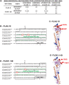
Figure 1. Epitope mapping of broadly neutralizing H5N1 human MAbs.
(A) End-point titers (mean of three replicates) using two H5N1 human MAbs (at 1 mg/ml) in a microneutralization assay performed with rgH5N1×PR8 (2∶6) reassorted viruses are shown. *, The information on in vivo protection of mice against wild-type A/Vietnam/1203/2004 and A/Indonesia/5/05 challenge was previously published . (B) HA segment [(-10)-233] was identified by GFPDL panning using MAb FLA5.10 (boxed). Amino acid number +1 corresponds to H3 (A/California/7/2004) amino acid -10. The critical contact residues for FLA5.10, identified using RPL are shown in red circles. (C) Alignment of the critical residues for MAb FLA5.10 binding on the 3-D structure of the HA monomer (Protein Data Base [PDB] identifier 21BX) with amino acid colors corresponding to (B). The predicted glycosylation sites (NXT/NXS) are shown in blue. (D) HA segment (32–320) identified by GFPDL-panning using MAb FLD21.140 (boxed). The putative contact residues identified using RPL, are shown in red circles. (E) Alignment of the critical residues for MAb FLD21.140 binding on the 3-D structure of the HA monomer (PDB Identifier 2IBX) with amino acid colors corresponding to (D). The predicted glycosylation sites (NXT/NXS) are shown in blue.
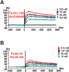
Figure 2. Steady-state binding equilibrium analysis of human MAbs to purified bacterially expressed H5 HA[(-10-223)] fragment.
Various concentrations of MAbs FLD21.140 (A) and FLA5.10 (B) were injected simultaneously onto recombinant HA [(-10)-223] (identified in Figure 1B) peptide, immobilized on a sensor chip through the free amine group, and onto a blank flow cell, free of peptide. Binding was recorded using ProteOn system surface plasmon resonance biosensor instrument (BioRad Labs). As a control, anti-CCR5 MAb 2D7) was injected at the same concentrations on HA [(-10)-223] coupled chip. RU, resonance units.
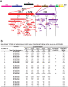
Figure 3. Elucidation of the epitope profile in HA and NA proteins recognized by antibodies in individuals that survived H5N1 infections in Vietnam.
(A) Alignment of peptides recognized by pooled sera from H5N1-infected individuals identified using H5 (HA+NA) GFPDL. Bars with arrows represent the identified inserts in 5′–3′ orientation. Numbered segments represent inserts that were selected with high frequencies (≥5; Table S1). These peptides were expressed and purified from E .coli or were chemically synthesized and used in ELISA. The sequences of the influenza encoded fragments are numbered according to the intact complete proteome (Figure S1). Peptide ID numbers are the same in (A) and (B). (B) ELISA reactivity of sera from individual H5N1-infected patients (Viet1–5) or pooled sera from 20 healthy Vietnamese adults (75% had neutralizing titers against either H3N2 influenza strains, H1N1 strains, or both) with H5N1 HA1 peptides (1–8), HA2 peptides (9–14), and NA peptides (15–21) (localization indicated in Figure 4A). An initial serum dilution (1∶100) was followed by serial 5-fold dilutions. End-point titers are reported. Days postadmission represent the time of serum collection for each patient. Six antigenic clusters in HA (cluster-I, 2,359–2,453; cluster-II, 2,454–2,621; cluster-III, 2,627–2,670; cluster-IV, 2,682–2,703; cluster-V, 2,706–2,814; cluster-VI, 2,823–2,816) recognized by the convalescent sera are shown.
Figure 4. Main antigenic clusters in the structures of HA and NA recognized by antibodies from H5N1 virus infected individuals.
(A) Antigenic clusters in HA, as identified in Figure 3B, are shown as surface exposed colored patches on one HA monomer within the HA trimer structure (PDB identifier 2IBX). The antigenic clusters (I–III) in HA1 cover the Antigenic Sites a, b, c, d, and e that have been described in the H3 HA1 based on mouse MAbs , . (B) The immunodominant conformational epitope in the NA (NA-3676-3854, peptide 15 in Figure 3B) is shown in green on the tetrameric NA structure (PDB Identifier 2HTY) with the predicted site of bound sialic acid shown in red. Side view and bird-eye views are shown.
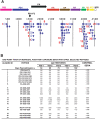
Figure 5. Antibody epitopes in H5N1 internal proteins (FLU-6) recognized by pooled sera from H5N1 infected individuals.
(A) Schematic alignment of the peptides identified using GFPDL (H5N1-FLU-6) expressing all internal proteins of influenza A/Vietnam/1203/2004. The predicted influenza encoded proteins are numbered according to the complete proteome (Figure S1). Bars with arrows indicate identified inserts in the 5′–3′ orientation. Numbered segments represent high frequency clones (≥5; Table S1). These peptides were expressed and purified from E. coli or were chemically synthesized and the numbers correspond to the peptide identifiers in the ELISA assay in (B). (B) Reactivities of sera from individual H5N1-infected patients (Viet 1–5) or sera from healthy Vietnamese adults against peptides derived from: PB2 (1); PB1 (2–3); PB1-F2 (4–7); PA (8); NP (9–12); M1 (13–16); M2e (17); M2 (18); NS1 (19–20); and NS2 (21).
Comment in
- Mapping antibody epitopes of the avian H5N1 influenza virus.
Yen HL, Peiris JS. Yen HL, et al. PLoS Med. 2009 Apr 21;6(4):e1000064. doi: 10.1371/journal.pmed.1000064. Epub 2009 Apr 21. PLoS Med. 2009. PMID: 19381281 Free PMC article.
Similar articles
- Antigenic Fingerprinting of Antibody Response in Humans following Exposure to Highly Pathogenic H7N7 Avian Influenza Virus: Evidence for Anti-PA-X Antibodies.
Khurana S, Chung KY, Coyle EM, Meijer A, Golding H. Khurana S, et al. J Virol. 2016 Sep 29;90(20):9383-93. doi: 10.1128/JVI.01408-16. Print 2016 Oct 15. J Virol. 2016. PMID: 27512055 Free PMC article. - H5N1 virus-like particle vaccine elicits cross-reactive neutralizing antibodies that preferentially bind to the oligomeric form of influenza virus hemagglutinin in humans.
Khurana S, Wu J, Verma N, Verma S, Raghunandan R, Manischewitz J, King LR, Kpamegan E, Pincus S, Smith G, Glenn G, Golding H. Khurana S, et al. J Virol. 2011 Nov;85(21):10945-54. doi: 10.1128/JVI.05406-11. Epub 2011 Aug 24. J Virol. 2011. PMID: 21865396 Free PMC article. Clinical Trial. - Cross-Protection by Inactivated H5 Prepandemic Vaccine Seed Strains against Diverse Goose/Guangdong Lineage H5N1 Highly Pathogenic Avian Influenza Viruses.
Criado MF, Sá E Silva M, Lee DH, Salge CAL, Spackman E, Donis R, Wan XF, Swayne DE. Criado MF, et al. J Virol. 2020 Nov 23;94(24):e00720-20. doi: 10.1128/JVI.00720-20. Print 2020 Nov 23. J Virol. 2020. PMID: 32999029 Free PMC article. - The antigenic architecture of the hemagglutinin of influenza H5N1 viruses.
Velkov T, Ong C, Baker MA, Kim H, Li J, Nation RL, Huang JX, Cooper MA, Rockman S. Velkov T, et al. Mol Immunol. 2013 Dec;56(4):705-19. doi: 10.1016/j.molimm.2013.07.010. Epub 2013 Aug 7. Mol Immunol. 2013. PMID: 23933511 Review. - [Tetravaccine--new fundamental approach to prevention of influenza pandemic].
Onishchenko GG, Zverev VV, Katlinskiĭ AV, Semchenko AV, Korovkin SA, Mel'nikov SIa, Mironov AN. Onishchenko GG, et al. Zh Mikrobiol Epidemiol Immunobiol. 2007 Jul-Aug;(4):15-9. Zh Mikrobiol Epidemiol Immunobiol. 2007. PMID: 17882832 Review. Russian.
Cited by
- Cross-protection of newly emerging HPAI H5 viruses by neutralizing human monoclonal antibodies: A viable alternative to oseltamivir.
Ren H, Wang G, Wang S, Chen H, Chen Z, Hu H, Cheng G, Zhou P. Ren H, et al. MAbs. 2016 Aug-Sep;8(6):1156-66. doi: 10.1080/19420862.2016.1183083. Epub 2016 May 11. MAbs. 2016. PMID: 27167234 Free PMC article. - Heterosubtypic neutralizing antibodies are produced by individuals immunized with a seasonal influenza vaccine.
Corti D, Suguitan AL Jr, Pinna D, Silacci C, Fernandez-Rodriguez BM, Vanzetta F, Santos C, Luke CJ, Torres-Velez FJ, Temperton NJ, Weiss RA, Sallusto F, Subbarao K, Lanzavecchia A. Corti D, et al. J Clin Invest. 2010 May;120(5):1663-73. doi: 10.1172/JCI41902. Epub 2010 Apr 12. J Clin Invest. 2010. PMID: 20389023 Free PMC article. - AS03-adjuvanted H5N1 vaccine promotes antibody diversity and affinity maturation, NAI titers, cross-clade H5N1 neutralization, but not H1N1 cross-subtype neutralization.
Khurana S, Coyle EM, Manischewitz J, King LR, Gao J, Germain RN, Schwartzberg PL, Tsang JS, Golding H; and the CHI Consortium. Khurana S, et al. NPJ Vaccines. 2018 Oct 1;3:40. doi: 10.1038/s41541-018-0076-2. eCollection 2018. NPJ Vaccines. 2018. PMID: 30302282 Free PMC article. - Identification of Conserved Peptides Comprising Multiple T Cell Epitopes of Matrix 1 Protein in H1N1 Influenza Virus.
Lohia N, Baranwal M. Lohia N, et al. Viral Immunol. 2015 Dec;28(10):570-9. doi: 10.1089/vim.2015.0060. Epub 2015 Sep 23. Viral Immunol. 2015. PMID: 26398199 Free PMC article. - Human monoclonal antibodies targeting the haemagglutinin glycoprotein can neutralize H7N9 influenza virus.
Chen Z, Wang J, Bao L, Guo L, Zhang W, Xue Y, Zhou H, Xiao Y, Wang J, Wu F, Deng Y, Qin C, Jin Q. Chen Z, et al. Nat Commun. 2015 Mar 30;6:6714. doi: 10.1038/ncomms7714. Nat Commun. 2015. PMID: 25819694
References
- Gambotto A, Barratt-Boyes SM, de Jong MD, Neumann G, Kawaoka Y. Human infection with highly pathogenic H5N1 influenza virus. Lancet. 2008;371:1464–1475. - PubMed
- Luke TC, Kilbane EM, Jackson JL, Hoffman SL. Meta-analysis: convalescent blood products for Spanish influenza pneumonia: a future H5N1 treatment? Ann Intern Med. 2006;145:599–609. - PubMed
Publication types
MeSH terms
Substances
LinkOut - more resources
Full Text Sources
Other Literature Sources
Medical
Molecular Biology Databases
Miscellaneous


