Immunohistochemical localization of phosphohistidine phosphatase PHPT1 in mouse and human tissues - PubMed (original) (raw)
Immunohistochemical localization of phosphohistidine phosphatase PHPT1 in mouse and human tissues
Xiau-Qun Zhang et al. Ups J Med Sci. 2009.
Abstract
Protein histidine phosphorylation accounts for about 6% of the total protein phosphorylation in eukaryotic cells; still details concerning histidine phosphorylation and dephosphorylation are limited. A mammalian 14-kDa phosphohistidine phosphatase, also denominated PHPT1, was found 6 years ago that provided a new tool in the study of phosphohistidine phosphorylation. The localization of PHPT1 mRNA by Northern blot analysis revealed high expression in heart and skeletal muscle. The main object of the present study was to determine the PHPT1 expression on protein level in mouse tissues in order to get further information on the physiological role of the enzyme. Tissue samples from adult mice and 14.5-day-old mouse embryos were processed for immunostaining using a PHPT1-specific polyclonal antibody. The same antibody was also provided to the Swedish human protein atlas project (HPR) (http://www.proteinatlas.org/index.php). The results from both studies were essentially consistent with the previously reported expression of mRNA of a few human tissues. In addition, several other tissues, including testis, displayed a high protein expression. A salient result of the present investigation was the ubiquitous expression of the PHPT1 protein and its high expression in continuously dividing epithelial cells.
Figures
Figure 1.
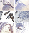
Immunohistochemical staining for PHPT1. A: An E14.5 mouse embryo sagittal section shows PHPT1 signal in the epithelium of the choroid plexus in the fourth ventricle of brain (arrow). B: Adjacent section of A, using preimmune serum as a negative control. C: The same expression pattern of PHPT1 was found in the epithelium layer of the choroid plexus in an adult mouse brain (arrow). Arrowheads point at the ependymal cells in the ventricle. D: Purkinje cell (arrow) in the cerebellum expressing PHPT1. E: PHPT1 expression in the E14.5 embryonic heart muscle. F: PHPT1 expression (arrow) in the epithelium layer of the developing gut of an E14.5 sagittal section. Amplifications were 100× for A, B, and E, 400× for C and D, and 25× for F.
Figure 2.
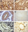
Immunohistochemical staining for PHPT1. A: A section of adult mouse kidney shows PHPT1 expression in the distal convoluted tubules (arrowhead) but not in the glomeruli (arrow) and a weak expression in the proximal convoluted tubule. B: PHPT1 is expressed in the Henle's loops of adult kidney. C and D: Sections of seminiferous tubule of adult mouse testis. Arrows in C point to the spermatogonium of seminiferous tubule. D: An absorption test shows that PHPT1 signals were abolished in the mouse testis. E: PHPT1 is expressed in the epithelium of interlobular duct of pancreas (arrow). F: PHPT1 is expressed in the epithelium of bronchiole (arrow). Arrowheads point at macrophages in alveoli. Amplifications were 400× for A–F.
Similar articles
- Phosphohistidine phosphatase 1 (PHPT1) also dephosphorylates phospholysine of chemically phosphorylated histone H1 and polylysine.
Ek P, Ek B, Zetterqvist Ö. Ek P, et al. Ups J Med Sci. 2015 Mar;120(1):20-7. doi: 10.3109/03009734.2014.996720. Epub 2015 Jan 9. Ups J Med Sci. 2015. PMID: 25574816 Free PMC article. - A splice variant of the human phosphohistidine phosphatase 1 (PHPT1) is degraded by the proteasome.
Inturi R, Wäneskog M, Vlachakis D, Ali Y, Ek P, Punga T, Bjerling P. Inturi R, et al. Int J Biochem Cell Biol. 2014 Dec;57:69-75. doi: 10.1016/j.biocel.2014.10.009. Epub 2014 Oct 14. Int J Biochem Cell Biol. 2014. PMID: 25450458 - Nuclear expression and clinical significance of phosphohistidine phosphatase 1 in clear-cell renal cell carcinoma.
Shen H, Yang P, Liu Q, Tian Y. Shen H, et al. J Int Med Res. 2015 Dec;43(6):747-57. doi: 10.1177/0300060515587576. Epub 2015 Nov 3. J Int Med Res. 2015. PMID: 26537769 - Histidine Phosphorylation: Protein Kinases and Phosphatases.
Ning J, Sala M, Reina J, Kalagiri R, Hunter T, McCullough BS. Ning J, et al. Int J Mol Sci. 2024 Jul 21;25(14):7975. doi: 10.3390/ijms25147975. Int J Mol Sci. 2024. PMID: 39063217 Free PMC article. Review. - Reversible phosphorylation of histidine residues in vertebrate proteins.
Klumpp S, Krieglstein J. Klumpp S, et al. Biochim Biophys Acta. 2005 Dec 30;1754(1-2):291-5. doi: 10.1016/j.bbapap.2005.07.035. Epub 2005 Sep 9. Biochim Biophys Acta. 2005. PMID: 16194631 Review.
Cited by
- Regulation of glucose- and mitochondrial fuel-induced insulin secretion by a cytosolic protein histidine phosphatase in pancreatic beta-cells.
Kamath V, Kyathanahalli CN, Jayaram B, Syed I, Olson LK, Ludwig K, Klumpp S, Krieglstein J, Kowluru A. Kamath V, et al. Am J Physiol Endocrinol Metab. 2010 Aug;299(2):E276-86. doi: 10.1152/ajpendo.00091.2010. Epub 2010 May 25. Am J Physiol Endocrinol Metab. 2010. PMID: 20501872 Free PMC article. - Kleefstra syndrome in Hungarian patients: additional symptoms besides the classic phenotype.
Hadzsiev K, Komlosi K, Czako M, Duga B, Szalai R, Szabo A, Postyeni E, Szabo T, Kosztolanyi G, Melegh B. Hadzsiev K, et al. Mol Cytogenet. 2016 Feb 25;9:22. doi: 10.1186/s13039-016-0231-2. eCollection 2016. Mol Cytogenet. 2016. PMID: 26918030 Free PMC article. - A salt bridge of the C-terminal carboxyl group regulates PHPT1 substrate affinity and catalytic activity.
Zavala E, Dansereau S, Burke MJ, Lipchock JM, Maschietto F, Batista V, Loria JP. Zavala E, et al. Protein Sci. 2024 Jun;33(6):e5009. doi: 10.1002/pro.5009. Protein Sci. 2024. PMID: 38747379 Free PMC article. - Phosphohistidine phosphatase 1 (PHPT1) also dephosphorylates phospholysine of chemically phosphorylated histone H1 and polylysine.
Ek P, Ek B, Zetterqvist Ö. Ek P, et al. Ups J Med Sci. 2015 Mar;120(1):20-7. doi: 10.3109/03009734.2014.996720. Epub 2015 Jan 9. Ups J Med Sci. 2015. PMID: 25574816 Free PMC article. - Alterations in reversible protein histidine phosphorylation as intracellular signals in cardiovascular disease.
Wieland T, Attwood PV. Wieland T, et al. Front Pharmacol. 2015 Aug 21;6:173. doi: 10.3389/fphar.2015.00173. eCollection 2015. Front Pharmacol. 2015. PMID: 26347652 Free PMC article. Review.
References
- Cohen P. The role of protein phosphorylation in human health and disease. The Sir Hans Krebs Medal Lecture. Eur J Biochem. 2001;268:5001–10. - PubMed
- Hunter T. Signalling—2000 and beyond. Cell. 2000;100:113–27. - PubMed
- Boyer PD, DeLuca M, Ebner KE, Hultquist DE, Peter JB. Identification of phosphohistidine in digests from a probable intermediate of oxidative phosphorylation. J Biol Chem. 1962;237:3306–8. - PubMed
- Matthews HR. Protein kinases and phosphatases that act on histidine, lysine, or arginine residues in eukaryotic proteins: a possible regulator of the mitogen-activated protein kinase cascade. Pharmacol Ther. 1995;67:323–50. - PubMed
- Besant PG, Attwood PV. Mammalian histidine kinases. Biochim Biophys Acta. 2005;1754:281–90. - PubMed
MeSH terms
Substances
LinkOut - more resources
Full Text Sources
Other Literature Sources
Molecular Biology Databases
Research Materials
Miscellaneous

