Impaired small-world efficiency in structural cortical networks in multiple sclerosis associated with white matter lesion load - PubMed (original) (raw)
Impaired small-world efficiency in structural cortical networks in multiple sclerosis associated with white matter lesion load
Yong He et al. Brain. 2009 Dec.
Abstract
White matter tracts, which play a crucial role in the coordination of information flow between different regions of grey matter, are particularly vulnerable to multiple sclerosis. Many studies have shown that the white matter lesions in multiple sclerosis are associated with focal abnormalities of grey matter, but little is known about the alterations in the coordinated patterns of cortical morphology among regions in the disease. Here, we used cortical thickness measurements from structural magnetic resonance imaging to investigate the relationship between the white matter lesion load and the topological efficiency of structural cortical networks in multiple sclerosis. Network efficiency was defined using a 'small-world' network model that quantifies the effectiveness of information transfer within brain networks. In this study, we first classified patients (n = 330) into six subgroups according to their total white matter lesion loads, and identified structural brain networks for each multiple sclerosis group by thresholding the corresponding inter-regional cortical thickness correlation matrix, followed by a network efficiency analysis with graph theoretical approaches. The structural cortical networks in multiple sclerosis demonstrated efficient small-world architecture regardless of the lesion load, an organization that maximizes the information processing at a relatively low wiring cost. However, we found that the overall small-world network efficiency in multiple sclerosis was significantly disrupted in a manner proportional to the extent of total white matter lesions. Moreover, regional efficiency was also significantly decreased in specific brain regions, including the insula and precentral gyrus as well as regions of prefrontal and temporal association cortices. Finally, we showed that the lesions also altered many cortical thickness correlations in the frontal, temporal and parietal lobes. Our results suggest that the white matter lesions in multiple sclerosis might be associated with aberrant neuronal connectivity among widely distributed brain regions, and provide structural (morphological) evidence for the notion of multiple sclerosis as a disconnection syndrome.
Figures
Figure 1
A flowchart for the construction of structural cortical networks in multiple sclerosis. (A) A representative cortical thickness map obtained from anatomical MRI (MacDonald et al., ; Kim et al., 2005). The colour bar indicates thickness. (B) The entire cerebral cortex was segmented into 54 areas according to a prior surface parcellation from the ICBM152 dataset (He et al., 2007). The cortical areas are displayed on the average surface, each colour representing a single region. (C) The inter-regional correlation matrix for each multiple sclerosis group was obtained by calculating Pearson's correlations between regional cortical thicknesses across subjects within the group. The colour bar indicates the correlation coefficient between regions. The correlation matrices were further thresholded into a set of binarized matrices to construct the multiple sclerosis structural cortical networks. For details, see Materials and methods section.
Figure 2
The local and global efficiency of the random and regular, and actual brain networks of each multiple sclerosis group as a function of cost. The multiple sclerosis brain networks showed higher local efficiency than the matched random networks (A), and higher global efficiency than the matched regular networks (B) at a wide range of cost thresholds. Thus, the multiple sclerosis brain networks for each group exhibited small-world properties regardless of disease severity. The brain networks were also found to be economical since both the local and global efficiency were much higher than the required cost. Note that the regular and random networks in the plots had the same number of nodes and edges as the real networks.
Figure 3
Changes in integrated absolute and relative network efficiency with lesion load. (A) Plots showing the significant decreases of integrated absolute local and global efficiency with TWMLL. (B) Plots showing the significant decreases of integrated relative local efficiency and slight (non-significant) increases of integrated relative global efficiency with TWMLL.
Figure 4
Changes in regional nodal characteristics of the insula with lesion load. (A) Insular region used in the analysis mapped onto a cortical surface. (B) Plot showing the decrease of the correlation strength in the insula with TWMLL. (C) Plot showing the decrease of the integrated absolute regional efficiency in the insula with TWMLL. (D) Plot showing the decrease of the integrated relative regional efficiency in the insula with TWMLL.
Figure 5
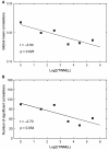
Changes in global cortical thickness correlations with lesion load. (A) Plot showing the decrease of the average correlations in multiple sclerosis with TWMLL. The average correlations were obtained by calculating the average values of inter-regional correlation matrices (Fig. 1). (B) Plot showing the decrease in the number of significant correlations with TWMLL.
Figure 6
Changes in regional cortical thickness correlations with lesion load. (A–D) Plots showing the decrease of the inter-regional positive correlations in multiple sclerosis with TWMLL. (E–G) Plots showing the decrease of the inter-regional negative correlations in multiple sclerosis with TWMLL. (H) Plot showing the increase of the inter-regional positive correlations in multiple sclerosis with TWMLL. Note that pairs of regions were only listed if (i) they were significantly non-zero in at least one group (q < 0.05, FDR-corrected); and (ii) they showed significant decrease with lesion load (P < 0.01, uncorrected). All regions involved were mapped onto a cortical surface.
Similar articles
- Abnormal topological organization in white matter structural networks revealed by diffusion tensor tractography in unmedicated patients with obsessive-compulsive disorder.
Zhong Z, Zhao T, Luo J, Guo Z, Guo M, Li P, Sun J, He Y, Li Z. Zhong Z, et al. Prog Neuropsychopharmacol Biol Psychiatry. 2014 Jun 3;51:39-50. doi: 10.1016/j.pnpbp.2014.01.005. Epub 2014 Jan 16. Prog Neuropsychopharmacol Biol Psychiatry. 2014. PMID: 24440373 - Structural insights into aberrant topological patterns of large-scale cortical networks in Alzheimer's disease.
He Y, Chen Z, Evans A. He Y, et al. J Neurosci. 2008 Apr 30;28(18):4756-66. doi: 10.1523/JNEUROSCI.0141-08.2008. J Neurosci. 2008. PMID: 18448652 Free PMC article. - Disconnection mechanism and regional cortical atrophy contribute to impaired processing of facial expressions and theory of mind in multiple sclerosis: a structural MRI study.
Mike A, Strammer E, Aradi M, Orsi G, Perlaki G, Hajnal A, Sandor J, Banati M, Illes E, Zaitsev A, Herold R, Guttmann CR, Illes Z. Mike A, et al. PLoS One. 2013 Dec 13;8(12):e82422. doi: 10.1371/journal.pone.0082422. eCollection 2013. PLoS One. 2013. PMID: 24349280 Free PMC article. - Cortical pathology and cognitive impairment in multiple sclerosis.
Calabrese M, Rinaldi F, Grossi P, Gallo P. Calabrese M, et al. Expert Rev Neurother. 2011 Mar;11(3):425-32. doi: 10.1586/ern.10.155. Expert Rev Neurother. 2011. PMID: 21375447 Review. - Gray and normal-appearing white matter in multiple sclerosis: an MRI perspective.
Vrenken H, Geurts JJ. Vrenken H, et al. Expert Rev Neurother. 2007 Mar;7(3):271-9. doi: 10.1586/14737175.7.3.271. Expert Rev Neurother. 2007. PMID: 17341175 Review.
Cited by
- The thalamo-cortical complex network correlates of chronic pain.
Zippo AG, Valente M, Caramenti GC, Biella GE. Zippo AG, et al. Sci Rep. 2016 Oct 13;6:34763. doi: 10.1038/srep34763. Sci Rep. 2016. PMID: 27734895 Free PMC article. - Alterations of Gray and White Matter Networks in Patients with Obsessive-Compulsive Disorder: A Multimodal Fusion Analysis of Structural MRI and DTI Using mCCA+jICA.
Kim SG, Jung WH, Kim SN, Jang JH, Kwon JS. Kim SG, et al. PLoS One. 2015 Jun 3;10(6):e0127118. doi: 10.1371/journal.pone.0127118. eCollection 2015. PLoS One. 2015. PMID: 26038825 Free PMC article. Clinical Trial. - Anomalous gray matter structural networks in major depressive disorder.
Singh MK, Kesler SR, Hadi Hosseini SM, Kelley RG, Amatya D, Hamilton JP, Chen MC, Gotlib IH. Singh MK, et al. Biol Psychiatry. 2013 Nov 15;74(10):777-85. doi: 10.1016/j.biopsych.2013.03.005. Epub 2013 Apr 18. Biol Psychiatry. 2013. PMID: 23601854 Free PMC article. - Machine learning in major depression: From classification to treatment outcome prediction.
Gao S, Calhoun VD, Sui J. Gao S, et al. CNS Neurosci Ther. 2018 Nov;24(11):1037-1052. doi: 10.1111/cns.13048. Epub 2018 Aug 23. CNS Neurosci Ther. 2018. PMID: 30136381 Free PMC article. Review. - Education, and the balance between dynamic and stationary functional connectivity jointly support executive functions in relapsing-remitting multiple sclerosis.
Lin SJ, Vavasour I, Kosaka B, Li DKB, Traboulsee A, MacKay A, McKeown MJ. Lin SJ, et al. Hum Brain Mapp. 2018 Dec;39(12):5039-5049. doi: 10.1002/hbm.24343. Epub 2018 Sep 21. Hum Brain Mapp. 2018. PMID: 30240533 Free PMC article.
References
- Au Duong MV, Audoin B, Boulanouar K, Ibarrola D, Malikova I, Confort-Gouny S, et al. Altered functional connectivity related to white matter changes inside the working memory network at the very early stage of MS. J Cereb Blood Flow Metab. 2005a;25:1245–53. - PubMed
- Au Duong MV, Boulanouar K, Audoin B, Treseras S, Ibarrola D, Malikova I, et al. Modulation of effective connectivity inside the working memory network in patients at the earliest stage of multiple sclerosis. Neuroimage. 2005b;24:533–8. - PubMed
- Audoin B, Au Duong MV, Malikova I, Confort-Gouny S, Ibarrola D, Cozzone PJ, et al. Functional magnetic resonance imaging and cognition at the very early stage of MS. J Neurol Sci. 2006a;245:87–91. - PubMed
Publication types
MeSH terms
LinkOut - more resources
Full Text Sources
Medical


