The neurobiology of sexual partner preferences in rams - PubMed (original) (raw)
Review
The neurobiology of sexual partner preferences in rams
Charles E Roselli et al. Horm Behav. 2009 May.
Abstract
The question of what causes a male animal to seek out and choose a female as opposed to another male mating partner is unresolved and remains an issue of considerable debate. The most developed biologic theory is the perinatal organizational hypothesis, which states that perinatal hormone exposure mediates sexual differentiation of the brain. Numerous animal experiments have assessed the contribution of perinatal testosterone and/or estradiol exposure to the development of a male-typical mate preference, but almost all have used hormonally manipulated animals. In contrast, variations in sexual partner preferences occur spontaneously in domestic rams, with as many as 8% of the population exhibiting a preference for same-sex mating partners (male-oriented rams). Thus, the domestic ram is an excellent experimental model to study possible links between fetal neuroendocrine programming of neural mechanisms and adult sexual partner preferences. In this review, we present an overview of sexual differentiation in relation to sexual partner preferences. We then summarize results that test the relevance of the organizational hypothesis to expression of same-sex sexual partner preferences in rams. Finally, we demonstrate that the sexual differentiation of brain and behavior in sheep does not depend critically on aromatization of testosterone to estradiol.
Figures
Figure 1
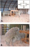
Photographs of the testing setup used to characterize sexual partner preferences in rams. A) Illustrates the position of the stimulus animals in a four-way stanchion (inset) placed in the center of a testing arena (10 m × 10 m). B) Shows a ram mounting a stimulus ram.
Figure 2
A)Schematic diagram of a coronal section through the sheep at the level of the optic chiasm (OC) and anterior commissure (AC). The position of the oSDN is shown bilateral to the third ventricle (V) in the central part of the medial preoptic nucleus (MPOA). The ventral paraventricular nucleus (vPV) is shown at the base of the ventricle. B) Micrograph of oSDN from the left hypothalamus of a female-oriented ram. The third ventricle is at the right of the figure. C) Section of a male-oriented ram comparable to that in (B). The photomicrograph are taken near the midpoint of the anterior - posterior extent of the oSDN. The scale bar is 1 mm.
Figure 3
Differences of the oSDN among female-oriented rams (n=8), male-oriented rams (n=9), and mid-luteal phase ewes (n=10). A, Differences in oSDN volume; B, Differences in neuron counts; C, Differences in neuron density; and D, Differences in oSDN length. Data are presented as means ± SEM. Bars with different letters differ significantly, P < 0.05. Reprinted from Roselli et al. (Roselli et al., 2004).
Figure 4
Sex comparisons of oSDN volumes in late gestational sheep fetuses (GD 135 ± 0.8 SEM). Data are presented as means ± SEM. Note that volume measurements were determined from autoradiograms from in situ hybridization for aromatase mRNA expression and correspond to similar measurements made in adults, see Fig. 3 in (Roselli et al., 2004). Reprinted from Roselli et al. (Roselli et al., 2007).
Figure 5
Effect of prenatal testosterone exposure on oSDN volume. Pregnant ewes received injections of testosterone propionate (100 mg) twice a week from days 30 to 90 of gestation so that their fetuses were exposed to elevated systemic levels of testosterone during the critical period for sexual differentiation. Control animals were not injected. Data are presented as means ± SEM, n = 4/group. Bars with different letters differ significantly, P < 0.05. Reprinted from Roselli et al. (Roselli et al., 2007).
Figure 6
Sex differences in the expression of progesterone receptor mRNA in the amygdala (AMYG) and hypothalamus-preoptic area (HPOA) of gestational day 64 fetal lambs. The levels of progesterone receptor mRNA were measured using a RNase protection assay that measured mRNA concentration using a standard curve of progesterone receptor sense RNA and normalized values to levels of cyclophilin mRNA in each sample. Data are presented as means ± SEM, n =3- 4/group. *, P < 0.5; **, P < 01 male versus female. Some fetuses were exposed to the aromatase ATD given by Silastic implants to their mothers as described in (Roselli, Resko, and Stormshak, 2003). This treatment had no effect on progesterone receptor mRNA expression. Reprinted from Roselli et al. (Roselli et al., 2006).
Similar articles
- Prenatal programming of sexual partner preference: the ram model.
Roselli CE, Stormshak F. Roselli CE, et al. J Neuroendocrinol. 2009 Mar;21(4):359-64. doi: 10.1111/j.1365-2826.2009.01828.x. J Neuroendocrinol. 2009. PMID: 19207819 Free PMC article. Review. - Depressing time: Waiting, melancholia, and the psychoanalytic practice of care.
Salisbury L, Baraitser L. Salisbury L, et al. In: Kirtsoglou E, Simpson B, editors. The Time of Anthropology: Studies of Contemporary Chronopolitics. Abingdon: Routledge; 2020. Chapter 5. In: Kirtsoglou E, Simpson B, editors. The Time of Anthropology: Studies of Contemporary Chronopolitics. Abingdon: Routledge; 2020. Chapter 5. PMID: 36137063 Free Books & Documents. Review. - "I've Spent My Whole Life Striving to Be Normal": Internalized Stigma and Perceived Impact of Diagnosis in Autistic Adults.
Huang Y, Trollor JN, Foley KR, Arnold SRC. Huang Y, et al. Autism Adulthood. 2023 Dec 1;5(4):423-436. doi: 10.1089/aut.2022.0066. Epub 2023 Dec 12. Autism Adulthood. 2023. PMID: 38116050 Free PMC article. - Falls prevention interventions for community-dwelling older adults: systematic review and meta-analysis of benefits, harms, and patient values and preferences.
Pillay J, Gaudet LA, Saba S, Vandermeer B, Ashiq AR, Wingert A, Hartling L. Pillay J, et al. Syst Rev. 2024 Nov 26;13(1):289. doi: 10.1186/s13643-024-02681-3. Syst Rev. 2024. PMID: 39593159 Free PMC article. - Antioxidants for female subfertility.
Showell MG, Mackenzie-Proctor R, Jordan V, Hart RJ. Showell MG, et al. Cochrane Database Syst Rev. 2020 Aug 27;8(8):CD007807. doi: 10.1002/14651858.CD007807.pub4. Cochrane Database Syst Rev. 2020. PMID: 32851663 Free PMC article.
Cited by
- The genetics of sex differences in brain and behavior.
Ngun TC, Ghahramani N, Sánchez FJ, Bocklandt S, Vilain E. Ngun TC, et al. Front Neuroendocrinol. 2011 Apr;32(2):227-46. doi: 10.1016/j.yfrne.2010.10.001. Epub 2010 Oct 15. Front Neuroendocrinol. 2011. PMID: 20951723 Free PMC article. Review. - Prenatal influence of an androgen agonist and antagonist on the differentiation of the ovine sexually dimorphic nucleus in male and female lamb fetuses.
Roselli CE, Reddy RC, Estill CT, Scheldrup M, Meaker M, Stormshak F, Montilla HJ. Roselli CE, et al. Endocrinology. 2014 Dec;155(12):5000-10. doi: 10.1210/en.2013-2176. Epub 2014 Sep 12. Endocrinology. 2014. PMID: 25216387 Free PMC article. - Developmental and Functional Effects of Steroid Hormones on the Neuroendocrine Axis and Spinal Cord.
Zubeldia-Brenner L, Roselli CE, Recabarren SE, Gonzalez Deniselle MC, Lara HE. Zubeldia-Brenner L, et al. J Neuroendocrinol. 2016 Jul;28(7):10.1111/jne.12401. doi: 10.1111/jne.12401. J Neuroendocrinol. 2016. PMID: 27262161 Free PMC article. Review. - Effects of Long-Term Flutamide Treatment During Development on Sexual Behaviour and Hormone Responsiveness in Rams.
Roselli CE, Meaker M, Stormshak F, Estill CT. Roselli CE, et al. J Neuroendocrinol. 2016 May;28(5):10.1111/jne.12389. doi: 10.1111/jne.12389. J Neuroendocrinol. 2016. PMID: 27005749 Free PMC article. - Ontogeny of cytochrome p450 aromatase mRNA expression in the developing sheep brain.
Roselli CE, Stormshak F. Roselli CE, et al. J Neuroendocrinol. 2012 Mar;24(3):443-52. doi: 10.1111/j.1365-2826.2011.02260.x. J Neuroendocrinol. 2012. PMID: 22128891 Free PMC article.
References
- Adkins-Regan E. Sex hormones and sexual orientation in animals. Psychobiol. 1988;16:335–347.
- Alexander BM, Rose JD, Stellflug JN, Fitzgerald JA, Moss GE. Fos-like immunoreactivity in brain regions of domestic rams following exposure to rams or ewes. Physiol. Behav. 2001a;73:75–80. - PubMed
- Alexander BM, Rose JD, Stellflug JN, Fitzgerald JA, Moss GE. Low-sexually performing rams but not male-oriented rams can be discriminated by cell size in the amygdala and preoptic area: a morphometric study. Behav. Brain Res. 2001b;119:15–21. - PubMed
- Alexander BM, Stellflug JN, Rose JD, Fitzgerald JA, Moss GE. Behavior and endocrine changes in high-performing, low-performing, and male-oriented domestic rams following exposure to rams and ewes in estrus when copulation is precluded. J. Anim. Sci. 1999;77:1869–1874. - PubMed
Publication types
MeSH terms
Substances
LinkOut - more resources
Full Text Sources


