Brain atrophy associated with baseline and longitudinal measures of cognition - PubMed (original) (raw)
Brain atrophy associated with baseline and longitudinal measures of cognition
V A Cardenas et al. Neurobiol Aging. 2011 Apr.
Abstract
The overall goal was to identify patterns of brain atrophy associated with cognitive impairment and future cognitive decline in non-demented elders. Seventy-one participants were studied with structural MRI and neuropsychological testing at baseline and 1-year follow-up. Deformation-based morphometry was used to examine the relationship between regional baseline brain tissue volume with baseline and longitudinal measures of delayed verbal memory, semantic memory, and executive function. Smaller right hippocampal and entorhinal cortex (ERC) volumes at baseline were associated with worse delayed verbal memory performance at baseline while smaller left ERC volume was associated with greater longitudinal decline. Smaller left superior temporal cortex at baseline was associated with worse semantic memory at baseline, while smaller left temporal white and gray matter volumes were associated with greater semantic memory decline. Increased CSF and smaller frontal lobe volumes were associated with impaired executive function at baseline and greater longitudinal executive decline. These findings suggest that baseline volumes of prefrontal and temporal regions may underlie continuing cognitive decline due to aging, pathology, or both in non-demented elderly individuals.
Copyright © 2009 Elsevier Inc. All rights reserved.
Conflict of interest statement
Disclosure Statement for Authors
The authors have no actual or potential conflicts of interest to disclose.
Figures
Figure 1
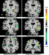
The T-statistic map is overlaid on the spatially normalized average MRI. The contours delineate statistically significant clusters of association between baseline brain volume and either baseline (left panels) or change (right panels) in cognitive scores. The top panel shows results for delayed memory, the middle for object naming, and the bottom for executive function. Voxels shaded red/yellow indicate smaller baseline tissue volumes associated with either worse baseline or greater declines in cognition, and blue shaded voxels indicate larger baseline tissue volumes associated with worse baseline or greater declines in cognition.
Figure 2
The T-statistic map is overlaid on the spatially normalized MRI. The contours delineate statistically significant clusters of association between baseline brain volume and age. Red and pink contours (encompassing blue shaded voxels) show regions where decreased tissue volume was associated with increasing age; green contours (encompassing red/yellow voxels) show regions of increased CSF volume with increasing age.
Similar articles
- The impact of glucose disorders on cognition and brain volumes in the elderly: the Sydney Memory and Ageing Study.
Samaras K, Lutgers HL, Kochan NA, Crawford JD, Campbell LV, Wen W, Slavin MJ, Baune BT, Lipnicki DM, Brodaty H, Trollor JN, Sachdev PS. Samaras K, et al. Age (Dordr). 2014 Apr;36(2):977-93. doi: 10.1007/s11357-013-9613-0. Epub 2014 Jan 9. Age (Dordr). 2014. PMID: 24402401 Free PMC article. - Longitudinal volumetric MRI change and rate of cognitive decline.
Mungas D, Harvey D, Reed BR, Jagust WJ, DeCarli C, Beckett L, Mack WJ, Kramer JH, Weiner MW, Schuff N, Chui HC. Mungas D, et al. Neurology. 2005 Aug 23;65(4):565-71. doi: 10.1212/01.wnl.0000172913.88973.0d. Neurology. 2005. PMID: 16116117 Free PMC article. - A decade of changes in brain volume and cognition.
Aljondi R, Szoeke C, Steward C, Yates P, Desmond P. Aljondi R, et al. Brain Imaging Behav. 2019 Apr;13(2):554-563. doi: 10.1007/s11682-018-9887-z. Brain Imaging Behav. 2019. PMID: 29744801 - A 10-year follow-up of hippocampal volume on magnetic resonance imaging in early dementia and cognitive decline.
den Heijer T, van der Lijn F, Koudstaal PJ, Hofman A, van der Lugt A, Krestin GP, Niessen WJ, Breteler MM. den Heijer T, et al. Brain. 2010 Apr;133(Pt 4):1163-72. doi: 10.1093/brain/awq048. Brain. 2010. PMID: 20375138 - Acute respiratory distress syndrome, sepsis, and cognitive decline: a review and case study.
Jackson JC, Hopkins RO, Miller RR, Gordon SM, Wheeler AP, Ely EW. Jackson JC, et al. South Med J. 2009 Nov;102(11):1150-7. doi: 10.1097/SMJ.0b013e3181b6a592. South Med J. 2009. PMID: 19864995 Free PMC article. Review.
Cited by
- Estimated maximal and current brain volume predict cognitive ability in old age.
Royle NA, Booth T, Valdés Hernández MC, Penke L, Murray C, Gow AJ, Maniega SM, Starr J, Bastin ME, Deary IJ, Wardlaw JM. Royle NA, et al. Neurobiol Aging. 2013 Dec;34(12):2726-33. doi: 10.1016/j.neurobiolaging.2013.05.015. Epub 2013 Jul 11. Neurobiol Aging. 2013. PMID: 23850342 Free PMC article. - Greater regional brain atrophy rate in healthy elderly subjects with a history of cigarette smoking.
Durazzo TC, Insel PS, Weiner MW; Alzheimer Disease Neuroimaging Initiative. Durazzo TC, et al. Alzheimers Dement. 2012 Nov;8(6):513-9. doi: 10.1016/j.jalz.2011.10.006. Alzheimers Dement. 2012. PMID: 23102121 Free PMC article. - The contributions of MRI-based measures of gray matter, white matter hyperintensity, and white matter integrity to late-life cognition.
He J, Wong VS, Fletcher E, Maillard P, Lee DY, Iosif AM, Singh B, Martinez O, Roach AE, Lockhart SN, Beckett L, Mungas D, Farias ST, Carmichael O, DeCarli C. He J, et al. AJNR Am J Neuroradiol. 2012 Oct;33(9):1797-803. doi: 10.3174/ajnr.A3048. Epub 2012 Apr 26. AJNR Am J Neuroradiol. 2012. PMID: 22538073 Free PMC article. - Imaging Techniques in Alzheimer's Disease: A Review of Applications in Early Diagnosis and Longitudinal Monitoring.
van Oostveen WM, de Lange ECM. van Oostveen WM, et al. Int J Mol Sci. 2021 Feb 20;22(4):2110. doi: 10.3390/ijms22042110. Int J Mol Sci. 2021. PMID: 33672696 Free PMC article. Review. - Brain Morphometry and Cognitive Performance in Normal Brain Aging: Age- and Sex-Related Structural and Functional Changes.
Statsenko Y, Habuza T, Smetanina D, Simiyu GL, Uzianbaeva L, Neidl-Van Gorkom K, Zaki N, Charykova I, Al Koteesh J, Almansoori TM, Belghali M, Ljubisavljevic M. Statsenko Y, et al. Front Aging Neurosci. 2022 Jan 26;13:713680. doi: 10.3389/fnagi.2021.713680. eCollection 2021. Front Aging Neurosci. 2022. PMID: 35153713 Free PMC article.
References
- Adlam AL, Bozeat S, Arnold R, Watson P, Hodges JR. Semantic knowledge in mild cognitive impairment and mild Alzheimer’s disease. Cortex. 2006;42(5):675–84. - PubMed
- Alvarez JA, Emory E. Executive function and the frontal lobes: a meta-analytic review. Neuropsychol Rev. 2006;16(1):17–42. - PubMed
- American Psychiatric Association. Diagnostic and Statistical Manual of Mental Disorders. 4. American Psychiatric Association; Washington, DC: 2000.
- Ashburner J, Friston KJ. Voxel-based morphometry--the methods. Neuroimage. 2000;11(6 Pt 1):805–21. - PubMed
- Crook TH, Bartus RT, Ferris SH, Whitehouse P, Cohen GD, Gershon S. Age Associated memory impairment: proposed diagnostic criteria and measures of clinical change: report of a National Institute of Mental Health Work Group. Dev Neuropsychol. 1986;2:261–76.
Publication types
MeSH terms
Grants and funding
- R01 AG010220/AG/NIA NIH HHS/United States
- R01 MH065392-01/MH/NIMH NIH HHS/United States
- R01 AG10897/AG/NIA NIH HHS/United States
- R01 AG010897-20/AG/NIA NIH HHS/United States
- R01 AG010897-22/AG/NIA NIH HHS/United States
- R01 AG010897/AG/NIA NIH HHS/United States
- R01 AG010897-21/AG/NIA NIH HHS/United States
- R01 MH65392/MH/NIMH NIH HHS/United States
- R03 EB008136-01/EB/NIBIB NIH HHS/United States
- P01 AG019724/AG/NIA NIH HHS/United States
- R01 MH065392/MH/NIMH NIH HHS/United States
- R03EB00813/EB/NIBIB NIH HHS/United States
- R01 MH065392-03/MH/NIMH NIH HHS/United States
- R01 MH065392-02/MH/NIMH NIH HHS/United States
- R01 AG010897-19/AG/NIA NIH HHS/United States
- R03 EB008136/EB/NIBIB NIH HHS/United States
- R01 AG010220-16/AG/NIA NIH HHS/United States
- P30 AG010129-129001/AG/NIA NIH HHS/United States
- R01 AG010897-18/AG/NIA NIH HHS/United States
- P30 AG010129/AG/NIA NIH HHS/United States
LinkOut - more resources
Full Text Sources
Other Literature Sources

