HIV-1 and influenza antibodies: seeing antigens in new ways - PubMed (original) (raw)
Review
HIV-1 and influenza antibodies: seeing antigens in new ways
Peter D Kwong et al. Nat Immunol. 2009 Jun.
Abstract
New modes of humoral recognition have been identified by studies of antibodies that neutralize human immunodeficiency virus type 1 and influenza A viruses. Understanding how such modes of antibody-antigen recognition can occur in the context of sophisticated mechanisms of humoral evasion has implications for the development of effective vaccines. Here we describe eight modes of antibody recognition first observed with human immunodeficiency virus type 1. Similarities to four of these modes have been identified with antibodies to a conserved 'stem' epitope on influenza A viruses. We outline how each of these different modes of antibody recognition is particularly suited to overcoming a specific viral evasion tactic and assess potential routes of re-elicitation in vaccine settings.
Figures
Figure 1
New modes of antibody recognition demonstrated by HIV-1-reactive antibodies. (a) Domain swapping: exchange of the heavy-chain variable (VH) domains on adjacent antigen-binding arms of the antibody 2G12 (left) rigidifies the interface between the arms, which permits recognition of three glycan moieties of gp120 (right). (b) Sequence-limited recognition: binding of the hypervariable V3 loop of gp120 (yellow) to the antibody 447-52D (light chain, salmon; heavy chain, sky blue), which uses mainly sequence-independent interactions with main-chain atoms. (c) Drug mimicry: binding of the C34 peptide (left), a slightly smaller version of the drug Fuzeon, and binding of the CDR H2 of antibody D5 (right) to gp41 (gray). (d) Germline-encoded recognition: binding of the VH1-69 encoded regions (red) of various CD4-induced antibodies to gp120 (gray) and CD4 (yellow). Despite recognition of the similar regions on gp120, the relative position of the VH1-69-encoded region varies. (e) Precise post-translational mimicry: the cleft between gp120 core (gray) and V3 loop (orange) specifically recognizes _O_-sulfated tyrosine on CCR5 (left) and the antibody 412d (right). (f) Two-step recognition: the epitope for the antibody 2F5 is thought to be only transiently available; corecognition of this epitope in the context of membrane increases binding affinity. (g) Quaternary-specific recognition: the antibody 2909 does not recognize gp120 or gp41 as separate units but only as the assembled functional Env viral spike. (h) Heavy chain–only recognition: binding of the antibody b12 to its gp120 epitope using only its heavy chain (green).
Figure 2
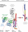
Features of HIV-1 antibody recognition used by influenza hemagglutinin-reactive antibodies. Several of the features of the HIV-1 antibodies are recapitulated in the antibodies CR6261 and F10, including heavy chain–only binding, germline-encoded recognition through a VH1-69 response, binding to recessed hydrophobic pockets that could be potential drug targets and sequence-independent recognition, although mainly on the antibody side for the formation of main-chain hydrogen bonds. The actual recognition mode, stabilization of the prefusion conformation that prevents the conformational rearrangement of HA2 required for membrane fusion, has been observed in other contexts: with HIV-1, the antibody D5 binds to a fusion intermediate, which prevents subsequent conformational changes for fusion; and with Ebola virus, the antibody KZ52 binds to the Ebola viral stem and locks the spike into its prefusion conformation.
Figure 3
Antibody-directed strategies of vaccine design. Unusual features of antibody recognition may provide clues to the immunological characteristics that allow elicitation of antibodies to a particular site. For example, precise immune focusing may be needed to elicit antibodies to the epitopes recognized by the HIV-1 and influenza antibodies b12, F10 and CR6261, which use heavy chain–only recognition. Immunofocusing techniques, such as glycan masking, in which all surfaces other than the site of vulnerability (‘bulls-eye’) are covered by N-linked glycosylation, and truncation, in which surfaces other than the site are deleted,, may have utility in this context (electron tomogram image of the native HIV-1 spike adapted from ref. 61). With the influenza antibodies, additional clues, such as VH1-69 use with low somatic mutation, suggest that a portion of the target site is recessed and hydrophobic in nature and that elicitation of appropriate antibodies in humans will be enhanced by the direct genomic encoding of recognition elements. Elicitation in vivo may require spacing of spikes far enough apart to increase access of antibody to the conserved stem target, although truncation and masking strategies of immunofocusing should also have utility.
Similar articles
- The challenges of eliciting neutralizing antibodies to HIV-1 and to influenza virus.
Karlsson Hedestam GB, Fouchier RA, Phogat S, Burton DR, Sodroski J, Wyatt RT. Karlsson Hedestam GB, et al. Nat Rev Microbiol. 2008 Feb;6(2):143-55. doi: 10.1038/nrmicro1819. Nat Rev Microbiol. 2008. PMID: 18197170 Review. - [Specific immunoglobulins from egg yolks of Japanese quail immunized with influenza and HIV-1 viruses].
Kovgan AA, Isaeva EI, Rovnova ZI, Shilov AA. Kovgan AA, et al. Dokl Akad Nauk SSSR. 1989 Jul-Aug;307(1):229-33. Dokl Akad Nauk SSSR. 1989. PMID: 2806062 Russian. No abstract available. - Abnormal humoral immune response to influenza vaccination in pediatric type-1 human immunodeficiency virus infected patients receiving highly active antiretroviral therapy.
Montoya CJ, Toro MF, Aguirre C, Bustamante A, Hernandez M, Arango LP, Echeverry M, Arango AE, Prada MC, Alarcon Hdel P, Rojas M. Montoya CJ, et al. Mem Inst Oswaldo Cruz. 2007 Jun;102(4):501-8. doi: 10.1590/s0074-02762007005000055. Mem Inst Oswaldo Cruz. 2007. PMID: 17612772 - A novel H6N1 virus-like particle vaccine induces long-lasting cross-clade antibody immunity against human and avian H6N1 viruses.
Yang JR, Chen CY, Kuo CY, Cheng CY, Lee MS, Cheng MC, Yang YC, Wu CY, Wu HS, Liu MT, Hsiao PW. Yang JR, et al. Antiviral Res. 2016 Feb;126:8-17. doi: 10.1016/j.antiviral.2015.10.019. Epub 2015 Nov 22. Antiviral Res. 2016. PMID: 26593980 - Influenza virus hemagglutinin stalk-based antibodies and vaccines.
Krammer F, Palese P. Krammer F, et al. Curr Opin Virol. 2013 Oct;3(5):521-30. doi: 10.1016/j.coviro.2013.07.007. Epub 2013 Aug 24. Curr Opin Virol. 2013. PMID: 23978327 Free PMC article. Review.
Cited by
- Reshaping antibody diversity.
Wang F, Ekiert DC, Ahmad I, Yu W, Zhang Y, Bazirgan O, Torkamani A, Raudsepp T, Mwangi W, Criscitiello MF, Wilson IA, Schultz PG, Smider VV. Wang F, et al. Cell. 2013 Jun 6;153(6):1379-93. doi: 10.1016/j.cell.2013.04.049. Cell. 2013. PMID: 23746848 Free PMC article. - Structure of hepatitis C virus envelope glycoprotein E2 antigenic site 412 to 423 in complex with antibody AP33.
Kong L, Giang E, Nieusma T, Robbins JB, Deller MC, Stanfield RL, Wilson IA, Law M. Kong L, et al. J Virol. 2012 Dec;86(23):13085-8. doi: 10.1128/JVI.01939-12. Epub 2012 Sep 12. J Virol. 2012. PMID: 22973046 Free PMC article. - Outsmarting Pathogens with Antibody Engineering.
Qerqez AN, Silva RP, Maynard JA. Qerqez AN, et al. Annu Rev Chem Biomol Eng. 2023 Jun 8;14:217-241. doi: 10.1146/annurev-chembioeng-101121-084508. Epub 2023 Mar 14. Annu Rev Chem Biomol Eng. 2023. PMID: 36917814 Free PMC article. Review. - 5' Rapid Amplification of cDNA Ends and Illumina MiSeq Reveals B Cell Receptor Features in Healthy Adults, Adults With Chronic HIV-1 Infection, Cord Blood, and Humanized Mice.
Waltari E, Jia M, Jiang CS, Lu H, Huang J, Fernandez C, Finzi A, Kaufmann DE, Markowitz M, Tsuji M, Wu X. Waltari E, et al. Front Immunol. 2018 Mar 26;9:628. doi: 10.3389/fimmu.2018.00628. eCollection 2018. Front Immunol. 2018. PMID: 29632541 Free PMC article. - The challenge of developing universal vaccines.
Rappuoli R. Rappuoli R. F1000 Med Rep. 2011;3:16. doi: 10.3410/M3-16. Epub 2011 Aug 1. F1000 Med Rep. 2011. PMID: 21904572 Free PMC article.
References
- Wei X, et al. Antibody neutralization and escape by HIV-1. Nature. 2003;422:307–312. - PubMed
- Weiss RA, et al. Neutralization of human T-lymphotropic virus type III by sera of AIDS and AIDS-risk patients. Nature. 1985;316:69–72. - PubMed
Publication types
MeSH terms
Substances
LinkOut - more resources
Full Text Sources
Other Literature Sources


