Role of TRPV3 in immune response to development of dermatitis - PubMed (original) (raw)
Role of TRPV3 in immune response to development of dermatitis
Kinichi Imura et al. J Inflamm (Lond). 2009.
Abstract
Background: Recently, it has been reported that the Gly573Ser substitution of transient receptor potential V3 (TRPV3) leads to increased ion-channel activity in keratinocytes. Our previous studies have indicated that the spontaneous hairless and dermatitis phenotypes of DS-Nh mice, which were newly established as an animal model of atopic dermatitis (AD), are caused by TRPV3Gly573Ser. Although this substitution causes hairlessness in several kinds of rodents, in our investigations, dermatitis developed in only a few animals. Here, we generated NC/Nga-Nh mice to elucidate the role of TRPV3Gly573Ser in NC/Nga mice, which is one of the most studied animal models of AD.
Methods: To establish and validate the new AD animal model, NC/Nga-Nh mice were generated using NC/Nga and DS-Nh mice, and their clinical features were compared. Next, T-cell receptor (TCR) Vbeta usage in splenocytes, evaluation of bacterial colonization, and serological and histological analyses were carried out. Finally, repeated-hapten-application dermatitis was induced in these mice.
Results: NC/Nga-Nh mice did not develop spontaneous dermatitis, whereas DS-Nh mice displayed this phenotype when maintained under the same conditions. Serological analysis indicated that there really was a phenotypic difference between these mice, and TCR repertoire analysis indicated that TCRVbeta haplotypes played an important role in the development of dermatitis. Artificial dermatitis developed in DS and NC/Nga-Nh mice, but not in DS-Nh and NC/Nga mice. Histological and serological analyses indicated that mouse strains were listed in descending order of number of skin mast cells: DS-Nh > DS approximately NC/Nga-Nh > NC/Nga, and serum IgE levels were increased after 2,4,6 trinitrochlorobenzene application in these mice. Serum IgE level in DS-Nh mice was lower than that mesured in other strains.
Conclusion: Our results confirm the contribution of the TRPV3Gly573Ser gene to the development of repeated hapten dermatitis, but not spontaneous dermatitis in NC/Nga mice.
Figures
Figure 1
Disease symptoms in DS-Nh and NC/Nga-Nh mice. (A) Clinical features of DS-Nh and NC/Nga-Nh mice at 20 weeks of age kept under conventional or SPF conditions. (B) Evaluation of scratching and rubbing behavior in both strains at 20 weeks of age (n = 8) (SPF, kept under SPF conditions; Conv, kept under conventional conditions). White bar represents mean value of the grouped mice. (**, ##: significant differences at p < 0.01).
Figure 2
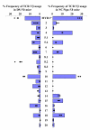
TCRBV repertoire analysis. The TCRBV repertoires in spleen cells from five DS-Nh or NC/Nga-Nh mice without in vitro stimulation.Bar indicates mean +2 SD of the TCRBV repertoires in spleen cells from five DS-Nh or NC/Nga-Nh mice without in vitro stimulation. Each dot indicates the percentage frequency of _TCRBV_-bearing T cells in spleen cells stimulated in vitro with SEC.
Figure 3
Bacterial colonization of skin lesions. (A) Isolation and identification of staphylococcal strains on the skin surface in both strains at 20 weeks of age (n = 5). (B) Measurement of serum levels of antibody to PGN in both strains at 20 weeks of age (n = 5).
Figure 4
Repeated application of TNCB in DS, DS-Nh, NC/Nga and NC/Nga-Nh mice. (A) Evaluation of dermatitis in these mice. Each value represents mean ± SD of four or five mice. (B and C) Clinical features of skin in these mice. (D) Clinical features of joints in DS-Nh mice. Arrows indicate arthritis-like lesions.
Figure 5
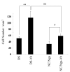
Number of mast cells in skin from DS-Nh, NC/Nga-Nh and control mice. Data represent the mean ± SD of six fields in six tissue samples. (**, ##: significant differences at p < 0.01), #: significant differences at p < 0.05).
Figure 6
Total serum IgE levels. Data are expressed as means ± SD of four or five mice. (*: significant differences at p < 0.05).
Similar articles
- Effects of TNCB sensitization in DS-Nh mice, serving as a model of atopic dermatitis, in comparison with NC/Nga mice.
Matsukura S, Aihara M, Hirasawa T, Ikezawa Z. Matsukura S, et al. Int Arch Allergy Immunol. 2005 Feb;136(2):173-80. doi: 10.1159/000083326. Epub 2005 Jan 12. Int Arch Allergy Immunol. 2005. PMID: 15650316 - Impact of the Gly573Ser substitution in TRPV3 on the development of allergic and pruritic dermatitis in mice.
Yoshioka T, Imura K, Asakawa M, Suzuki M, Oshima I, Hirasawa T, Sakata T, Horikawa T, Arimura A. Yoshioka T, et al. J Invest Dermatol. 2009 Mar;129(3):714-22. doi: 10.1038/jid.2008.245. Epub 2008 Aug 28. J Invest Dermatol. 2009. PMID: 18754035 - Association of T-cell receptor Vbeta haplotypes with dry skin in DS-Nh mice.
Imura K, Yoshioka T, Hikita I, Hirasawa T, Sakata T, Matsutani T, Horikawa T, Arimura A. Imura K, et al. Clin Exp Dermatol. 2009 Jan;34(1):61-7. doi: 10.1111/j.1365-2230.2008.02921.x. Clin Exp Dermatol. 2009. PMID: 19018787 - Analysis of T cell receptor (TCR) BV-gene clonotypes in NC/Nga mice developing dermatitis resembling human atopic dermatitis.
Matsuoka A, Kato T, Soma Y, Takahama H, Nakamura M, Matsuoka H, Mizoguchi M. Matsuoka A, et al. J Dermatol Sci. 2005 Apr;38(1):17-24. doi: 10.1016/j.jdermsci.2004.11.011. Epub 2005 Jan 12. J Dermatol Sci. 2005. PMID: 15795120 - Transforming growth factor-beta1 suppresses atopic dermatitis-like skin lesions in NC/Nga mice.
Sumiyoshi K, Nakao A, Ushio H, Mitsuishi K, Okumura K, Tsuboi R, Ra C, Ogawa H. Sumiyoshi K, et al. Clin Exp Allergy. 2002 Feb;32(2):309-14. doi: 10.1046/j.1365-2222.2002.01221.x. Clin Exp Allergy. 2002. PMID: 11929498
Cited by
- TRPV3 and Itch: The Role of TRPV3 in Chronic Pruritus according to Clinical and Experimental Evidence.
Um JY, Kim HB, Kim JC, Park JS, Lee SY, Chung BY, Park CW, Kim HO. Um JY, et al. Int J Mol Sci. 2022 Nov 29;23(23):14962. doi: 10.3390/ijms232314962. Int J Mol Sci. 2022. PMID: 36499288 Free PMC article. Review. - Targeting TRPV3 for the Development of Novel Analgesics.
Huang SM, Chung MK. Huang SM, et al. Open Pain J. 2013;6(Spec Iss 1):119-126. doi: 10.2174/1876386301306010119. Open Pain J. 2013. PMID: 25285178 Free PMC article. - Abnormal Somatosensory Behaviors Associated With a Gain-of-Function Mutation in TRPV3 Channels.
Fatima M, Slade H, Horwitz L, Shi A, Liu J, McKinstry D, Villani T, Xu H, Duan B. Fatima M, et al. Front Mol Neurosci. 2022 Jan 4;14:790435. doi: 10.3389/fnmol.2021.790435. eCollection 2021. Front Mol Neurosci. 2022. PMID: 35058747 Free PMC article. - Advances in TRP channel drug discovery: from target validation to clinical studies.
Koivisto AP, Belvisi MG, Gaudet R, Szallasi A. Koivisto AP, et al. Nat Rev Drug Discov. 2022 Jan;21(1):41-59. doi: 10.1038/s41573-021-00268-4. Epub 2021 Sep 15. Nat Rev Drug Discov. 2022. PMID: 34526696 Free PMC article. Review. - "ThermoTRP" Channel Expression in Cancers: Implications for Diagnosis and Prognosis (Practical Approach by a Pathologist).
Szallasi A. Szallasi A. Int J Mol Sci. 2023 May 22;24(10):9098. doi: 10.3390/ijms24109098. Int J Mol Sci. 2023. PMID: 37240443 Free PMC article. Review.
References
LinkOut - more resources
Full Text Sources
Other Literature Sources


