Rapid prototyping of 3D DNA-origami shapes with caDNAno - PubMed (original) (raw)
Rapid prototyping of 3D DNA-origami shapes with caDNAno
Shawn M Douglas et al. Nucleic Acids Res. 2009 Aug.
Abstract
DNA nanotechnology exploits the programmable specificity afforded by base-pairing to produce self-assembling macromolecular objects of custom shape. For building megadalton-scale DNA nanostructures, a long 'scaffold' strand can be employed to template the assembly of hundreds of oligonucleotide 'staple' strands into a planar antiparallel array of cross-linked helices. We recently adapted this 'scaffolded DNA origami' method to producing 3D shapes formed as pleated layers of double helices constrained to a honeycomb lattice. However, completing the required design steps can be cumbersome and time-consuming. Here we present caDNAno, an open-source software package with a graphical user interface that aids in the design of DNA sequences for folding 3D honeycomb-pleated shapes A series of rectangular-block motifs were designed, assembled, and analyzed to identify a well-behaved motif that could serve as a building block for future studies. The use of caDNAno significantly reduces the effort required to design 3D DNA-origami structures. The software is available at http://cadnano.org/, along with example designs and video tutorials demonstrating their construction. The source code is released under the MIT license.
Figures
Figure 1.
caDNAno Interface and design pipeline. (a) Screenshot of caDNAno interface. Left, Slice panel displays a cross-sectional view of the honeycomb lattice where helices can be added to the design. Middle, Path panel provides an interface to edit an unrolled 2D schematic of the scaffold and staple paths. Right, Render panel provides a real-time 3D model of the design. (b) Exported SVG schematic of example design from a, with scaffold (blue) and staple (multi-color) sequences. (c) Path panel snapshot during first step of the design process. Short stretches of scaffold are inserted into the Path panel as helices are added via the Slice panel. (d) The Path panel editing tools are used to stitch together a continuous scaffold path. (e) The auto-staple button is used to generate a default set of continuous staple paths, including crossovers. The breakpoint tool is subsequently used to split the staple paths into lengths between 18 and 49 bases. Finally, the scaffold sequence is applied to generate the list of staple sequences. (f) Exported X3D model from the Render panel.
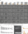
Figure 2.
Transmission electron microscopy (TEM) and agarose-gel analysis of DNA-origami blocks. The nomenclature of the designs is m × n, where m is the number of _x_-raster rows, and n is the number of helices per _x_-raster row. (i), 15 × 4 motif; (ii) 10 × 6 motif; (iii) 8 × 8 motif; (iv) 6 × 10 motif; (v) 4 × 16 motif; (vi) 3 × 20 motif; (vii) 2 × 30 motif. (a) Cylinder-model projections and transmission-electron micrographs for rectangular-block designs. (b) Partially folded models, which do not represent the actual folding pathway, are displayed above fully folded models. Scaffold crossovers only occur between helices that are neighbors in the partially folded models. Thus, these models capture an important feature of the design: the path of the scaffold stays within a 2D surface. (c) Agarose-gel analysis of folding of blocks. Marker is a 1 kb ladder. Red boxes indicate the region of each lane that was counted as the fastest-migrating monomeric species for yield estimates in option d and that was physically extracted from the gel during purification before TEM imaging. The 6 × 10 design displays the fastest gel mobility. (d) Fraction of scaffold incorporated into fastest-migrating monomeric species, as estimated by ethidium-bromide-fluorescence intensity. (e) Fraction of well-folded species after gel purification, as estimated by image analysis of 100 randomly selected particles for each shape. Scale bars: 25 nm.
Similar articles
- Self-assembly of DNA into nanoscale three-dimensional shapes.
Douglas SM, Dietz H, Liedl T, Högberg B, Graf F, Shih WM. Douglas SM, et al. Nature. 2009 May 21;459(7245):414-8. doi: 10.1038/nature08016. Nature. 2009. PMID: 19458720 Free PMC article. - Multilayer DNA origami packed on a square lattice.
Ke Y, Douglas SM, Liu M, Sharma J, Cheng A, Leung A, Liu Y, Shih WM, Yan H. Ke Y, et al. J Am Chem Soc. 2009 Nov 4;131(43):15903-8. doi: 10.1021/ja906381y. J Am Chem Soc. 2009. PMID: 19807088 Free PMC article. - Rapid prototyping of arbitrary 2D and 3D wireframe DNA origami.
Jun H, Wang X, Parsons MF, Bricker WP, John T, Li S, Jackson S, Chiu W, Bathe M. Jun H, et al. Nucleic Acids Res. 2021 Oct 11;49(18):10265-10274. doi: 10.1093/nar/gkab762. Nucleic Acids Res. 2021. PMID: 34508356 Free PMC article. - DNA Origami: Scaffolds for Creating Higher Order Structures.
Hong F, Zhang F, Liu Y, Yan H. Hong F, et al. Chem Rev. 2017 Oct 25;117(20):12584-12640. doi: 10.1021/acs.chemrev.6b00825. Epub 2017 Jun 12. Chem Rev. 2017. PMID: 28605177 Review. - Increasing Complexity in Wireframe DNA Nanostructures.
Piskunen P, Nummelin S, Shen B, Kostiainen MA, Linko V. Piskunen P, et al. Molecules. 2020 Apr 16;25(8):1823. doi: 10.3390/molecules25081823. Molecules. 2020. PMID: 32316126 Free PMC article. Review.
Cited by
- Enzymatic production of 'monoclonal stoichiometric' single-stranded DNA oligonucleotides.
Ducani C, Kaul C, Moche M, Shih WM, Högberg B. Ducani C, et al. Nat Methods. 2013 Jul;10(7):647-52. doi: 10.1038/nmeth.2503. Epub 2013 Jun 2. Nat Methods. 2013. PMID: 23727986 Free PMC article. - DNA nanostructures: a shift from assembly to applications.
Lanier LA, Bermudez H. Lanier LA, et al. Curr Opin Chem Eng. 2015 Feb 1;7:93-100. doi: 10.1016/j.coche.2015.01.001. Curr Opin Chem Eng. 2015. PMID: 25729640 Free PMC article. - DNA origami book biosensor for multiplex detection of cancer-associated nucleic acids.
Domljanovic I, Loretan M, Kempter S, Acuna GP, Kocabey S, Ruegg C. Domljanovic I, et al. Nanoscale. 2022 Oct 27;14(41):15432-15441. doi: 10.1039/d2nr03985k. Nanoscale. 2022. PMID: 36219167 Free PMC article. - Mechanistic Aspects for the Modulation of Enzyme Reactions on the DNA Scaffold.
Lin P, Yang H, Nakata E, Morii T. Lin P, et al. Molecules. 2022 Sep 24;27(19):6309. doi: 10.3390/molecules27196309. Molecules. 2022. PMID: 36234845 Free PMC article. Review. - Designing a bio-responsive robot from DNA origami.
Ben-Ishay E, Abu-Horowitz A, Bachelet I. Ben-Ishay E, et al. J Vis Exp. 2013 Jul 8;(77):e50268. doi: 10.3791/50268. J Vis Exp. 2013. PMID: 23893007 Free PMC article.
References
- Seeman N.C. Nucleic acid junctions and lattices. J. Theor. Biol. 1982;99:237–247. - PubMed
- Seeman N.C. DNA in a material world. Nature. 2003;421:427–431. - PubMed
- Chen J.H., Seeman N.C. Synthesis from DNA of a molecule with the connectivity of a cube. Nature. 1991;350:631–633. - PubMed
- Fu T.J., Seeman N.C. DNA double-crossover molecules. Biochemistry. 1993;32:3211–3220. - PubMed
- Li X.J., Yang X.P., Qi J., Seeman N.C. Antiparallel DNA double crossover molecules as components for nanoconstruction. J. Am. Chem. Soc. 1996;118:6131–6140.
Publication types
MeSH terms
Substances
LinkOut - more resources
Full Text Sources
Other Literature Sources

