Measurement of human immunodeficiency virus type 1 preintegration transcription by using Rev-dependent Rev-CEM cells reveals a sizable transcribing DNA population comparable to that from proviral templates - PubMed (original) (raw)
Measurement of human immunodeficiency virus type 1 preintegration transcription by using Rev-dependent Rev-CEM cells reveals a sizable transcribing DNA population comparable to that from proviral templates
Subashini R Iyer et al. J Virol. 2009 Sep.
Abstract
Preintegration transcription is an early process in human immunodeficiency virus type 1 infection and has been suggested to occur at a low level. The templates have also been suggested to represent a small population of nonintegrated viral DNA, particularly the two-long-terminal-repeat (2-LTR) circles. However, these determinations were made by either using PCR amplification of viral transcripts in bulk cell populations or utilizing the LTR-driving reporter cells that measure the synthesis of Tat. The intrinsic leakiness of LTR often makes the measurement of low-level viral transcription inaccurate. Since preintegration transcription also generates Rev, to eliminate the nonspecificity associated with the use of LTR alone we have developed a novel Rev-dependent indicator cell, Rev-CEM, to measure preintegration transcription based on the amount of Rev generated. In this report, using Rev-CEM cells, we demonstrate that preintegration transcription occurs on a much larger scale than expected. The transcribing population derived from nonintegrated viral DNA was comparable (at approximately 70%) to that derived from provirus in a productive viral replication cycle. Nevertheless, each nonintegrated viral DNA template exhibited a significant reduction in the level of transcriptional activity in the absence of integration. We also performed flow cytometry sorting of infected cells to identify viral templates. Surprisingly, our results suggest that the majority of 2-LTR circles are not active in directing transcription. It is likely that the nonintegrated templates are from the predominant DNA species, such as the full-length, linear DNA. Our results also suggest that a nonintegrating lentiviral vector can be as effective as an integrating vector in directing gene expression in nondividing cells, with the proper choice of an internal promoter.
Figures
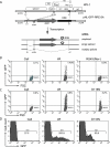
FIG. 1.
Application of the Rev-CEM Rev-dependent cell in measurement of preintegration transcription. (A) Rev-CEM carries a Rev-dependent lentiviral vector, pNL-GFP-RRE-SA. Landmarks from left to right are as follows: D1, HIV splicing donor 1; ψ, the HIV RNA genome packaging signal sequence; A5 and D4, HIV splicing acceptor 5 and donor 4; A7, HIV splicing acceptor 7. Viral transcripts are represented below the construct. Only the unspliced and singly spliced transcripts expressed GFP. (B) Rev-CEM cells were infected with equal p24 levels of HIV-1NL4-3 (Wt) or a Rev mutant, RS4X(Rev-). The percentages of GFP-positive cells were measured by flow cytometry at 48 h. (C) Rev-CEM cells were infected with the Wt virus (1.4 × 10⁶ cpm per million cells [reverse transcriptase unit]) or an integrase mutant, D116N (1.1 × 10⁶ cpm per million cells). The percentages of GFP-positive cells were measured at 48 h with flow cytometry. (D) Histoplot results for the infection described for panel C, showing the average GFP intensities of the GFP-positive cells. M-1, GFP-positive cell region.
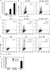
FIG. 2.
Characterization of preintegration transcription using Rev-CEM. (A and B) Preintegration transcription does not result from plasmid DNA contaminating virion particles. (A) HIV-1NL4-3/D116N (D116N) particles were treated with Benzonase (Benz; 125 U/ml) or subjected to mock treatment for 15 min at 37°C. Virion DNA was then purified and quantified by real-time PCR as described in Materials and Methods. (B) The same viruses, with or without Benzonase treatment, were also used to infect Rev-CEM cells, and the percentages of GFP-positive cells were measured by flow cytometry at 48 h. (C and D) Preintegration transcription is dependent on the presence of newly synthesized viral DNA. (C) Rev-CEM cells were left untreated or treated with the reverse transcriptase inhibitor AZT (50 μM) for 12 h and then infected with D116N. The percentages and average GFP intensities (M) of GFP-positive cells were measured at 48 h with flow cytometry. (D) As controls, cells were also infected with the Wt virus to demonstrate inhibition by AZT. (E) Preintegration transcription from nonintegrated viral DNA templates. CEM-SS cells were infected with equal p24 levels of the VSV-G pseudotyped Wt or D116N virus. Cells were lysed 48 h after infection, and total cellular DNA was extracted and quantified with Alu/real-time PCR for integrated viral DNA as described in Materials and Methods. Controls for background amplification of nonintegrated viral DNA were prepared by PCR in the presence (Alu-gag) or absence (gag only) of the Alu forward primer.
FIG. 3.
Effects of VSV-G pseudotyping on preintegration transcription. Single-cycle replicating HIV-1 particles were produced by cotransfection of an envelope-negative construct, pNL4-3(KFS) or pNL4-3(KFS)(D116N), with an HIV-1 envelope construct, pNL-ΔΨ-Env, or with a VSV-G construct, pHCMV-G. Equal p24 levels of the viral particles carrying either the HIV-1 envelope or VSV-G were used to infect Rev-CEM cells. The percentages of GFP-positive cells were measured by flow cytometry at 48 h.
FIG. 4.
The population of transcribing templates in D116N infection in comparison with Wt infection populations. Rev-CEM cells were infected with equal p24 levels of the VSV-G-pseudotyped Wt or D116N viruses. The percentages and average GFP intensities of GFP-positive cells were measured by flow cytometry at 48 h. Uninfected Rev-CEM cells were used as a control. Data represent results from three independent infections. D116N-infected cells had a much lower GFP intensity than Wt-infected cells (52.29 ± 4.18 in D116N versus 468.99 ± 61 in Wt cells [averages of the results of three infections]), whereas the percentage of the GFP-positive population remained comparable (9.5% in D116N versus 14.6% in Wt cells [averages of the results of three infections]). Similar results were obtained with viral particles carrying the envelope of HIV-1NL4-3 (data not shown).
FIG. 5.
Effects of the integrase inhibitor 118-D-24 on HIV-1 transcription in Rev-CEM cells. (A) The integrase inhibitor 118-D-24 (50 μM) was used to treat Rev-CEM cells for 4 h prior to HIV-1 infection. Cells treated with 118-D-24 or left untreated were then infected with equal p24 levels of the Wt (VSV-G pseudotyped) virus for 2 h; following infection and washing, cells were cultured in the continuous presence or absence of 118-D-24, respectively. The percentages and average GFP intensities (M) were measured by flow cytometry at 48 h. Uninfected, drug-treated Rev-CEM cells were used as a control. (B) Repeat of the same infection with a lower viral dosage. For additional controls, cells were also treated with AZT (50 μM) or 118-D-24 plus AZT and then identically infected. (C) Alu-HIV PCR measurement of 118-D-24 inhibition of HIV integration. CEM-SS cells were infected exactly as described for panel B in the presence or absence of 118-D-24. Total cellular DNA was purified at 48 h postinfection and then amplified with Alu-HIV PCR as described in Materials and Methods. The cellular β-actin pseudogene was also amplified to ensure that equal amounts of DNA were used.
FIG. 6.
Flow cytometry sorting and PCR quantification of 2-LTR circles. (A) Schematic representation of flow cytometry cell sorting of D116N-infected Rev-CEM cells and subsequent quantification of 2-LTR circles by PCR. (B) An example of one sorting experiment. Cells were infected with D116N, washed, cultured for 48 h, and then analyzed by flow cytometry. Cells within the red frames were sorted into GFP-positive and GFP-negative populations. Sorted cells were reanalyzed by flow cytometer to check cell purity. (C) PCR amplification of 2-LTR circles following sorting and DNA extraction. In this sorting experiment, Rev-CEM cells were infected with D116N. The GFP-positive cells represented 7% of the cell population. (D) To facilitate real-time PCR quantification of 2-LTR circles, the LTR region in mutant D116N (HIV-1NL4-3/D116N) was replaced with the LTR of HIV-189.6 (see Materials and Methods). Rev-CEM cells were infected with the resulting 89.6-LTR-D116N virus, washed, cultured for 48 h, and then sorted. The levels of GFP-positive cells in sortings 2 and 3 were 27.3% and 23.0%, respectively.
Similar articles
- Inhibition of human immunodeficiency virus type 1 in human T cells by a potent Rev response element decoy consisting of the 13-nucleotide minimal Rev-binding domain.
Lee SW, Gallardo HF, Gilboa E, Smith C. Lee SW, et al. J Virol. 1994 Dec;68(12):8254-64. doi: 10.1128/JVI.68.12.8254-8264.1994. J Virol. 1994. PMID: 7966618 Free PMC article. - HIV Preintegration Transcription and Host Antagonism.
Wu Y. Wu Y. Curr HIV Res. 2023;21(3):160-171. doi: 10.2174/1570162X21666230621122637. Curr HIV Res. 2023. PMID: 37345240 Free PMC article. - Mode of action of SDZ NIM 811, a nonimmunosuppressive cyclosporin A analog with activity against human immunodeficiency virus type 1 (HIV-1): interference with early and late events in HIV-1 replication.
Steinkasserer A, Harrison R, Billich A, Hammerschmid F, Werner G, Wolff B, Peichl P, Palfi G, Schnitzel W, Mlynar E, et al. Steinkasserer A, et al. J Virol. 1995 Feb;69(2):814-24. doi: 10.1128/JVI.69.2.814-824.1995. J Virol. 1995. PMID: 7815548 Free PMC article. - Episomal HIV-1 DNA and its relationship to other markers of HIV-1 persistence.
Martinez-Picado J, Zurakowski R, Buzón MJ, Stevenson M. Martinez-Picado J, et al. Retrovirology. 2018 Jan 30;15(1):15. doi: 10.1186/s12977-018-0398-1. Retrovirology. 2018. PMID: 29378611 Free PMC article. Review. - Distribution and fate of HIV-1 unintegrated DNA species: a comprehensive update.
Hamid FB, Kim J, Shin CG. Hamid FB, et al. AIDS Res Ther. 2017 Feb 16;14(1):9. doi: 10.1186/s12981-016-0127-6. AIDS Res Ther. 2017. PMID: 28209198 Free PMC article. Review.
Cited by
- Expression of Nef from unintegrated HIV-1 DNA downregulates cell surface CXCR4 and CCR5 on T-lymphocytes.
Sloan RD, Donahue DA, Kuhl BD, Bar-Magen T, Wainberg MA. Sloan RD, et al. Retrovirology. 2010 May 13;7:44. doi: 10.1186/1742-4690-7-44. Retrovirology. 2010. PMID: 20465832 Free PMC article. - Discovery of Novel Small-Molecule Inhibitors of LIM Domain Kinase for Inhibiting HIV-1.
Yi F, Guo J, Dabbagh D, Spear M, He S, Kehn-Hall K, Fontenot J, Yin Y, Bibian M, Park CM, Zheng K, Park HJ, Soloveva V, Gharaibeh D, Retterer C, Zamani R, Pitt ML, Naughton J, Jiang Y, Shang H, Hakami RM, Ling B, Young JAT, Bavari S, Xu X, Feng Y, Wu Y. Yi F, et al. J Virol. 2017 Jun 9;91(13):e02418-16. doi: 10.1128/JVI.02418-16. Print 2017 Jul 1. J Virol. 2017. PMID: 28381571 Free PMC article. - Development of a nonintegrating Rev-dependent lentiviral vector carrying diphtheria toxin A chain and human TRAF6 to target HIV reservoirs.
Wang Z, Tang Z, Zheng Y, Yu D, Spear M, Iyer SR, Bishop B, Wu Y. Wang Z, et al. Gene Ther. 2010 Sep;17(9):1063-76. doi: 10.1038/gt.2010.53. Epub 2010 Apr 22. Gene Ther. 2010. PMID: 20410930 Free PMC article. - An HIV-1 replication pathway utilizing reverse transcription products that fail to integrate.
Trinité B, Ohlson EC, Voznesensky I, Rana SP, Chan CN, Mahajan S, Alster J, Burke SA, Wodarz D, Levy DN. Trinité B, et al. J Virol. 2013 Dec;87(23):12701-20. doi: 10.1128/JVI.01939-13. Epub 2013 Sep 18. J Virol. 2013. PMID: 24049167 Free PMC article. - Different Pathways Conferring Integrase Strand-Transfer Inhibitors Resistance.
Richetta C, Tu NQ, Delelis O. Richetta C, et al. Viruses. 2022 Nov 22;14(12):2591. doi: 10.3390/v14122591. Viruses. 2022. PMID: 36560595 Free PMC article. Review.
References
- Aguilar-Cordova, E., J. Chinen, L. Donehower, D. E. Lewis, and J. W. Belmont. 1994. A sensitive reporter cell line for HIV-1 tat activity, HIV-1 inhibitors, and T cell activation effects. AIDS Res. Hum. Retrovir. 10295-301. - PubMed
- Benkirane, M., R. F. Chun, H. Xiao, V. V. Ogryzko, B. H. Howard, Y. Nakatani, and K. T. Jeang. 1998. Activation of integrated provirus requires histone acetyltransferase. p300 and P/CAF are coactivators for HIV-1 Tat. J. Biol. Chem. 27324898-24905. - PubMed
- Brown, P. O., B. Bowerman, H. E. Varmus, and J. M. Bishop. 1987. Correct integration of retroviral DNA in vitro. Cell 49347-356. - PubMed
Publication types
MeSH terms
Substances
LinkOut - more resources
Full Text Sources


