Robust, high-throughput solution structural analyses by small angle X-ray scattering (SAXS) - PubMed (original) (raw)
doi: 10.1038/nmeth.1353. Epub 2009 Jul 20.
Angeli L Menon, Michal Hammel, Robert P Rambo, Farris L Poole 2nd, Susan E Tsutakawa, Francis E Jenney Jr, Scott Classen, Kenneth A Frankel, Robert C Hopkins, Sung-Jae Yang, Joseph W Scott, Bret D Dillard, Michael W W Adams, John A Tainer
Affiliations
- PMID: 19620974
- PMCID: PMC3094553
- DOI: 10.1038/nmeth.1353
Robust, high-throughput solution structural analyses by small angle X-ray scattering (SAXS)
Greg L Hura et al. Nat Methods. 2009 Aug.
Abstract
We present an efficient pipeline enabling high-throughput analysis of protein structure in solution with small angle X-ray scattering (SAXS). Our SAXS pipeline combines automated sample handling of microliter volumes, temperature and anaerobic control, rapid data collection and data analysis, and couples structural analysis with automated archiving. We subjected 50 representative proteins, mostly from Pyrococcus furiosus, to this pipeline and found that 30 were multimeric structures in solution. SAXS analysis allowed us to distinguish aggregated and unfolded proteins, define global structural parameters and oligomeric states for most samples, identify shapes and similar structures for 25 unknown structures, and determine envelopes for 41 proteins. We believe that high-throughput SAXS is an enabling technology that may change the way that structural genomics research is done.
Figures
Figure 1
High-throughput SAXS pipeline. (a) Configuration of the SAXS endstation shows X-ray beam path, sample position, pipetting robot, and area detector. (b) Schematic of the sample area showing how the sample is loaded by the robot into a temperature-controlled cell. Positive helium pressure reduces air scatter and oxidative damage. (c) SAXS analysis tree for rapid and robust data processing and analysis. Proteins are first categorized as aggregated (using either the scattering curve itself or dynamic light scattering (DLS)), mixtures (based on native gel electrophoresis or multi-angle light scattering (MALS)), or mono-disperse samples. For monodisperse samples, SAXS data next defines global solution structural parameters radius of gyration, maximum dimension, and calculated mass. Sequence-based homology search discovers existing structures that can be used to analyze both mixtures and monodisperse samples. Approximate time scales are noted in each step. Perl scripts are used to collect information and begin processes for dashed paths. Both primary data and derived shapes are stored at the BioIsis internet accessible utility.
Figure 2
SAXS analysis provides feedback on challenging samples that are polydisperse or inhomogeneous. (a) PF0230 and PF1548 were mixtures by native gel electrophoresis. Overlaying the SAXS-predicted PF0230 envelope with a close homolog (PDB 2CWE) revealed consistency to the homolog dimer with additional density indicating a larger species. (b) SAXS results directly discerned aggregation based on low angle Guinier regions (insert) for three protein samples PF0418 (red), PF1733 (blue) and PF1281 (green). Features (oscillations) in the SAXS scattering curve for PF0418 and PF1281 suggest that small adjustments in sample preparation may yield workable data, e.g. PF1281 was markedly improved after passing through a filter (purple). (c) Probable multimers may be identified when atomic resolution results are available of the protein or a homolog. Here, multimers in crystal lattices (PF0094 homolog PDB 1J08, PF0380 PDB 1VK1, PF0930 homolog PDB 1V7L, and PF1090 PDB 1SJ1) are used to identify a best fit to the SAXS data.
Figure 3
SAXS provides accurate shape and assembly in solution for most samples. (a) For the ten proteins with structural homologs or existing structures, the experimental scattering data (colors) were compared with the scattering curve calculated for the matching structure (black). (b) For monodisperse samples, the envelope determinations (colored as in a) were overlaid with the existing structures (ribbons). All monomeric units had a seven amino-acid His-tag attached. (c) For the 9 proteins with no pre-existing structural information, envelope predictions from two independent programs were compared and generally agree. The DAMMIN results (black mesh) were generated without symmetry. The GASBOR results used 2-fold symmetry for PF0014/0015, PF0965/0966/0967/0971, PF1911 (dimer), PF00716 (dimer), PF0699 (dimer) and PF1950 (dimer). Four-fold symmetry was imposed on tetrameric PF1291 and PF1372. (d) Plotting the SAXS data as I*q2 vs. q (Kratky plot) highlights proteins with large unfolded regions. The Kratky plot of PF0715 is shown for comparison of a folded protein and shows characteristic parabolic behavior at wide angles. In contrast PF0706.1, PF2047.1, and PF1282/1205 have SAXS data consistent with unfolded regions as reflected in the non-parabolic wide-angle properties.
Figure 4
SAXS determines accurate assembly state in solution, as shown for acetyl-CoA synthetase subunit (PF1787). The experimental scattering curve for PF1787 (black) is shown with calculated scattering curves for monomeric (magenta dots) and dimeric (green dashes) atomic resolution structures of homologs. The best fit (red) to the experimental SAXS data is calculated from a 3-fold symmetric trimer derived from a monomeric homologue (PDB 1WR2). The trimeric form of PF1787 was confirmed using I(0), the extrapolated intensity at 0 scattering angle, normalized for concentration (inset). Proteins standards lysozyme (Lys), xylanase (Xyl), PF1281, bovine serum albumin (BSA) and glucose isomerase (GI) were used to place the data on a relative scale. Relevant structures from analysis of PF1787 are shown on the right. The crystallographic dimer (green) is a flexibly-linked 2-domain protein. Models with 3-fold symmetry enforced (blue) match the SAXS results.
Figure 5
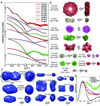
SAXS defines accurate shape and assembly in solution for unknown structures and can uncover unsuspected structural similarity. Experimental scattering curves for proteins with no known structural homolog (left, color) were compared with calculated scattering (black curves on left) from PDB structures identified by DARA, a database of scattering curves calculated from the PDB database. Results from the shape reconstruction program GASBOR (colored envelopes) are overlaid onto the structures identified by DARA (ribbon models, right). In addition, PF1674 and PF1281 with known structures show a limitation in the DARA search (see text) and the need for better comparative algorithms.
Similar articles
- Determination of protein oligomeric structure from small-angle X-ray scattering.
Korasick DA, Tanner JJ. Korasick DA, et al. Protein Sci. 2018 Apr;27(4):814-824. doi: 10.1002/pro.3376. Epub 2018 Feb 10. Protein Sci. 2018. PMID: 29352739 Free PMC article. Review. - High-throughput studies of protein shapes and interactions by synchrotron small-angle X-ray scattering.
Jeffries CM, Svergun DI. Jeffries CM, et al. Methods Mol Biol. 2015;1261:277-301. doi: 10.1007/978-1-4939-2230-7_15. Methods Mol Biol. 2015. PMID: 25502205 - Structural characterization of proteins and complexes using small-angle X-ray solution scattering.
Mertens HD, Svergun DI. Mertens HD, et al. J Struct Biol. 2010 Oct;172(1):128-41. doi: 10.1016/j.jsb.2010.06.012. Epub 2010 Jun 15. J Struct Biol. 2010. PMID: 20558299 Review. - A practical guide to small angle X-ray scattering (SAXS) of flexible and intrinsically disordered proteins.
Kikhney AG, Svergun DI. Kikhney AG, et al. FEBS Lett. 2015 Sep 14;589(19 Pt A):2570-7. doi: 10.1016/j.febslet.2015.08.027. Epub 2015 Aug 29. FEBS Lett. 2015. PMID: 26320411 Review. - Small-angle X-ray scattering on biological macromolecules and nanocomposites in solution.
Blanchet CE, Svergun DI. Blanchet CE, et al. Annu Rev Phys Chem. 2013;64:37-54. doi: 10.1146/annurev-physchem-040412-110132. Epub 2012 Dec 3. Annu Rev Phys Chem. 2013. PMID: 23216378 Review.
Cited by
- Rigid monoclonal antibodies improve detection of SARS-CoV-2 nucleocapsid protein.
Hodge CD, Rosenberg DJ, Grob P, Wilamowski M, Joachimiak A, Hura GL, Hammel M. Hodge CD, et al. MAbs. 2021 Jan-Dec;13(1):1905978. doi: 10.1080/19420862.2021.1905978. MAbs. 2021. PMID: 33843452 Free PMC article. - Multiple conformations of SAM-II riboswitch detected with SAXS and NMR spectroscopy.
Chen B, Zuo X, Wang YX, Dayie TK. Chen B, et al. Nucleic Acids Res. 2012 Apr;40(7):3117-30. doi: 10.1093/nar/gkr1154. Epub 2011 Dec 1. Nucleic Acids Res. 2012. PMID: 22139931 Free PMC article. - Upgraded ESRF BM29 beamline for SAXS on macromolecules in solution.
Pernot P, Round A, Barrett R, De Maria Antolinos A, Gobbo A, Gordon E, Huet J, Kieffer J, Lentini M, Mattenet M, Morawe C, Mueller-Dieckmann C, Ohlsson S, Schmid W, Surr J, Theveneau P, Zerrad L, McSweeney S. Pernot P, et al. J Synchrotron Radiat. 2013 Jul;20(Pt 4):660-4. doi: 10.1107/S0909049513010431. Epub 2013 May 18. J Synchrotron Radiat. 2013. PMID: 23765312 Free PMC article. - Dengue virus nonstructural protein 5 adopts multiple conformations in solution.
Bussetta C, Choi KH. Bussetta C, et al. Biochemistry. 2012 Jul 31;51(30):5921-31. doi: 10.1021/bi300406n. Epub 2012 Jul 16. Biochemistry. 2012. PMID: 22757685 Free PMC article. - Envisioning the dynamics and flexibility of Mre11-Rad50-Nbs1 complex to decipher its roles in DNA replication and repair.
Lafrance-Vanasse J, Williams GJ, Tainer JA. Lafrance-Vanasse J, et al. Prog Biophys Mol Biol. 2015 Mar;117(2-3):182-193. doi: 10.1016/j.pbiomolbio.2014.12.004. Epub 2015 Jan 7. Prog Biophys Mol Biol. 2015. PMID: 25576492 Free PMC article. Review.
References
- Robinson CV, Sali A, Baumeister W. The molecular sociology of the cell. Nature. 2007;450:973. - PubMed
- Green BD, Keller M. Capturing the uncultivated majority. Curr. Opin. Biotechnol. 2006;17:236. - PubMed
- Wilmes P, Bond PL. Metaproteomics: studying functional gene expression in microbial ecosystems. Trends Microbiol. 2006;14:92. - PubMed
- Putnam CD, Hammel M, Hura GL, Tainer JA. X-ray solution scattering (SAXS) combined with crystallography and computation: defining accurate macromolecular structures, conformations and assemblies in solution. Q. Rev. Biophys. 2007;40:191. - PubMed
- Fox BG, et al. Structural genomics: from genes to structures with valuable materials and many questions in between. Nat. Methods. 2008;5:129. - PubMed
Publication types
MeSH terms
Substances
Grants and funding
- R01 CA117638/CA/NCI NIH HHS/United States
- R01 CA117638-06/CA/NCI NIH HHS/United States
- R01 CA117638-07/CA/NCI NIH HHS/United States
- R01 CA117638-08/CA/NCI NIH HHS/United States
LinkOut - more resources
Full Text Sources
Other Literature Sources


