SDH5, a gene required for flavination of succinate dehydrogenase, is mutated in paraganglioma - PubMed (original) (raw)
. 2009 Aug 28;325(5944):1139-42.
doi: 10.1126/science.1175689. Epub 2009 Jul 23.
Oleh Khalimonchuk, Margit Schraders, Noah Dephoure, Jean-Pierre Bayley, Henricus Kunst, Peter Devilee, Cor W R J Cremers, Joshua D Schiffman, Brandon G Bentz, Steven P Gygi, Dennis R Winge, Hannie Kremer, Jared Rutter
Affiliations
- PMID: 19628817
- PMCID: PMC3881419
- DOI: 10.1126/science.1175689
SDH5, a gene required for flavination of succinate dehydrogenase, is mutated in paraganglioma
Huai-Xiang Hao et al. Science. 2009.
Abstract
Mammalian mitochondria contain about 1100 proteins, nearly 300 of which are uncharacterized. Given the well-established role of mitochondrial defects in human disease, functional characterization of these proteins may shed new light on disease mechanisms. Starting with yeast as a model system, we investigated an uncharacterized but highly conserved mitochondrial protein (named here Sdh5). Both yeast and human Sdh5 interact with the catalytic subunit of the succinate dehydrogenase (SDH) complex, a component of both the electron transport chain and the tricarboxylic acid cycle. Sdh5 is required for SDH-dependent respiration and for Sdh1 flavination (incorporation of the flavin adenine dinucleotide cofactor). Germline loss-of-function mutations in the human SDH5 gene, located on chromosome 11q13.1, segregate with disease in a family with hereditary paraganglioma, a neuroendocrine tumor previously linked to mutations in genes encoding SDH subunits. Thus, a mitochondrial proteomics analysis in yeast has led to the discovery of a human tumor susceptibility gene.
Figures
Fig. 1
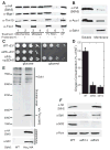
Sdh5 is required for respiration and interacts with Sdh1. (A) Mitochondria, mitoplasts generated by hypotonic swelling, and 1% Triton X-100–solubilized mitochondria from a strain expressing Sdh5-HA were treated with (+) or without (−) proteinase K and analyzed by immunoblotting with an untreated mitochondria control (UT). Mge1, Tim10, and Fzo1 are matrix, intermembrane space, and outer membrane proteins, respectively. (B) Soluble and membrane fractions of purified mitochondria (4) as in (A) were immunoblotted. Aco1, soluble matrix protein; Sdh1, membrane-associated matrix protein. (C) Serial dilutions of WT and _sdh5_Δ strains containing empty vector (EV) or a plasmid expressing Sdh5-HA were spotted on glucose or glycerol medium and grown at 30°C for 2 or 3 days, respectively. (D) Oxygen consumption in WT, _sdh5_Δ, and _sdh1_Δ strains grown to mid-log phase in raffinose media (±SD, n = 3 biological replicates). Similar results were obtained in glucose medium. (E) Tandem purification eluates (4) from a WT strain and a strain expressing Sdh5-His6/HA were resolved with SDS-PAGE and visualized by silver staining (top panel) or immunoblot with antibodies to Sdh1 and HA (lower panels). (F) Immunoblot of purified mitochondria from WT, _sdh1_Δ, or _sdh2_Δ strains expressing Sdh5-HA. Porin, mitochondrial loading control.
Fig. 2
Sdh5 is required for SDH activity and stability. (A) SDH and malate dehydrogenase activity (4) from WT and _sdh5_Δ mitochondria, normalized to total protein (±SD, n = 3 biological replicates). (B) In-gel activity assay of ETC complexes after BN-PAGE of mitochondrial membranes from WT and _sdh5_Δ strains. V2, complex V dimer. (C) Coomassiestained BN-PAGE of mitochondrial membranes from WT and _sdh5_Δ strains. (D) Immunoblot of BN-PAGE–separated complex II/SDH using an antibody to Myc to show Myc-tagged Sdh3 in WT and _sdh5_Δ mitochondria. (E) Immunoblot of BN-PAGE–separated WT and Sdh5-TAP mitochondria using an antibody to TAP, without and with 1% SDS pretreatment. Porin is an ~440-kD control. (F) Immunoblot of mitochondria from WT and _sdh5_Δ strains in which Sdh3 or Sdh4 was Myc-tagged, separated into soluble (sol) and membrane (mem) fractions (4), or unfractionated (total). Aco1, soluble matrix protein. The indicated percentage is the amount remaining in _sdh5_Δ mitochondria relative to WT mitochondria.
Fig. 3
Sdh5 is necessary and sufficient for Sdh1 flavination. (A) WT, _sdh5_Δ, and _sdh1_Δ mitochondria were resolved by SDS-PAGE and imaged (4) for covalent FAD (top panel) or immunoblotted (lower panels). (B) Fluorescence gel image (top panel) and immunoblot (lower panels) as in (A), with whole-cell extract from WT or _flx1_Δ _sdh5_Δ strains containing EV, CEN plasmid SDH5 (flx1Δ: ~1 copy per cell), or 2μ plasmid SDH5 (O/E: ~10 copies per cell). The bar graph shows normalized FAD fluorescence (±SD, n = 3 biological replicates) (bottom panel). (C) His-tagged yeast Sdh1 was expressed alone or with Sdh5 or Sdh2 in E. coli, purified, and analyzed for FAD fluorescence as in (A) and by Coomassie blue staining.
Fig. 4
A human SDH5 loss-of-function mutation in PGL2. (A) The heterozygous c.232G>A mutation segregates with disease in the PGL2 lineage (17). Black symbols, affected persons; white symbols, unaffected persons; +, heterozygous mutation; NT, not tested. Diamonds with the number 4 represent four unaffected individuals. Individuals who are not affected because they carry the mutation on their maternal chromosome 11 are marked by an m. One healthy maternal mutation carrier and five non-affected paternal mutation carriers are not shown in the pedigree for privacy reasons. (B) Fluorescence gel image (top panel) and immunoblotting (lower panels) of samples from human tumors, cell lines, and mouse tissues. Lanes 1 and 2, sporadic PGL tumors; lanes 3 to 5, PGL2 tumors (hSDH5 G78R); lanes 6 and 7, HEK293 and HepG2 human cell lines; lanes 8 and 9, normal mouse skeletal muscle (skM) and liver. (C) Lysate from HEK293 cells containing EV or expressing WT or G78R hSDH5-Myc were immunoprecipitated with agarose beads conjugated with antibody to Myc. Lysate, eluate, and unbound fraction were FAD-imaged (top panel) and immunoblotted (lower three panels). (D) Serial dilutions of WT and _sdh5_Δ strains containing EV or plasmids expressing yeast Sdh5-Myc, WT human SDH5-Myc, or G78R hSDH5-Myc were spotted on glucose or glycerol medium and grown at 30°C for 2 or 3 days, respectively. (E) Fluorescence gel image (top panel) and immunoblotting of whole-cell extract from the five strains in (D) (lower panels). PGK, loading control. FAD fluorescence was normalized to Sdh1 protein level and expressed as a percentage relative to WT (Flavo%, ±SD, n = 3 biological replicates).
Similar articles
- Solution NMR structure of yeast succinate dehydrogenase flavinylation factor Sdh5 reveals a putative Sdh1 binding site.
Eletsky A, Jeong MY, Kim H, Lee HW, Xiao R, Pagliarini DJ, Prestegard JH, Winge DR, Montelione GT, Szyperski T. Eletsky A, et al. Biochemistry. 2012 Oct 30;51(43):8475-7. doi: 10.1021/bi301171u. Epub 2012 Oct 19. Biochemistry. 2012. PMID: 23062074 Free PMC article. - SDH5 mutations and familial paraganglioma: somewhere Warburg is smiling.
Kaelin WG Jr. Kaelin WG Jr. Cancer Cell. 2009 Sep 8;16(3):180-2. doi: 10.1016/j.ccr.2009.08.013. Cancer Cell. 2009. PMID: 19732718 - Mitochondrial matrix proteostasis is linked to hereditary paraganglioma: LON-mediated turnover of the human flavinylation factor SDH5 is regulated by its interaction with SDHA.
Bezawork-Geleta A, Saiyed T, Dougan DA, Truscott KN. Bezawork-Geleta A, et al. FASEB J. 2014 Apr;28(4):1794-804. doi: 10.1096/fj.13-242420. Epub 2014 Jan 10. FASEB J. 2014. PMID: 24414418 - Emerging concepts in the flavinylation of succinate dehydrogenase.
Kim HJ, Winge DR. Kim HJ, et al. Biochim Biophys Acta. 2013 May;1827(5):627-36. doi: 10.1016/j.bbabio.2013.01.012. Epub 2013 Feb 1. Biochim Biophys Acta. 2013. PMID: 23380393 Free PMC article. Review. - Succinate dehydrogenase (SDH) and mitochondrial driven neoplasia.
Gill AJ. Gill AJ. Pathology. 2012 Jun;44(4):285-92. doi: 10.1097/PAT.0b013e3283539932. Pathology. 2012. PMID: 22544211 Review.
Cited by
- A case report - Volatile metabolomic signature of malignant melanoma using matching skin as a control.
Abaffy T, Möller M, Riemer DD, Milikowski C, Defazio RA. Abaffy T, et al. J Cancer Sci Ther. 2011;3(6):140-144. doi: 10.4172/1948-5956.1000076. J Cancer Sci Ther. 2011. PMID: 22229073 Free PMC article. - Over-representation of the G12S polymorphism of the SDHD gene in patients with MEN2A syndrome.
Lendvai N, Tóth M, Valkusz Z, Bekő G, Szücs N, Csajbók E, Igaz P, Kriszt B, Kovács B, Rácz K, Patócs A. Lendvai N, et al. Clinics (Sao Paulo). 2012;67 Suppl 1(Suppl 1):85-9. doi: 10.6061/clinics/2012(sup01)15. Clinics (Sao Paulo). 2012. PMID: 22584711 Free PMC article. - Yeast as a system for modeling mitochondrial disease mechanisms and discovering therapies.
Lasserre JP, Dautant A, Aiyar RS, Kucharczyk R, Glatigny A, Tribouillard-Tanvier D, Rytka J, Blondel M, Skoczen N, Reynier P, Pitayu L, Rötig A, Delahodde A, Steinmetz LM, Dujardin G, Procaccio V, di Rago JP. Lasserre JP, et al. Dis Model Mech. 2015 Jun;8(6):509-26. doi: 10.1242/dmm.020438. Dis Model Mech. 2015. PMID: 26035862 Free PMC article. Review. - Mutation profiling in eight cases of vagal paragangliomas.
Kudryavtseva AV, Kalinin DV, Pavlov VS, Savvateeva MV, Fedorova MS, Pudova EA, Kobelyatskaya AA, Golovyuk AL, Guvatova ZG, Razmakhaev GS, Demidova TB, Simanovsky SA, Slavnova EN, Poloznikov AА, Polyakov AP, Melnikova NV, Dmitriev AA, Krasnov GS, Snezhkina AV. Kudryavtseva AV, et al. BMC Med Genomics. 2020 Sep 18;13(Suppl 8):115. doi: 10.1186/s12920-020-00763-4. BMC Med Genomics. 2020. PMID: 32948195 Free PMC article. - Pheochromocytoma and paraganglioma: understanding the complexities of the genetic background.
Fishbein L, Nathanson KL. Fishbein L, et al. Cancer Genet. 2012 Jan-Feb;205(1-2):1-11. doi: 10.1016/j.cancergen.2012.01.009. Cancer Genet. 2012. PMID: 22429592 Free PMC article. Review.
References
- Lin MT, Beal MF. Nature. 2006;443:787. - PubMed
- See supporting material on Science Online.
- Andersson SG, et al. Nature. 1998;396:133. - PubMed
Publication types
MeSH terms
Substances
LinkOut - more resources
Full Text Sources
Other Literature Sources
Medical
Molecular Biology Databases
Miscellaneous


