TBP2 is a substitute for TBP in Xenopus oocyte transcription - PubMed (original) (raw)
TBP2 is a substitute for TBP in Xenopus oocyte transcription
Waseem Akhtar et al. BMC Biol. 2009.
Abstract
Background: TATA-box-binding protein 2 (TBP2/TRF3) is a vertebrate-specific paralog of TBP that shares with TBP a highly conserved carboxy-terminal domain and the ability to bind the TATA box. TBP2 is highly expressed in oocytes whereas TBP is more abundant in embryos.
Results: We find that TBP2 is proteolytically degraded upon meiotic maturation; after germinal vesicle breakdown relatively low levels of TBP2 expression persist. Furthermore, TBP2 localizes to the transcriptionally active loops of lampbrush chromosomes and is recruited to a number of injected promoters in oocyte nuclei. Using an altered binding specificity mutant reporter system we show that TBP2 promotes RNA polymerase II transcription in vivo. Intriguingly, TBP, which in oocytes is undetectable at the protein level, can functionally replace TBP2 when ectopically expressed in oocytes, showing that switching of initiation factors can be driven by changes in their expression. Proteolytic degradation of TBP2 is not required for repression of transcription during meiotic maturation, suggesting a redundant role in this repression or a role in initiation factor switching between oocytes and embryos.
Conclusion: The expression and transcriptional activity of TBP2 in oocytes show that TBP2 is the predominant initiation factor in oocytes, which is substituted by TBP on a subset of promoters in embryos as a result of proteolytic degradation of TBP2 during meiotic maturation.
Figures
Figure 1
TBP2 is actively down regulated upon meiotic maturation. (A) Western blot analysis of TBP expression in oocytes, eggs and late blastula (stage 9) embryos with four different antibodies; 58C9, SL27, SL30 and SL33. Asterisk indicates non-specific bands. (B) Western blot analysis of TBP2 expression in oocyte germinal vesicles and embryonic nuclei with a TBP2-specific monoclonal antibody 3E6. (C) Stage VI Xenopus oocytes were incubated in MBSH buffer containing 1 μg/ml progesterone. Batches of 15 oocytes were collected at indicated time intervals and analyzed by western blotting. (D) Stage VI Xenopus oocytes injected with TBP and HA-TBP2 mRNA were incubated in MBSH buffer containing 1 μg/ml progesterone. Batches of 15 oocytes were collected at indicated time intervals and analyzed by western blotting. (E) Oocytes expressing exogenous TBP were treated with either progesterone (1 μg/ml) or cycloheximide (15 μg/ml). Batches of 15 oocytes were collected 4 hr and 16 hr after the treatment and analyzed by western blotting. Asterisk indicates non-specific bands.
Figure 2
TBP2 is recruited to transcriptionally active loops of lampbrush chromosomes. Germinal vesical (GV) spreads were probed with TBP2 specific antibodies 1G6 (top panel) and 3E6 (bottom panel) as described in Methods. The inset shows a magnified chromosomal loop. Cajal bodies (CB) and B-snurposomes (BS) are indicated.
Figure 3
TBP2 is associated with active promoters in oocytes. (A) Chromatin immunoprecipitation (ChIP) assay on germinal vesical (GV)-injected promoters and endogenous 5S rRNA promoters using TBP2 and pol-II antibodies. Enrichment is the signal relative to background which in this case is the ChIP recovery from a non-promoter region (intron construct). (B) ChIP assay on GV-injected pol-II promoters and endogenous 5S rRNA promoter from oocytes injected with HA-TBP2 mRNA. Enrichment is the signal relative to background which in this case is the ChIP signal from oocytes not expressing HA-TBP2 but injected with the same promoter construct. Error bars in (A) and (B) represent the standard error of the mean from three independent experiments.
Figure 4
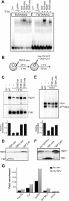
Both TBP and TBP2 can promote transcription from TATA box containing promoters. (A) EMSA showing the binding of in vitro translated wild-type and abs-mutant TBP and TBP2 to a wild-type and a mutant TATA box probe. (B) A scheme of the experiment shown in (C) is presented. TBP-abs or TBP2-abs mRNA was injected into stage VI oocytes. After 4 hr oocytes were injected with either wild type or TGTA mutant hsp-70 promoter. pCMV-CAT was injected as an internal control. (C) Primer extension was performed to determine the expression from the hsp70 (hsp-70) and CMV promoter (CMV). Ctrl: RNA from un-injected oocytes. The bar graph shows the quantification of transcription signal from hsp-70 promoter normalized with that from CMV. The error bars represent standard error of mean from three independent experiments. (D) Western blot analysis of TBP2-wt, TBP-abs and TBP2-abs expression in oocytes for the experiment in (C). (E) Expression from the ZFP36L2 promoter with regular or mutant TATA box in the presence of TBP and TBP2 abs-mutants was analyzed by primer extension. The bar graph shows the quantification of transcription signal from ZFP36L2 promoter normalized with that from CMV from two independent experiments. (F) Western blot analysis of TBP-abs and TBP2-abs expression in oocytes for the experiment in (E). (G) Chromatin immunoprecipitation (ChIP) assay on germinal vesical-injected promoters from oocytes injected with HA-TBP or HA-TBP2 mRNA. Enrichment is the signal relative to the ChIP signal from oocytes not expressing HA-tagged protein.
Figure 5
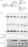
Transcription repression occurs in the presence of abundant TBP2. (A) A scheme of the experiment shown in (B) is presented. Either 2 ng or 4 ng of TBP2 mRNA was injected into stage VI oocytes. After a delay of 12 hr oocytes were injected in the nucleus with 1.0 ng of hsp70 and 0.5 ng of Cyr61 promoter constructs either with or without 18 ng of competitor DNA. One hour after the nuclear injections oocytes were divided into two groups. One group was treated with progesterone (final concentration 2 μg/ml). After a further incubation of 12 hr groups of 20 healthy oocytes were collected for RNA and protein analysis. (B) For transcription analysis, a primer extension was performed as described in Methods. The positions of accurately initiated transcripts from the hsp70 (hsp-70) and Cyr61 promoter (Cyr61) are indicated. The conditions used for each lane are described below the gel, and at the bottom expression of TBP2 has been shown for the corresponding lane. Phospho-MAPK was used as a marker for maturation while TFIIF served as a loading control.
Figure 6
TBP2 degradation takes place after the establishment of transcriptional shut down. (A) A scheme of the experiment shown in (B) is presented. Oocyte nuclei were injected with 0.6 ng of hsp70 and 0.3 ng of Cyr61 promoter constructs. After a delay of 12 hr oocytes were divided into two groups and one group was treated with progesterone (final concentration 2 μg/ml). Oocytes were synchronized at germinal vesicle breakdown (GVBD). Some were processed immediately for chromatin immunoprecipitation (ChIP) and immunoblotting along with control oocytes (not treated with progesterone) whereas others were analyzed 5 hr after GVBD. (B) Recruitment of pol-II (upper graph) and TBP2 (lower graph) to hsp70 and Cyr61 promoters in control oocytes versus oocytes at GVBD and 5 hr after GVBD was assayed by ChIP. Below the graphs expression of TBP2 has been shown corresponding to each sample. TFIIF served as a loading control.
Figure 7
A model of the regulation of TATA-box binding proteins during early stages of embryogenesis. Oocytes express TBP2 which is involved in initiation of transcription. Upon meiotic maturation, global repression of transcription is established and TBP2 is actively degraded. During early cleavages after fertilization, maternal stores of TBP mRNA are translated [15] and by the mid-blastula transition (MBT) both TBP and residual TBP2 contribute to zygotic transcription [8,9].
Similar articles
- Specialized and redundant roles of TBP and a vertebrate-specific TBP paralog in embryonic gene regulation in Xenopus.
Jallow Z, Jacobi UG, Weeks DL, Dawid IB, Veenstra GJ. Jallow Z, et al. Proc Natl Acad Sci U S A. 2004 Sep 14;101(37):13525-30. doi: 10.1073/pnas.0405536101. Epub 2004 Sep 2. Proc Natl Acad Sci U S A. 2004. PMID: 15345743 Free PMC article. - Regulated expression of TATA-binding protein-related factor 3 (TRF3) during early embryogenesis.
Yang Y, Cao J, Huang L, Fang HY, Sheng HZ. Yang Y, et al. Cell Res. 2006 Jul;16(7):610-21. doi: 10.1038/sj.cr.7310064. Cell Res. 2006. PMID: 16721357 - What defines the maternal transcriptome?
Tora L, Vincent SD. Tora L, et al. Biochem Soc Trans. 2021 Nov 1;49(5):2051-2062. doi: 10.1042/BST20201125. Biochem Soc Trans. 2021. PMID: 34415300 Free PMC article. Review. - TBP2 is a general transcription factor specialized for female germ cells.
Müller F, Tora L. Müller F, et al. J Biol. 2009;8(11):97. doi: 10.1186/jbiol196. Epub 2009 Nov 30. J Biol. 2009. PMID: 19951399 Free PMC article. Review.
Cited by
- TBPL2/TFIIA complex establishes the maternal transcriptome through oocyte-specific promoter usage.
Yu C, Cvetesic N, Hisler V, Gupta K, Ye T, Gazdag E, Negroni L, Hajkova P, Berger I, Lenhard B, Müller F, Vincent SD, Tora L. Yu C, et al. Nat Commun. 2020 Dec 22;11(1):6439. doi: 10.1038/s41467-020-20239-4. Nat Commun. 2020. PMID: 33353944 Free PMC article. - The epigenome in early vertebrate development.
Bogdanović O, van Heeringen SJ, Veenstra GJ. Bogdanović O, et al. Genesis. 2012 Mar;50(3):192-206. doi: 10.1002/dvg.20831. Epub 2011 Dec 27. Genesis. 2012. PMID: 22139962 Free PMC article. Review. - TBP-related factors: a paradigm of diversity in transcription initiation.
Akhtar W, Veenstra GJ. Akhtar W, et al. Cell Biosci. 2011 Jun 27;1(1):23. doi: 10.1186/2045-3701-1-23. Cell Biosci. 2011. PMID: 21711503 Free PMC article. - Hierarchical molecular events driven by oocyte-specific factors lead to rapid and extensive reprogramming.
Jullien J, Miyamoto K, Pasque V, Allen GE, Bradshaw CR, Garrett NJ, Halley-Stott RP, Kimura H, Ohsumi K, Gurdon JB. Jullien J, et al. Mol Cell. 2014 Aug 21;55(4):524-36. doi: 10.1016/j.molcel.2014.06.024. Epub 2014 Jul 24. Mol Cell. 2014. PMID: 25066233 Free PMC article. - TBP facilitates RNA Polymerase I transcription following mitosis.
Kwan JZJ, Nguyen TF, Teves SS. Kwan JZJ, et al. RNA Biol. 2024 Jan;21(1):42-51. doi: 10.1080/15476286.2024.2375097. Epub 2024 Jul 3. RNA Biol. 2024. PMID: 38958280 Free PMC article.
References
Publication types
MeSH terms
Substances
LinkOut - more resources
Full Text Sources


