Ethanol disrupts chondrification of the neurocranial cartilages in medaka embryos without affecting aldehyde dehydrogenase 1A2 (Aldh1A2) promoter methylation - PubMed (original) (raw)
Ethanol disrupts chondrification of the neurocranial cartilages in medaka embryos without affecting aldehyde dehydrogenase 1A2 (Aldh1A2) promoter methylation
Yuhui Hu et al. Comp Biochem Physiol C Toxicol Pharmacol. 2009 Nov.
Abstract
Medaka (Oryzias latipes) embryos at different developmental stages were exposed to ethanol for 48 h, then allowed to hatch. Teratogenic effects were evaluated in hatchlings after examining chondrocranial cartilage deformities. Ethanol disrupted cartilage development in medaka in a dose and developmental stage-specific manner. Compared to controls, the linear length of the neurocranium and other cartilages were reduced in ethanol-treated groups. Moreover, the chondrification in cartilages, specifically trabeculae and polar cartilages, were inhibited by ethanol. To understand the mechanism of ethanol teratogenesis, NAD(+): NADH status during embryogenesis and the methylation pattern of Aldh1A2 promoter in whole embryos and adult tissues (brain, eye, heart and liver) were analyzed. Embryos 6 dpf had higher NAD(+) than embryos 0 or 2 dpf. Ethanol (200 or 400 mM) was able to reduce NAD(+) content in 2 and 6 dpf embryos. However, in both cases reductions were not significantly different from the controls. Moreover, no significant difference in either NADH content or in NAD(+): NADH status of the ethanol-treated embryos, with regard to controls, was observed. The promoter of Aldh1A2 contains 31 CpG dinucleotides (-705 to +154, ATG=+1); none of which were methylated. Compared to controls, embryonic ethanol exposure (100 and 400 mM) was unable to alter Aldh1A2 promoter methylation in embryos or in the tissues of adults (breeding) developmentally exposed to ethanol (300 mM, 48 hpf). From these data we conclude that ethanol teratogenesis in medaka does not induce alteration in the methylation pattern of Aldh1A2 promoter, but does change cartilage development.
Figures
Figure 1. Representative photomicrograph of the neurocranium (A, D =dorsal side, B lateral side) and splanchnocranium (C= dorsal side) of a medaka hatchlings 10 dpf
The hatchlings were stained with alcian blue showing the different regions of the head skeleton considered for morphometric analysis.
Figure 2. Representative photomicrographs of the neurocranium (dorsal view) of medaka hatchlings showing the disruption in chondrification of neurocranial cartilages by ethanol during development
The embryos were exposed to ethanol (300 mM) 48 hpf and transferred to fresh hatching solution. The controls were maintained in hatching solution only. The hatchlings (10 dpf) were stained with alcian blue. It was observed that chondrification of trabecular cartilages (TC) and polar cartilages (PC) are disrupted by ethanol (B-D). A= control (no ethanol); B, C, and D= 300 mM ethanol (0-48 hpf).In A, a normal TC and PC are seen; in B, C, and D various levels of disrupted chondrification in TC and PC are seen (B= reduced EP with tiny TC and unequal PC; C=reduced EP with short and unequal TC and absence of *PC; D=reduced EP with no TC* and unequal PC).
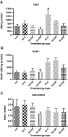
Figure 3. Effect of ethanol on NAD+ and NADH status of medaka embryo during development
Embryos were exposed to 200 and 400 mM ethanol 48 hpf and either used for coenzyme assay or maintained in hatching solution 6 dpf and used for the coenzyme assay. The corresponding controls were maintained in hatching solution only. To determine the basal value the fertilized eggs ~ 1 hpf were used. Each bar is the mean ±SD of 4-6 observations. The data were analyzed by one way ANOVA followed by post-hoc Tukey’s multiple comparison test; p<0.05 was considered as significant. Bar head with pound symbol (#) indicates that the results are significantly different from the samples of zero day. A= NAD+, B= NADH, C= NAD+: NADH.
Figure 4. The nucleotide sequences of Aldh1A2 promoter of Japanese medaka (Oryzias latipes)
The nucleotide sequence (NT) information are originally obtained from Ensemble (
www.ensemble.org/Oryzias\_latipes/gene
) and verified by PCR amplification, cloning and sequencing. The NT represented in upper case letters are the first exon (227 nt) where ATG is represented in bold letters. The underlined regions were used for designing primers for genomic DNA amplification. CpG dinucleotides are colored. There are 31 CpG dinucleotides present in the region of which 26 are considered for analysis. Numbers on the right represent NT number, starting with ATG as +1.
Similar articles
- Disruption of circulation by ethanol promotes fetal alcohol spectrum disorder (FASD) in medaka (Oryzias latipes) embryogenesis.
Hu Y, Khan IA, Dasmahapatra AK. Hu Y, et al. Comp Biochem Physiol C Toxicol Pharmacol. 2008 Sep;148(3):273-80. doi: 10.1016/j.cbpc.2008.06.006. Epub 2008 Jun 22. Comp Biochem Physiol C Toxicol Pharmacol. 2008. PMID: 18621148 Free PMC article. - Ethanol attenuates Aldh9 mRNA expression in Japanese medaka (Oryzias latipes) embryogenesis.
Wang X, Zhu S, Khan IA, Dasmahapatra AK. Wang X, et al. Comp Biochem Physiol B Biochem Mol Biol. 2007 Mar;146(3):357-63. doi: 10.1016/j.cbpb.2006.11.006. Epub 2006 Nov 21. Comp Biochem Physiol B Biochem Mol Biol. 2007. PMID: 17236798 - Japanese medaka (Oryzias latipes): developmental model for the study of alcohol teratology.
Wang X, Williams E, Haasch ML, Dasmahapatra AK. Wang X, et al. Birth Defects Res B Dev Reprod Toxicol. 2006 Feb;77(1):29-39. doi: 10.1002/bdrb.20072. Birth Defects Res B Dev Reprod Toxicol. 2006. PMID: 16496295 - Ethanol teratogenesis in Japanese medaka: effects at the cellular level.
Wu M, Chaudhary A, Khan IA, Dasmahapatra AK. Wu M, et al. Comp Biochem Physiol B Biochem Mol Biol. 2008 Jan;149(1):191-201. doi: 10.1016/j.cbpb.2007.09.008. Epub 2007 Sep 16. Comp Biochem Physiol B Biochem Mol Biol. 2008. PMID: 17913529 Free PMC article. - Modulation of ethanol toxicity by Asian ginseng (Panax ginseng) in Japanese ricefish (Oryzias latipes) embryogenesis.
Haron MH, Avula B, Khan IA, Mathur SK, Dasmahapatra AK. Haron MH, et al. Comp Biochem Physiol C Toxicol Pharmacol. 2013 Apr;157(3):287-97. doi: 10.1016/j.cbpc.2013.02.001. Epub 2013 Feb 9. Comp Biochem Physiol C Toxicol Pharmacol. 2013. PMID: 23402931
Cited by
- Towards incorporating epigenetic mechanisms into carcinogen identification and evaluation.
Herceg Z, Lambert MP, van Veldhoven K, Demetriou C, Vineis P, Smith MT, Straif K, Wild CP. Herceg Z, et al. Carcinogenesis. 2013 Sep;34(9):1955-67. doi: 10.1093/carcin/bgt212. Epub 2013 Jun 7. Carcinogenesis. 2013. PMID: 23749751 Free PMC article. Review. - Retinoic acid and meiosis induction in adult versus embryonic gonads of medaka.
Adolfi MC, Herpin A, Regensburger M, Sacquegno J, Waxman JS, Schartl M. Adolfi MC, et al. Sci Rep. 2016 Sep 28;6:34281. doi: 10.1038/srep34281. Sci Rep. 2016. PMID: 27677591 Free PMC article. - Sex-reversal and Histopathological Assessment of Potential Endocrine-Disrupting Effects of Graphene Oxide on Japanese medaka (Oryzias latipes) Larvae.
Myla A, Dasmahapatra AK, Tchounwou PB. Myla A, et al. Chemosphere. 2021 Sep;279:130768. doi: 10.1016/j.chemosphere.2021.130768. Epub 2021 May 17. Chemosphere. 2021. PMID: 34134430 Free PMC article. - Toxicity implications for early life stage Japanese medaka (Oryzias latipes) exposed to oxyfluorfen.
Powe DK, Dasmahapatra AK, Russell JL, Tchounwou PB. Powe DK, et al. Environ Toxicol. 2018 May;33(5):555-568. doi: 10.1002/tox.22541. Epub 2018 Jan 31. Environ Toxicol. 2018. PMID: 29385312 Free PMC article. - Epigenetic Mechanisms in Developmental Alcohol-Induced Neurobehavioral Deficits.
Basavarajappa BS, Subbanna S. Basavarajappa BS, et al. Brain Sci. 2016 Apr 8;6(2):12. doi: 10.3390/brainsci6020012. Brain Sci. 2016. PMID: 27070644 Free PMC article. Review.
References
- Blakley PM, Scott WJ., Jr. Determination of the proximate teratogen of the mouse fetal alcohol syndrome. 2. pharmacokinetics of the placental transfer of ethanol and acetaldehyde. Toxicol Appl Pharmacol. 1984;72:364–371. - PubMed
- Carvan MJ, III, Loucks E, Weber DN, Williams FE. Ethanol effects on the developing zebrafish: neurobehavior and skeletal morphogenesis. Neurotoxicol. Teratol. 2004;26:757–768. - PubMed
- Clagett-Dame M, DeLuca HF. The role of vitamin A in mammalian reproduction and embryonic development. Annu. Rev. Nutr. 2002;22:347–381. - PubMed
- Contractor RG, Foran CM, Li S, Willett KL. Evidence of geneder- and Tissue-specific promoter methylation and the potential for ethinylestradiol-induced changes in Japanese medaka (Oryzias latipes) estrogen receptor and aromatase genes. J Toxicol Environ. Health. 2004;67A:1–22. - PubMed
Publication types
MeSH terms
Substances
Grants and funding
- P20 RR016476/RR/NCRR NIH HHS/United States
- RR016476/RR/NCRR NIH HHS/United States
- R03 AA016915/AA/NIAAA NIH HHS/United States
- R03AA016915/AA/NIAAA NIH HHS/United States
- R03 AA016915-02/AA/NIAAA NIH HHS/United States
LinkOut - more resources
Full Text Sources


