miR-200 expression regulates epithelial-to-mesenchymal transition in bladder cancer cells and reverses resistance to epidermal growth factor receptor therapy - PubMed (original) (raw)
. 2009 Aug 15;15(16):5060-72.
doi: 10.1158/1078-0432.CCR-08-2245. Epub 2009 Aug 11.
Meng Zhong, Woonyoung Choi, Wei Qi, Milena Nicoloso, Ameeta Arora, George Calin, Hua Wang, Arlene Siefker-Radtke, David McConkey, Menashe Bar-Eli, Colin Dinney
Affiliations
- PMID: 19671845
- PMCID: PMC5938624
- DOI: 10.1158/1078-0432.CCR-08-2245
miR-200 expression regulates epithelial-to-mesenchymal transition in bladder cancer cells and reverses resistance to epidermal growth factor receptor therapy
Liana Adam et al. Clin Cancer Res. 2009.
Abstract
Purpose: The epithelial-to-mesenchymal transition (EMT) is a cell development-regulated process in which noncoding RNAs act as crucial modulators. Recent studies have implied that EMT may contribute to resistance to epidermal growth factor receptor (EGFR)-directed therapy. The aims of this study were to determine the potential role of microRNAs (miRNA) in controlling EMT and the role of EMT in inducing the sensitivity of human bladder cancer cells to the inhibitory effects of the anti-EGFR therapy.
Experimental design: miRNA array screening and real-time reverse transcription-PCR were used to identify and validate the differential expression of miRNAs involved in EMT in nine bladder cancer cell lines. A list of potential miR-200 direct targets was identified through the TargetScan database. The precursor of miR-200b and miR-200c was expressed in UMUC3 and T24 cells using a retrovirus or a lentivirus construct, respectively. Protein expression and signaling pathway modulation, as well as intracellular distribution of EGFR and ERRFI-1, were validated through Western blot analysis and confocal microscopy, whereas ERRFI-1 direct target of miR-200 members was validated by using the wild-type and mutant 3'-untranslated region/ERRFI-1/luciferse reporters.
Results: We identified a tight association between the expression of miRNAs of the miR-200 family, epithelial phenotype, and sensitivity to EGFR inhibitors-induced growth inhibition in bladder carcinoma cell lines. Stable expression of miR-200 in mesenchymal UMUC3 cells increased E-cadherin levels, decreased expression of ZEB1, ZEB2, ERRFI-1, and cell migration, and increased sensitivity to EGFR-blocking agents. The changes in EGFR sensitivity by silencing or forced expression of ERRFI-1 or by miR-200 expression have also been validated in additional cell lines, UMUC5 and T24. Finally, luciferase assays using 3'-untranslated region/ERRFI-1/luciferase and miR-200 cotransfections showed that the direct down-regulation of ERRFI-1 was miR-200-dependent because mutations in the two putative miR-200-binding sites have rescued the inhibitory effect.
Conclusions: Members of the miR-200 family appear to control the EMT process and sensitivity to EGFR therapy in bladder cancer cells and the expression of miR-200 is sufficient to restore EGFR dependency at least in some of the mesenchymal bladder cancer cells. The targets of miR-200 include ERRFI-1, which is a novel regulator of EGFR-independent growth.
Figures
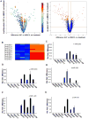
Figure 1. Identification of miRNAs that are differentially expressed in an epithelial versus a mesenchymal bladder cancer cell line
A) Volcano plots showing all miRNAs detected in the 2 cell lines (left panel) or only the miRNA with significantly different expression (right panel, red dots) as opposed to non significant levels of differential exression (right panel, blue dots). Bayesian log odds of differential expression are plotted against log2 (expression in UMUC5 divided by expression in KU7 cells). B) Heatmap of differentially expressed miRNA clustered by cell type (left, low-expressed miRNAs in KU7 cells; right, high expression of the same miRNAs in UMUC5 cells). C-G) Quantification of miRNAs in a panel of 9 bladder cancer cell lines as measured by TaqMan real-time RT-PCR. Data are means of triplicate RT-PCR assays.
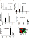
Figure 2. Identification of miR-200 direct targets in a panel of bladder cancer cell lines
A) The Renilla reporter plasmid harboring a miR-200c site with miR-200c precursor or the unrelated miR-26b (control) precursor were transiently co-transfected into KU7, UMUC3, UMUC5, and UMUC13 cells along with a firefly luciferase reporter (pGL3 control) for normalization. The data are mean ± standard error of mean of separate transfections (n=6), and are shown as the ratio of Renilla activity to firefly. B-E) Measurement by real-time RT-PCR of epithelial and mesenchymal markers: ZEB1, ZEB2, E-cadherin, and P-cadherin. The data are means of measurements from triplicate experiments. F) Heat-map of 6 differentially expressed putative targets of miR-200c in a panlel of 9 bladder cancer cell lines using the Illumina gene grofilling array platform.
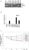
Figure 3. Relation between the differentially expressed ERRFI-1 and TGF-α and cell sensitivity to EGFR-targeted therapy
A) Measurement by real-time westernblot of ERRFI-1 and E-cadherin. B) Measurement by real-time RT-PCR of mRNA relative levels of TGF-α. The data are means of measurements from triplicate experiments. C) Cell proliferation measurement (DNA index) of 9 bladder cancer cell lines in the absence or presence of different cetuximab (C225) concentrations. Data are means of at least 3 triplicate experiments.
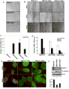
Figure 4. The miR-200c expression induces an EMT phenotype in UMUC3 bladder cancer cell line
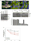
A) Phase contrast microscopy of UMUC3 cells transduced with a miR-200c containing retrovirus (UMUC3/200c) or with an empty, control, retroviral construct (UMUC3-E). Scale bars represent 100 μm. B) Measurement of in vitro cell migration by “wound healing” assay. Representative pictures for same single spot are shown. The experiment was performed twice in triplicate experiments. Scale bars represent 400 μm. C) Quantification of miR-200c in empty virus transduced (UMUC3-E) and miR-200c-transduced (UMUC3/200c) cells. The levels of miR-200c in UMUC5 cells are plotted for comparison. D) Measurement by real-time RT-PCR of the epithelial and mesenchymal markers E-cadherin, ZEB1 and ZEB2 in empty virus-transduced cells and miR-200c-transduced UMUC3 cells. E) Confocal microscopy analysis of the UMUC3 series co-stained for ZEB1 or ZEB2 (red pixels) and nuclear DNA (green pixels). Note downregulation of nuclear ZEB1 and ZEB2 in UMUC3 clones expressing the miR-200c. F) Measurement of ERRFI-1 and E-cadherin protein levels by western blot. Actin reprobing served as internal (loading) control. Lower panel, OD relative values expressed as ratios between actin (internal control) and ERRFI-1. Note up-regulation of E-cadherin and down-regulation of ERRFI-1 protein levels upon miR-200c expression.
Figure 5. The miR-200c expression reverses EGFR resistance in UMUC3 cells
A) Confocal microscopy analysis of the UMUC3 series costained for ERRFI-1 (red pixels) and EGFR (green pixels) and a DNA dye, showing the nucleus (blue pixels). Note the yellow pixels (left panel) as a result of red and green pixels co-localization. B) Immunoblot of autophosphorylated EGFR, total EGFR, and total ERRFI-1 in cells transduced with miR-200c from the experiment above. In this experiment, actin served as the internal control. C) Upper panels, Immunoblot of phosphorylated MAPKinase, total MAPKinase and total EGFR of the UMUC3 series. Cells grown in 2% serum-supplemented MEM were left untreated or were treated with increased concentrations of cetuximab (C225) for 3 hours. Lower panel, OD relative values expressed as ratios between EGFR (internal control) and pMAPKinase. D) Cell proliferation measurement of the UMUC3 series using radioactive thymidine incorporation. Each experiment was done in at least two different triplicates.
Figure 6
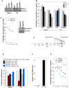
ERRFI-1 is a direct target of miR-200 and implicated in response to EGFR inhibitors. A). Western blot measurement of ERRFI-1 protein in UMUC3 and T24 cells after transfection with a non-targeting sh construct, a GAPDH or a ERRFI-1sh construct. Vinculin served as internal control. B) Cell proliferation measurement of the UMUC3 and T24 series using radioactive thymidine incorporation after stransfections described in the previous panel. Each experiment was done in at least two triplicates. C) Cell proliferation measurement of the UMUC5 series using radioactive thymidine incorporation after ERRFI-1 transfection. The level of ERRFI-1 protein is shown in the right panel. Vinculin served as internal control. D) Schematic representation of the 3′UTR region of ERRFI-1 displaying the miRNA potential binding sites as predicted by TargetScan. Red boxes represent miR-200b/c/429 potential binding sites. E) measurement of miR-200 repressive activity on wild-type and mutant 3′UTR/ERRFFI-1 reporters. F) Real-time RT-PCR of T24 series after miR-200b transduction using a lentiviral system. PEV, empty vector-transduced cells served as negative controls. G) Cell proliferation measurement of the T24 series using radioactive thymidine incorporation after miR-200b lentiviral transduction. Each experiment was done in at least two different triplicates. UMUC9 served as positive controls being highly-sensitive responders to EGFR blockers, in this case, Iressa.
Similar articles
- miR-200c inhibits invasion, migration and proliferation of bladder cancer cells through down-regulation of BMI-1 and E2F3.
Liu L, Qiu M, Tan G, Liang Z, Qin Y, Chen L, Chen H, Liu J. Liu L, et al. J Transl Med. 2014 Nov 4;12:305. doi: 10.1186/s12967-014-0305-z. J Transl Med. 2014. PMID: 25367080 Free PMC article. - The TGFβ-miR-499a-SHKBP1 pathway induces resistance to EGFR inhibitors in osteosarcoma cancer stem cell-like cells.
Wang T, Wang D, Zhang L, Yang P, Wang J, Liu Q, Yan F, Lin F. Wang T, et al. J Exp Clin Cancer Res. 2019 May 28;38(1):226. doi: 10.1186/s13046-019-1195-y. J Exp Clin Cancer Res. 2019. PMID: 31138318 Free PMC article. - Role of epithelial-to-mesenchymal transition (EMT) in drug sensitivity and metastasis in bladder cancer.
McConkey DJ, Choi W, Marquis L, Martin F, Williams MB, Shah J, Svatek R, Das A, Adam L, Kamat A, Siefker-Radtke A, Dinney C. McConkey DJ, et al. Cancer Metastasis Rev. 2009 Dec;28(3-4):335-44. doi: 10.1007/s10555-009-9194-7. Cancer Metastasis Rev. 2009. PMID: 20012924 Free PMC article. Review. - miR-200c: a versatile watchdog in cancer progression, EMT, and drug resistance.
Mutlu M, Raza U, Saatci Ö, Eyüpoğlu E, Yurdusev E, Şahin Ö. Mutlu M, et al. J Mol Med (Berl). 2016 Jun;94(6):629-44. doi: 10.1007/s00109-016-1420-5. Epub 2016 Apr 20. J Mol Med (Berl). 2016. PMID: 27094812 Review.
Cited by
- Reduced expression of miR-200 family members contributes to antiestrogen resistance in LY2 human breast cancer cells.
Manavalan TT, Teng Y, Litchfield LM, Muluhngwi P, Al-Rayyan N, Klinge CM. Manavalan TT, et al. PLoS One. 2013 Apr 23;8(4):e62334. doi: 10.1371/journal.pone.0062334. Print 2013. PLoS One. 2013. PMID: 23626803 Free PMC article. - Autophagy limits the cytotoxic effects of the AKT inhibitor AZ7328 in human bladder cancer cells.
Dickstein RJ, Nitti G, Dinney CP, Davies BR, Kamat AM, McConkey DJ. Dickstein RJ, et al. Cancer Biol Ther. 2012 Nov;13(13):1325-38. doi: 10.4161/cbt.21793. Epub 2012 Aug 16. Cancer Biol Ther. 2012. PMID: 22895070 Free PMC article. - Expression of serum miR-200a, miR-200b, and miR-200c as candidate biomarkers in epithelial ovarian cancer and their association with clinicopathological features.
Zuberi M, Mir R, Das J, Ahmad I, Javid J, Yadav P, Masroor M, Ahmad S, Ray PC, Saxena A. Zuberi M, et al. Clin Transl Oncol. 2015 Oct;17(10):779-87. doi: 10.1007/s12094-015-1303-1. Epub 2015 Jun 11. Clin Transl Oncol. 2015. PMID: 26063644 - EGFR-expression in primary urinary bladder cancer and corresponding metastases and the relation to HER2-expression. On the possibility to target these receptors with radionuclides.
Carlsson J, Wester K, De La Torre M, Malmström PU, Gårdmark T. Carlsson J, et al. Radiol Oncol. 2015 Mar 3;49(1):50-8. doi: 10.2478/raon-2014-0015. eCollection 2015 Mar. Radiol Oncol. 2015. PMID: 25810701 Free PMC article. - The Regulatory Roles of Non-coding RNAs in Angiogenesis and Neovascularization From an Epigenetic Perspective.
Hernández-Romero IA, Guerra-Calderas L, Salgado-Albarrán M, Maldonado-Huerta T, Soto-Reyes E. Hernández-Romero IA, et al. Front Oncol. 2019 Oct 24;9:1091. doi: 10.3389/fonc.2019.01091. eCollection 2019. Front Oncol. 2019. PMID: 31709179 Free PMC article. Review.
References
- Dinney CP, McConkey DJ, Millikan RE, Wu X, Bar-Eli M, Adam L, Kamat AM, Siefker-Radtke AO, Tuziak T, Sabichi AL, Grossman HB, Benedict WF, Czerniak B. Focus on bladder cancer. Cancer Cell. 2004 Aug;6(2):111–6. Review. - PubMed
- Dreicer R. Cancer. 2008. Jul 9, Advanced bladder cancer: So many drugs, so little progress : what's wrong with this picture? - PubMed
- Zhang X, Atala A, Godbey WT. Expression-targeted gene therapy for the treatment of transitional cell carcinoma. Cancer Gene Ther. 2008 Mar 7; - PubMed
- Agarwal PK, Black PC, McConkey DJ, Dinney CP. Emerging drugs for targeted therapy of bladder cancer. Expert Opin Emerg Drugs. 2007 Sep;12(3):435–48. Review. - PubMed
- Blaveri E, Brewer JL, Roydasgupta R, Fridlyand J, DeVries S, Koppie T, Pejavar S, Mehta K, Carroll P, Simko JP, Waldman FM. Bladder cancer stage and outcome by array-based comparative genomic hybridization. Clin Cancer Res. 2005 Oct 1;11(19 Pt 1):7012–22. - PubMed
Publication types
MeSH terms
Substances
LinkOut - more resources
Full Text Sources
Other Literature Sources
Medical
Research Materials
Miscellaneous
