Molecular imaging of pulmonary cancer and inflammation - PubMed (original) (raw)
Review
Molecular imaging of pulmonary cancer and inflammation
Chaitanya R Divgi. Proc Am Thorac Soc. 2009.
Abstract
Molecular imaging (MI) may be defined as imaging in vivo using molecules that report on biologic function. This review will focus on the clinical use of radioactive tracers (nonpharmacologic amounts of compounds labeled with a radioactive substance) that permit external imaging using single photon emission computed tomography (planar, SPECT) or positron emission tomography (PET) imaging. Imaging of lung cancer has been revolutionized with the use of fluorine-18-labeled fluorodeoxyglucose (18F-FDG), an analog of glucose that can be imaged using PET. The ability to carry out whole body imaging after intravenous injection of 18F-FDG allows accurate staging of disease, helping to determine regional and distant nodal and other parenchymal involvement. Glycolysis is increased in nonmalignant conditions, including inflammation (e.g., sarcoidosis), and 18F-FDG PET is a sensitive method for evaluation of active inflammatory disease. Inflammatory disease has been imaged, even before the advent of PET, with planar and SPECT imaging using gallium-67, a radiometal that binds to transferrin. Metabolic alteration in pulmonary pathology is currently being studied, largely in lung cancer, primarily with PET, with a variety of other radiotracers. Prominent among these is thymidine; fluorine-18-labeled thymidine PET is being increasingly used to evaluate proliferation rate in lung and other cancers. This overview will focus on the clinical utility of 18F-FDG PET in the staging and therapy evaluation of lung cancer as well as in imaging of nonmalignant pulmonary conditions. PET and SPECT imaging with other radiotracers of interest will also be reviewed. Future directions in PET imaging of pulmonary pathophysiology will also be explored.
Figures
Figure 1.
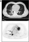
Patient with lung cancer, imaged with fluorodeoxyglucose (FDG) positron emission tomorgraphy (PET)/computed tomography (CT). The patient had known right lung cancer and hilar metastasis (A, arrows); right supraclavicular nodal metastasis (B, arrow) and left femoral osseous metastasis (C, arrow) were revealed on FDG PET/CT, changing management.
Figure 2.
Same patient as in Figure 1, before (left) and after (right) therapy. The bone lesion (arrows) is much less glycolytically active after therapy, though it appears unchanged on companion CT.
Figure 3.
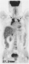
Chest FDG PET of a patient with active sarcoidosis. Bilateral inflamed mediastinal nodes (arrows) are a characteristic feature of this disease.
Figure 4.
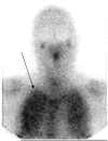
An anterior chest planar image of a patient with HIV and symptoms of Pneumocystis carinii pneumonia. There is uptake of gallium-67 in both lungs (arrow).
Figure 5.
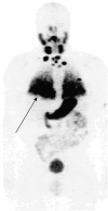
A chest FDG PET three-dimensional projection of patient with metastatic thyroid cancer, imaged 3 days after oral iodine-124. Diffuse lung metastases (arrow) are clearly visualized.
Similar articles
- More advantages in detecting bone and soft tissue metastases from prostate cancer using 18F-PSMA PET/CT.
Pianou NK, Stavrou PZ, Vlontzou E, Rondogianni P, Exarhos DN, Datseris IE. Pianou NK, et al. Hell J Nucl Med. 2019 Jan-Apr;22(1):6-9. doi: 10.1967/s002449910952. Epub 2019 Mar 7. Hell J Nucl Med. 2019. PMID: 30843003 - Nuclear medicine imaging in tuberculosis using commercially available radiopharmaceuticals.
Sathekge M, Maes A, D'Asseler Y, Vorster M, Van de Wiele C. Sathekge M, et al. Nucl Med Commun. 2012 Jun;33(6):581-90. doi: 10.1097/MNM.0b013e3283528a7c. Nucl Med Commun. 2012. PMID: 22422098 Review. - Utility of Molecular Imaging with 2-Deoxy-2-[Fluorine-18] Fluoro-DGlucose Positron Emission Tomography (18F-FDG PET) for Small Cell Lung Cancer (SCLC): A Radiation Oncology Perspective.
Sager O, Dincoglan F, Demiral S, Uysal B, Gamsiz H, Elcim Y, Gundem E, Dirican B, Beyzadeoglu M. Sager O, et al. Curr Radiopharm. 2019;12(1):4-10. doi: 10.2174/1874471012666181120162434. Curr Radiopharm. 2019. PMID: 30465520 Review. - Evaluation of thoracic tumors with 18F-fluorothymidine and 18F-fluorodeoxyglucose-positron emission tomography.
Yap CS, Czernin J, Fishbein MC, Cameron RB, Schiepers C, Phelps ME, Weber WA. Yap CS, et al. Chest. 2006 Feb;129(2):393-401. doi: 10.1378/chest.129.2.393. Chest. 2006. PMID: 16478857 - [Evaluation of fluorine-18-fluorodeoxyglucose whole body positron emission tomography imaging in the clinical diagnosis of lung cancer].
Demura Y, Mizuno S, Wakabayashi M, Totani Y, Okamura S, Ameshima S, Ishizaki T, Miyamori I, Ito H, Yonekura Y. Demura Y, et al. Nihon Kokyuki Gakkai Zasshi. 2000 Sep;38(9):676-81. Nihon Kokyuki Gakkai Zasshi. 2000. PMID: 11109804 Japanese.
Cited by
- The effect of high standard uptake value in lung cancer metastatic lesions on survival.
Aydin S, Balcı A, Dumanlı A, Nesrin Acar L, Ayşin M, Gencer A, Öz G, Demir H, Ece Davarcı S. Aydin S, et al. Croat Med J. 2023 Jun 30;64(3):179-185. doi: 10.3325/cmj.2023.64.179. Croat Med J. 2023. PMID: 37391915 Free PMC article. - 18F-Fluoromisonidazole in tumor hypoxia imaging.
Xu Z, Li XF, Zou H, Sun X, Shen B. Xu Z, et al. Oncotarget. 2017 Oct 7;8(55):94969-94979. doi: 10.18632/oncotarget.21662. eCollection 2017 Nov 7. Oncotarget. 2017. PMID: 29212283 Free PMC article. Review. - Retrospective analysis for the false positive diagnosis of PET-CT scan in lung cancer patients.
Feng M, Yang X, Ma Q, He Y. Feng M, et al. Medicine (Baltimore). 2017 Oct;96(42):e7415. doi: 10.1097/MD.0000000000007415. Medicine (Baltimore). 2017. PMID: 29049175 Free PMC article. - [Long-term survival of personalized surgical treatment of locally advanced non-small cell lung cancer based on molecular staging].
Zhou Q, Shi Y, Chen J, Liu B, Wang Y, Zhu D, Zhang HT, Xu P, Gong Y, Chen G, Wei S, Qiu X, Niu Z, Chen X, Lei Z, Duan L, Wu Z. Zhou Q, et al. Zhongguo Fei Ai Za Zhi. 2011 Feb;14(2):86-106. doi: 10.3779/j.issn.1009-3419.2011.02.15. Zhongguo Fei Ai Za Zhi. 2011. PMID: 21342639 Free PMC article. Chinese. - System a amino acid transport-targeted brain and systemic tumor PET imaging agents 2-amino-3-[(18)F]fluoro-2-methylpropanoic acid and 3-[(18)F]fluoro-2-methyl-2-(methylamino)propanoic acid.
Yu W, McConathy J, Olson JJ, Goodman MM. Yu W, et al. Nucl Med Biol. 2015 Jan;42(1):8-18. doi: 10.1016/j.nucmedbio.2014.07.002. Epub 2014 Aug 1. Nucl Med Biol. 2015. PMID: 25263130 Free PMC article.
References
- Bar-Shalom R, Kagna O, Israel O, Guralnik L. Noninvasive diagnosis of solitary pulmonary lesions in cancer patients based on 2-fluoro-2-deoxy-D-glucose avidity on positron emission tomography/computed tomography. Cancer 2008;113:3213–3221. - PubMed
- Schöder H, Moskowitz C. PET imaging for response assessment in lymphoma: potential and limitations. Radiol Clin North Am 2008;46:225–241. - PubMed
- Bading JR, Shields AF. Imaging of cell proliferation: status and prospects. J Nucl Med 2008;49:64S–80S. - PubMed
- Chang JY, Dong L, Liu H, Starkschall G, Balter P, Mohan R, Liao Z, Cox JD, Komaki R. Image-guided radiation therapy for non-small cell lung cancer. J Thorac Oncol 2008;3:177–186. - PubMed
Publication types
MeSH terms
Substances
LinkOut - more resources
Full Text Sources
Medical


