Septins: molecular partitioning and the generation of cellular asymmetry - PubMed (original) (raw)
Septins: molecular partitioning and the generation of cellular asymmetry
Michael A McMurray et al. Cell Div. 2009.
Abstract
During division, certain cellular contents can be distributed unequally; daughter cells with different fates have different needs. Septins are proteins that participate in the establishment and maintenance of asymmetry during cell morphogenesis, thereby contributing to the unequal partitioning of cellular contents during division. The septins themselves provide a paradigm for studying how elaborate multi-component structures are assembled, dynamically modified, and segregated through each cell division cycle and during development. Here we review our current understanding of the supramolecular organization of septins, the function of septins in cellular compartmentalization, and the mechanisms that control assembly, dynamics, and inheritance of higher-order septin structures, with particular emphasis on recent findings made in budding yeast (Saccharomyces cerevisiae).
Figures
Figure 1
Models for septin organization and diffusion barrier function in the collar and split rings assemblies at the yeast bud neck. This model, based on experimental observations and considerable speculation, illustrates views of the mother-bud neck from a position within the bud (approximated by the gray plane), showing the plasma membrane (orange), globular septin G domains (white balls), and non-septin proteins (blue, green) integral to the plasma membrane and restricted to discrete cortical domains via septin-based diffusion barriers (e.g., Sec3 [6,7]). Prior to cytokinesis, the septins at the bud neck comprise a filamentous collar (left view), retaining Sec3 in the bud (blue). The beginning of cytokinesis is marked by splitting of the collar into two discrete rings (right view), followed by septin-dependent accumulation of Sec3 (green) and other cytokinesis factors within a cortical neck compartment, concomitant with actomyosin ring contraction and growth of the chitinous septum. During this transition, the C-terminal extensions (wavy lines) projecting orthogonally from the filaments in the collar rotate 90°, allowing for greater side-by-side compaction of the filaments.
Figure 2
Model for major transitions in septin assembly and modification state during the yeast budding cycle. Subcellular septin localization (green) during the cycle cycle is accompanied by changes in the organization and covalent modification of septin subunits (grey and white balls). (1) In the G1 phase, hetero-octamers of septin subunits (gray balls) within the "old" ring persisting from the previous cell division are subject to phosphorylation (brown dots) by G1 cyclin-activated cyclin-dependent kinases (Cdks). This modification on certain subunits (e.g., Cdc3 [50]) promotes dissolution of the old ring, permitting relocalization to a new ring at the next budding site. Newly translated septin polypeptides fold, bind GTP, and assemble into sub-octameric complexes (both Cdc11—Cdc12—Cdc3—Cdc10 and Shs1—Cdc12—Cdc3—Cdc10 tetramers, in this model; white balls) that remain stably associated throughout the lifetime of the proteins. Co-incorporation of pre-existing and newly-synthesized subcomplexes precedes (2) phosphorylation by Cla4 (purple dots) of certain subunits (e.g., Cdc10 [10]), which promotes assembly into an organized array of filaments at the neck of the emerging bud. (3) Prior to cytokinesis, SUMO (blue hexagons) is attached to certain subunits in a Siz1- and Siz2-dependent manner only on the mother side of the neck [63,76,77]. (4) During mitosis, septin phosphorylation (orange dots) by mother-side Gin4 (and presumably by its sister protein kinase Kcc4 on the bud side) promotes splitting of the septin collar. (5) Following the completion of cytokinesis and cell separation, septin filaments disassemble into hetero-octamers; residual ring-like septin deposition may reflect persistent self-reinforcing organization of PtdIns4,5P2 and septin-binding transmembrane proteins at the cell cortex. Note that removal of each septin modification upon completion of the preceding transition is speculative, but consistent with the role ascribed, for example, to the action of the Rts1-containing isoform of PP2A [53], and with the ability of old and new subunits to populate all septin-containing structures in a given cell [42].
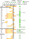
Figure 3
Spatial and temporal organization of protein-modifying enzymes at the bud neck of Saccharomyces cerevisiae. Gene products known or predicted to have the capacity to modify other proteins and that have been visualized at the bud neck by fluorescence microscopy are listed along with the stages of the mitotic division cycle at which they are found at the neck (orange bars), and the region of the bud neck to which they localize (green), where known. Also indicated are the time when emergence of the bud first becomes visible (dashed lined) and the time period corresponding to disassembly of the mitotic spindle and completion of the septum (grey bar). Smt3 is the yeast ortholog of SUMO. It should be noted that this list does not include certain enzymes known to act on septins with important functional consequences (e.g, Cla4 [10]) that do not stably associate with the bud neck, and instead localize to, but quickly depart from, the future site of bud emergence [78]. See Table 1 for citations of the appropriate supporting literature. Adapted from [60] with permission from Elsevier.
Similar articles
- Reuse, replace, recycle. Specificity in subunit inheritance and assembly of higher-order septin structures during mitotic and meiotic division in budding yeast.
McMurray MA, Thorner J. McMurray MA, et al. Cell Cycle. 2009 Jan 15;8(2):195-203. doi: 10.4161/cc.8.2.7381. Cell Cycle. 2009. PMID: 19164941 Free PMC article. Review. - Septin architecture and function in budding yeast.
Farkašovský M. Farkašovský M. Biol Chem. 2020 Jul 28;401(8):903-919. doi: 10.1515/hsz-2019-0401. Biol Chem. 2020. PMID: 31913844 Review. - The role of Bni5 in the regulation of septin higher-order structure formation.
Patasi C, Godočíková J, Michlíková S, Nie Y, Káčeriková R, Kválová K, Raunser S, Farkašovský M. Patasi C, et al. Biol Chem. 2015 Dec;396(12):1325-37. doi: 10.1515/hsz-2015-0165. Biol Chem. 2015. PMID: 26351911 - Septin-Associated Protein Kinases in the Yeast Saccharomyces cerevisiae.
Perez AM, Finnigan GC, Roelants FM, Thorner J. Perez AM, et al. Front Cell Dev Biol. 2016 Nov 1;4:119. doi: 10.3389/fcell.2016.00119. eCollection 2016. Front Cell Dev Biol. 2016. PMID: 27847804 Free PMC article. Review. - A novel septin-associated protein, Syp1p, is required for normal cell cycle-dependent septin cytoskeleton dynamics in yeast.
Qiu W, Neo SP, Yu X, Cai M. Qiu W, et al. Genetics. 2008 Nov;180(3):1445-57. doi: 10.1534/genetics.108.091900. Epub 2008 Sep 14. Genetics. 2008. PMID: 18791237 Free PMC article.
Cited by
- Mammalian SEPT9 isoforms direct microtubule-dependent arrangements of septin core heteromers.
Sellin ME, Stenmark S, Gullberg M. Sellin ME, et al. Mol Biol Cell. 2012 Nov;23(21):4242-55. doi: 10.1091/mbc.E12-06-0486. Epub 2012 Sep 5. Mol Biol Cell. 2012. PMID: 22956766 Free PMC article. - Three-dimensional ultrastructure of the septin filament network in Saccharomyces cerevisiae.
Bertin A, McMurray MA, Pierson J, Thai L, McDonald KL, Zehr EA, García G 3rd, Peters P, Thorner J, Nogales E. Bertin A, et al. Mol Biol Cell. 2012 Feb;23(3):423-32. doi: 10.1091/mbc.E11-10-0850. Epub 2011 Dec 7. Mol Biol Cell. 2012. PMID: 22160597 Free PMC article. - An efficient protocol for the purification and labeling of entire yeast septin rods from E.coli for quantitative in vitro experimentation.
Renz C, Johnsson N, Gronemeyer T. Renz C, et al. BMC Biotechnol. 2013 Jul 26;13:60. doi: 10.1186/1472-6750-13-60. BMC Biotechnol. 2013. PMID: 23889817 Free PMC article. - Cell cycle-independent integration of stress signals by Xbp1 promotes Non-G1/G0 quiescence entry.
Argüello-Miranda O, Marchand AJ, Kennedy T, Russo MAX, Noh J. Argüello-Miranda O, et al. J Cell Biol. 2022 Jan 3;221(1):e202103171. doi: 10.1083/jcb.202103171. Epub 2021 Oct 25. J Cell Biol. 2022. PMID: 34694336 Free PMC article. - Septin GTPases spatially guide microtubule organization and plus end dynamics in polarizing epithelia.
Bowen JR, Hwang D, Bai X, Roy D, Spiliotis ET. Bowen JR, et al. J Cell Biol. 2011 Jul 25;194(2):187-97. doi: 10.1083/jcb.201102076. J Cell Biol. 2011. PMID: 21788367 Free PMC article.
References
- McMurray MA, Thorner J. Biochemical properties and supramolecular architecture of septin hetero-oligomers and septin filaments. In: Hall PA, Russell SEG, Pringle JR, editor. The Septins. Chicester, West Sussex, UK: John Wiley & Sons, Ltd; 2008. pp. 49–100.
- Bertin A, McMurray MA, Grob P, Park SS, Garcia G, 3rd, Patanwala I, Ng HL, Alber T, Thorner J, Nogales E. Saccharomyces cerevisiae septins: supramolecular organization of heterooligomers and the mechanism of filament assembly. Proc Natl Acad Sci USA. 2008;105:8274–8279. doi: 10.1073/pnas.0803330105. - DOI - PMC - PubMed
LinkOut - more resources
Full Text Sources
Molecular Biology Databases


