Mesenchymal stem cells derived from dental tissues vs. those from other sources: their biology and role in regenerative medicine - PubMed (original) (raw)
Review
Mesenchymal stem cells derived from dental tissues vs. those from other sources: their biology and role in regenerative medicine
G T-J Huang et al. J Dent Res. 2009 Sep.
Abstract
To date, 5 different human dental stem/progenitor cells have been isolated and characterized: dental pulp stem cells (DPSCs), stem cells from exfoliated deciduous teeth (SHED), periodontal ligament stem cells (PDLSCs), stem cells from apical papilla (SCAP), and dental follicle progenitor cells (DFPCs). These postnatal populations have mesenchymal-stem-cell-like (MSC) qualities, including the capacity for self-renewal and multilineage differentiation potential. MSCs derived from bone marrow (BMMSCs) are capable of giving rise to various lineages of cells, such as osteogenic, chondrogenic, adipogenic, myogenic, and neurogenic cells. The dental-tissue-derived stem cells are isolated from specialized tissue with potent capacities to differentiate into odontogenic cells. However, they also have the ability to give rise to other cell lineages similar to, but different in potency from, that of BMMSCs. This article will review the isolation and characterization of the properties of different dental MSC-like populations in comparison with those of other MSCs, such as BMMSCs. Important issues in stem cell biology, such as stem cell niche, homing, and immunoregulation, will also be discussed.
Figures
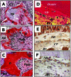
Figure 1.
Subcutaneous DPSC transplants in immunocompromised mice (A-C) and characterization of DPSC-mediated dentinogenesis in vivo (D-F). (A) Four wks after transplantation, DPSCs differentiated into odontoblasts (open arrows) responsible for new dentin (D) formation on the surface of the HA/TCP (HA). (B,C) At 8 and 16 wks post-transplantation, respectively. (D) Newly formed reparative dentin-like structure (ND) attached to the surfaces of human dentin in DPSC/dentin transplants. BV, blood vessels; CT, connective tissue; dentinogenic cells (black arrowheads). DPSCs formed reparative dentin-like structure containing entrapped cells (open arrowheads). (E) In DPSC/dentin transplants, dentinogenic cells (open arrows) and trapped cells (open arrowheads) within the newly formed reparative dentin-like structure (ND) were immunoreactive to human DSP antibody, as was the pre-existing dentin (black arrows). (F) Staining of human-specific anti-mitochondria antibody, showing the human origin of DPSCs (open arrows). Bar, 40 µm in A-C, 20 µm in D-F (adapted from Batouli et al., 2003).
Figure 2.
In vitro neurogenesis of SHED (A-D), transplanted SHED into immunocompromised mice (E-H) and into the mouse brain (I-K). (A,B) Toluidine blue staining of the altered morphology of SHED after neurogenic induction. (C,D) Immunopositive staining for MAP2 and Tau on dendrites and axons (arrows), respectively. (E,F) Eight wks after transplantation into the subcutaneous space, SHED differentiate into odontoblasts (open arrows) and form dentin-like structure (D) on the surfaces of HA. The same field is shown for human-specific alu in situ hybridization, indicating the human origin of odontoblasts (open arrows in F). (G) Immunohistochemical staining of DSPP on the regenerated dentin (black arrows). (H) Newly generated bone (B) by host cells in the same SHED transplant shows no reactivity to the DSPP antibody. (I-K) Neurogenically induced SHED injected into the dentate gyrus of the hippocampus of immunocompromised mice for 10 days. (I) NFM (red) and (J) human-specific anti-mitochondrial antibody (green) and (K) merged images showing co-localization of the two (adapted from Miura et al., 2003, with permission).
Figure 3.
The anatomy of the human apical papilla (A-C) and dentinogenesis of SCAP in immunocompromised mice (D-F). (A) An extracted human third molar depicting root attached to the root apical papilla (open arrows) at the developmental stage. (B) Hematoxylin and eosin staining of human developing root (R) depicting epithelial diaphragm (open arrows) and apical cell-rich zone (open arrowheads). (C) Harvested root apical papilla for stem cell isolation. (D) Eight wks after transplantation, SCAP differentiated into odontoblasts (arrows) that formed dentin (D) on the surfaces of a HA carrier. (E) SCAP differentiated into odontoblasts (arrows) are positive for anti-human specific mitochondria antibody staining. (F) Immunohistochemical staining of SCAP-generated dentin (D) showing positive anti-DSP antibody staining (arrows) (adapted from Sonoyama et al., 2006, , with permission).
Figure 4.
Generation of cementum-like structure and collagen fibers by PDLSCs in immunocompromised mice (A,B) and PDLSCs in periodontal tissue repair in immunocompromised rats (C-E). (A,B) Transplanted PDLSCs formed cementum-like structures (C) that connected to newly formed collagen fibers (dashed lines), similar to the structure of Sharpey’s fibers, and generated a substantial amount of collagen fibers (arrows in B). (C-E) Staining of human-specific anti-mitochondria antibody showing human PDLSCs located in the PDL compartment (arrowheads in C), involved in the attachment of PDL to the tooth surface (arrows in D), and participating in the repair of alveolar bone (arrows in E) and PDL (arrowhead in E) (adapted from Seo et al., 2004, with permission).
Figure 5.
Swine SCAP/PDLSC-mediated bio-root engineering**. (A)** Extracted minipig lower incisor and root-shaped HA/TCP carrier loaded with SCAP. (B) Gelfoam containing PDLSCs (open arrow) to cover the HA/SCAP (black arrow) and implanted into the lower incisor socket (open arrowhead). (C) HA/SCAP-Gelfoam/PDLSCs were implanted into a newly extracted incisor socket. A post channel was pre-created inside the root-shaped HA carrier (arrow). (D) Three months after implantation, the bio-root was exposed and a porcelain crown inserted. (E) Four wks after fixation, the porcelain crown was retained after normal tooth use. (F) After 3 months’ implantation, the HA/SCAP-Gelfoam/PDLSC implant formed a hard root structure (open arrows) in the mandibular incisor area, as shown by CT scan image. A clear PDL space was found between the implant and surrounding bony tissue (arrowhead). (G, H) H&E staining showed that implanted HA/SCAP-Gelfoam/PDLSC contains newly regenerated dentin (D) and PDL tissue (PDL) on the outside of the implant. (I) Compressive strength measurement showed that newly formed bio-roots have compressive strength much higher than that of the original HA/TCP carrier (*P = 0.0002), but lower than that in natural swine root dentin (*P = 0.003) (NR, natural minipig root; BR, newly formed bio-root; HA, original HA carrier) (adapted from Sonoyama et al., 2006, with permission).
Similar articles
- Mesenchymal stem cells and tooth engineering.
Peng L, Ye L, Zhou XD. Peng L, et al. Int J Oral Sci. 2009 Mar;1(1):6-12. doi: 10.4248/ijos.08032. Int J Oral Sci. 2009. PMID: 20690498 Free PMC article. Review. - Dental stem cells: recent progresses in tissue engineering and regenerative medicine.
Botelho J, Cavacas MA, Machado V, Mendes JJ. Botelho J, et al. Ann Med. 2017 Dec;49(8):644-651. doi: 10.1080/07853890.2017.1347705. Epub 2017 Jul 12. Ann Med. 2017. PMID: 28649865 Review. - Stem Cells Derived from Dental Tissues.
Aydin S, Şahin F. Aydin S, et al. Adv Exp Med Biol. 2019;1144:123-132. doi: 10.1007/5584_2018_333. Adv Exp Med Biol. 2019. PMID: 30635857 Review. - Dental stem cells and their sources.
Sedgley CM, Botero TM. Sedgley CM, et al. Dent Clin North Am. 2012 Jul;56(3):549-61. doi: 10.1016/j.cden.2012.05.004. Epub 2012 Jun 23. Dent Clin North Am. 2012. PMID: 22835537 Review. - Mesenchymal stem cells in the dental tissues: perspectives for tissue regeneration.
Estrela C, Alencar AH, Kitten GT, Vencio EF, Gava E. Estrela C, et al. Braz Dent J. 2011;22(2):91-8. doi: 10.1590/s0103-64402011000200001. Braz Dent J. 2011. PMID: 21537580 Review.
Cited by
- Stem cell homing in periodontal tissue regeneration.
Meng L, Wei Y, Liang Y, Hu Q, Xie H. Meng L, et al. Front Bioeng Biotechnol. 2022 Oct 13;10:1017613. doi: 10.3389/fbioe.2022.1017613. eCollection 2022. Front Bioeng Biotechnol. 2022. PMID: 36312531 Free PMC article. Review. - METTL3-mediated m6A modification regulates cell cycle progression of dental pulp stem cells.
Luo H, Liu W, Zhang Y, Yang Y, Jiang X, Wu S, Shao L. Luo H, et al. Stem Cell Res Ther. 2021 Mar 1;12(1):159. doi: 10.1186/s13287-021-02223-x. Stem Cell Res Ther. 2021. PMID: 33648590 Free PMC article. - Neural crest stem cells: discovery, properties and potential for therapy.
Achilleos A, Trainor PA. Achilleos A, et al. Cell Res. 2012 Feb;22(2):288-304. doi: 10.1038/cr.2012.11. Epub 2012 Jan 10. Cell Res. 2012. PMID: 22231630 Free PMC article. Review. - Exploring Various Transfection Approaches and Their Applications in Studying the Regenerative Potential of Dental Pulp Stem Cells.
Alkharobi H. Alkharobi H. Curr Issues Mol Biol. 2023 Dec 13;45(12):10026-10040. doi: 10.3390/cimb45120626. Curr Issues Mol Biol. 2023. PMID: 38132472 Free PMC article. Review. - Bone repair by periodontal ligament stem cellseeded nanohydroxyapatite-chitosan scaffold.
Ge S, Zhao N, Wang L, Yu M, Liu H, Song A, Huang J, Wang G, Yang P. Ge S, et al. Int J Nanomedicine. 2012;7:5405-14. doi: 10.2147/IJN.S36714. Epub 2012 Oct 10. Int J Nanomedicine. 2012. PMID: 23091383 Free PMC article.
References
- Abe S, Yamaguchi S, Amagasa T. (2007). Multilineage cells from apical pulp of human tooth with immature apex. Oral Sci Int 4:45-58
- About I, Bottero MJ, de Denato P, Camps J, Franquin JC, Mitsiadis TA. (2000). Human dentin production in vitro. Exp Cell Res 258:33-41 - PubMed
- Abukawa H, Kaban LB, Williams WB, Terada S, Vacanti JP, Troulis MJ. (2006). Effect of interferon-alpha-2b on porcine mesenchymal stem cells. J Oral Maxillofac Surg 64:1214-1220 - PubMed
Publication types
MeSH terms
Grants and funding
- R01 DE17449/DE/NIDCR NIH HHS/United States
- R01 DE017449/DE/NIDCR NIH HHS/United States
- R01 DE019156-05/DE/NIDCR NIH HHS/United States
- R21 DE017632/DE/NIDCR NIH HHS/United States
- R21 DE017632-02/DE/NIDCR NIH HHS/United States
- R01 DE019156/DE/NIDCR NIH HHS/United States
- R01 DE019156-01/DE/NIDCR NIH HHS/United States
- R01 DE017449-03/DE/NIDCR NIH HHS/United States
LinkOut - more resources
Full Text Sources
Other Literature Sources


