Koilocytes indicate a role for human papilloma virus in breast cancer - PubMed (original) (raw)
Koilocytes indicate a role for human papilloma virus in breast cancer
J S Lawson et al. Br J Cancer. 2009.
Abstract
Background: High-risk human papilloma viruses (HPVs) are candidates as causal viruses in breast cancer. The scientific challenge is to determine whether HPVs are causal and not merely passengers or parasites. Studies of HPV-related koilocytes in breast cancer offer an opportunity to address this crucial issue. Koilocytes are epithelial cells characterised by perinuclear haloes surrounding condensed nuclei and are commonly present in cervical intraepithelial neoplasia. Koilocytosis is accepted as pathognomonic (characteristic of a particular disease) of HPV infection. The aim of this investigation is to determine whether putative koilocytes in normal and malignant breast tissues are because of HPV infection.
Methods: Archival formalin-fixed normal and malignant breast specimens were investigated by histology, in situ PCR with confirmation of the findings by standard PCR and sequencing of the products, plus immunohistochemistry to identify HPV E6 oncoproteins.
Results: human papilloma virus-associated koilocytes were present in normal breast skin and lobules and in the breast skin and cancer tissue of patients with ductal carcinoma in situ (DCIS) and invasive ductal carcinomas (IDCs).
Interpretation: As koilocytes are known to be the precursors of some HPV-associated cervical cancer, it follows that HPVs may be causally associated with breast cancer.
Figures
Figure 1
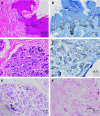
Normal breast specimen. Breast lobules and breast skin showing HPV-associated koilocytes, HPV E6 oncoprotein expression and HPV type 18 by in situ PCR in koilocyte nuclei. (A) Breast skin with koilocytes (H & E stain), (B) breast skin from the same subject showing koilocytes and HPV E6 oncoprotein expression in the basal layers of the skin (immunohistochemistry), (C) breast lobules from the same subject with koilocytes (H & E stain), (D) breast lobules from the same specimen with HPV E6 oncoprotein expression plus koilocytes (immunohistochemistry), (E) positive HPV type 18 expression in the nuclei of koilocytes in the same specimen by in situ PCR, (F) negative HPV expression in the same specimen by in situ PCR with primers omitted (control analysis). The arrows indicate selected putative koilocytes.
Figure 2
Putative koilocytes in a ductal carcinoma in situ breast cancer specimen. (A) H & E stain, (B) HPV E6 oncoprotein by immunohistochemistry, (C) in situ PCR for HPV type 18 showing positive staining in the putative koilocytes, (D) negative control omitting primers from the in situ PCR. The arrows indicate selected putative koilocytes.
Figure 3
Human papilloma virus (HPV) 16/18 E6 staining in normal and ductal carcinoma in situ (DCIS) breast cancer specimens. (A) H & E stain of normal breast skin specimen (enlarged version of Figure 1A), (B) HPV-E6 oncoprotein by immunohistochemistry of normal breast skin specimen (enlarged version of Figure 1B), (C) haematoxylin stain, with eosin omitted, of DCIS specimen (enlarged version of Figure 2A), (D) HPV-E6 oncoprotein by immunohistochemistry of DCIS specimen (enlarged version of Figure 2B). The HPV E6 staining (orange colour) in panels B and D appears in the intercellular spaces, some nuclei, cytoplasm and possibly the surface of the cell membranes of the koilocytes. The arrows indicate selected putative koilocytes.
Comment in
- Regarding: Koilocytes indicate a role for human papilloma virus in breast cancer.
Sandstrom RE. Sandstrom RE. Br J Cancer. 2010 Feb 16;102(4):786-7; author reply 788. doi: 10.1038/sj.bjc.6605549. Epub 2010 Feb 2. Br J Cancer. 2010. PMID: 20125157 Free PMC article. No abstract available. - Is HPV-18 present in human breast cancer cell lines?
Peran I, Riegel A, Dai Y, Schlegel R, Liu X. Peran I, et al. Br J Cancer. 2010 May 11;102(10):1549-50; author reply 1551-2. doi: 10.1038/sj.bjc.6605671. Epub 2010 Apr 20. Br J Cancer. 2010. PMID: 20407441 Free PMC article. No abstract available.
Similar articles
- Human papilloma virus is associated with breast cancer.
Heng B, Glenn WK, Ye Y, Tran B, Delprado W, Lutze-Mann L, Whitaker NJ, Lawson JS. Heng B, et al. Br J Cancer. 2009 Oct 20;101(8):1345-50. doi: 10.1038/sj.bjc.6605282. Epub 2009 Sep 1. Br J Cancer. 2009. PMID: 19724278 Free PMC article. - Hypothetic association between human papillomavirus infection and breast carcinoma.
Liang W, Tian H. Liang W, et al. Med Hypotheses. 2008;70(2):305-7. doi: 10.1016/j.mehy.2007.05.032. Epub 2007 Jul 25. Med Hypotheses. 2008. PMID: 17656036 - Epstein-Barr virus, human papillomavirus and mouse mammary tumour virus as multiple viruses in breast cancer.
Glenn WK, Heng B, Delprado W, Iacopetta B, Whitaker NJ, Lawson JS. Glenn WK, et al. PLoS One. 2012;7(11):e48788. doi: 10.1371/journal.pone.0048788. Epub 2012 Nov 19. PLoS One. 2012. PMID: 23183846 Free PMC article. - Nature of cervical cancer and other HPV - associated cancers.
Georgieva S, Iordanov V, Sergieva S. Georgieva S, et al. J BUON. 2009 Jul-Sep;14(3):391-8. J BUON. 2009. PMID: 19810128 Review. - Catching viral breast cancer.
Lawson JS, Glenn WK. Lawson JS, et al. Infect Agent Cancer. 2021 Jun 10;16(1):37. doi: 10.1186/s13027-021-00366-3. Infect Agent Cancer. 2021. PMID: 34108009 Free PMC article. Review.
Cited by
- Presence of human papilloma virus in a series of breast carcinoma from Argentina.
Pereira Suarez AL, Lorenzetti MA, Gonzalez Lucano R, Cohen M, Gass H, Martinez Vazquez P, Gonzalez P, Preciado MV, Chabay P. Pereira Suarez AL, et al. PLoS One. 2013 Apr 25;8(4):e61613. doi: 10.1371/journal.pone.0061613. Print 2013. PLoS One. 2013. PMID: 23637866 Free PMC article. - The viral origins of breast cancer.
Lawson JS, Glenn WK. Lawson JS, et al. Infect Agent Cancer. 2024 Aug 26;19(1):39. doi: 10.1186/s13027-024-00595-2. Infect Agent Cancer. 2024. PMID: 39187871 Free PMC article. Review. - High risk human papillomavirus and Epstein Barr virus in human breast milk.
Glenn WK, Whitaker NJ, Lawson JS. Glenn WK, et al. BMC Res Notes. 2012 Sep 1;5:477. doi: 10.1186/1756-0500-5-477. BMC Res Notes. 2012. PMID: 22937830 Free PMC article. - Oncogenic Viruses and Breast Cancer: Mouse Mammary Tumor Virus (MMTV), Bovine Leukemia Virus (BLV), Human Papilloma Virus (HPV), and Epstein-Barr Virus (EBV).
Lawson JS, Salmons B, Glenn WK. Lawson JS, et al. Front Oncol. 2018 Jan 22;8:1. doi: 10.3389/fonc.2018.00001. eCollection 2018. Front Oncol. 2018. PMID: 29404275 Free PMC article. Review. - Tracing human papillomavirus in skin and mucosal squamous cell carcinoma: a histopathological retrospective survey.
Nili A, Daneshpazhooh M, Mahmoudi H, Kamyab K, Jamshidi ST, Soleiman-Meigooni S, Darvishi M. Nili A, et al. Dermatol Reports. 2024 Feb 1;16(2):9833. doi: 10.4081/dr.2024.9833. eCollection 2024 Jun 14. Dermatol Reports. 2024. PMID: 38979521 Free PMC article.
References
- Abadi MA, Ho GY, Burk RD, Romney SL, Kadish AS (1998) Stringent criteria for histological diagnosis of koilocytosis fail to eliminate overdiagnosis of human papillomavirus infection and cervical intraepithelial neoplasia grade 1. Hum Pathol 29: 54–59 - PubMed
- Aggarwal S, Arora VK, Gupta S, Singh N, Bhatia A (2009) Koilocytosis: correlations with high-risk HPV and its comparison on tissue sections and cytology, urothelial carcinoma. Diagn Cytopathol 37: 174–177 - PubMed
- Ayre JE (1951) Cancer cytology of the uterus. Grune Stratton, New York, 1951 quoted by Hajdu SI The link between koilocytosis and human papillomaviruses. Ann Clin Lab Sci 36: 485–487 - PubMed
- Boon ME, Kok LP (1985) Koilocytotic lesions of the cervix: the interrelation of morphometric features, the presence of papilloma-virus antigens, and the degree of koilocytosis. Histopathology 9: 751–763 - PubMed
- Bryan JT, Brown DR (2001) Transmission of human papillomavirus type 11 infection by desquamated cornified cells. Virology 281: 35–42 - PubMed
Publication types
MeSH terms
LinkOut - more resources
Full Text Sources
Other Literature Sources
Medical
Research Materials


