CBP and p300 are cytoplasmic E4 polyubiquitin ligases for p53 - PubMed (original) (raw)
CBP and p300 are cytoplasmic E4 polyubiquitin ligases for p53
Dingding Shi et al. Proc Natl Acad Sci U S A. 2009.
Erratum in
- Proc Natl Acad Sci U S A. 2009 Oct 27;106(43):18427. Kung, Andrew [corrected to Kung, Andrew L]
Abstract
p300 and CREB-binding protein (CBP) act as multifunctional regulators of p53 via acetylase and polyubiquitin ligase (E4) activities. Prior work in vitro has shown that the N-terminal 595 aa of p300 encode both generic ubiquitin ligase (E3) and p53-directed E4 functions. Analysis of p300 or CBP-deficient cells revealed that both coactivators were required for endogenous p53 polyubiquitination and the normally rapid turnover of p53 in unstressed cells. Unexpectedly, p300/CBP ubiquitin ligase activities were absent in nuclear extracts and exclusively cytoplasmic. Consistent with the cytoplasmic localization of its E3/E4 activity, CBP deficiency specifically stabilized cytoplasmic, but not nuclear p53. The N-terminal 616 aa of CBP, which includes the conserved Zn(2+)-binding C/H1-TAZ1 domain, was the minimal domain sufficient to destabilize p53 in vivo, and it included within an intrinsic E3 autoubiquitination activity and, in a two-step E4 assay, exhibited robust E4 activity for p53. Cytoplasmic compartmentalization of p300/CBP's ubiquitination function reconciles seemingly opposed functions and explains how a futile cycle is avoided-cytoplasmic p300/CBP E4 activities ubiquitinate and destabilize p53, while physically separate nuclear p300/CBP activities, such as p53 acetylation, activate p53.
Conflict of interest statement
The authors declare no conflict of interest.
Figures
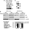
Fig. 1.
CBP and p300 regulate p53 stability and polyubiquitination. (A and B) p53 abundance and stability in U2OS cells after CBP or p300 siRNA treatment. (A) U2OS cells transiently transfected with the indicated siRNAs for 72 h were harvested for immunoblotting with anti-CBP, -p300, -hsp70 (loading control), and -p53 antibodies. (B) (Top) U2OS cells transfected with the indicated siRNAs for 72 h were treated with cycloheximide, and lysates were harvested at the indicated times for analysis by p53 immunoblot. (Bottom) p53 levels were quantitated on a blot scanner (LiCOR), and half-life calculated based on the decay of normalized (to GAPDH loading control) p53 levels to 50% of their original level. Values are an average of three independent experiments. Error bars, ±1 standard deviation (S.D.). (C and D) CBP and p300 both drive p53 polyubiquitination in vivo. U2OS cells were treated with the indicated siRNAs for 48 h and transfected with HA-Ub expression vector 24 h before harvest. MG132 (10 μM) was added for 4 h before harvest as indicated. Cell lysates were IP'd with anti-p53 Ab, followed by anti-HA or anti-p53 immunoblot. PolyUb indicates those p53 species >100 kDa that are larger than the largest MUM species as determined by use of non-chain-forming Ub moieties (14). Input lysates were blotted with the indicated antibodies.
Fig. 2.
The CBP N terminus regulates p53 polyubiquitination and stability. (A) CBP N-terminus regulates p53 stability. (Top) CBP-shA cells were transfected with vector or the indicated CBP rescue alleles, treated with cycloheximide 48 h after transfection, and lysates analyzed by p53 immunoblot. (Bottom) p53 levels were quantitated by densitometry, and half-life calculated based on decay of normalized (to GAPDH loading control) p53 levels to 50% of their original level. Values are an average of three independent experiments. Error bars, ±1 S.D. (B) Purified recombinant CBP protein catalyzes p53 polyubiquitination. (Top) Purified recombinant p53-MDM2 protein complexes were preincubated with ubiquitin reaction components at 37 °C for 30 min. Purified FLAG-CBP (1, 10, or 100 ng) was then added, followed by further incubation for 1 h at 37 °C. Reactions were analyzed by p53 immunoblot. A shorter exposure (middle) shows the unmodified p53 protein level. (Bottom) The ratio of high molecular weight (>100 kDa) p53 species: Total p53 abundance was plotted at 20-kDa intervals for the indicated conditions. (C) The CBP N terminus is an active E4 in vivo. H1299 cells were transfected with His-Ub vector, p53 or p53-UbF expression vectors, along with the indicated CBP plasmids or empty vector. (Top) Forty-eight hours after transfection, lysates were purified on Ni-NTA beads, and eluted products immunoblotted with anti-p53 antibody. (Bottom) Input lysates were immunoblotted with anti-FLAG and p53 antibodies. The abundance of high molecular weight p53-Ub conjugates was determined by scanning densitometry and normalized to total amount of p53UbF in the input lysate. Experiment shown is representative of three independent experiments. (D) Sequential E4 assay. Purified FLAG-MDM2/HA-p53 complexes were exposed to E1, E2, Ub, and ATP to generate mono-Ub/MUM p53. HA-p53-Ub conjugates were purified by anti-HA IP and stringent washing, and then exposed to E2 and ATP a second time with or without E1, along with FLAG-Ub and affinity purified CBP (1–616) or (1–200). After further washes, HA-p53-Ub conjugates were analyzed by anti-FLAG immunoblot.
Fig. 3.
Subcellular localization of CBP/p300 E3 activity. (A) p53 localization in CBP-deficient cells. Nuclear or cytoplasmic fractions of control or CBP-shA cells were analyzed by immunoblotting with Rb (nuclear marker), actin (cytoplasmic marker), and p53 antibodies. (B) CBP regulation of p53 turnover in the cytoplasm and nucleus. Control and CBP-shA U2OS cells treated with cycloheximide were fractionated into nuclear and cytoplasmic fractions at the indicated time points. (Top three panels) The fractions were immunoblotted for p53, PARP (nuclear marker), and actin (cytoplasmic marker). (Lower left) Subcellular fractions (time = 0) from control and CBP-shA cells were immunoblotted with anti-CBP, anti-PARP, anti-α-tubulin, and anti-p53 antibodies. (Lower right) Determination of nuclear and cytoplasmic p53 half-life from Cont-sh and CBP-shA cells. Result is the average half-life from four separate experiments. * indicates significant P = 0.01 for difference in cytoplasmic p53 t1/2 between Cont-sh and CBP-shA. (C) Subcellular localization of p300 and CBP. U2OS cells were fractionated into nuclear and cytoplasmic fractions, and the fractions were immunoblotted for CBP, p300, Rb (nuclear marker), and actin (cytoplasmic marker).
Fig. 4.
p300/CBP E3 activities are predominantly cytoplasmic. (A) (Top and Middle) Nuclear and cytoplasmic fractions of U2OS cells were immunoprecipitated with anti-CBP antibody, and the washed IPs incubated with E1/E2, Ub, and ATP as indicated, followed by anti-Ub and CBP immunoblotting of the reactions. (Bottom) Immunoblot of CBP and Rb (nuclear marker) in nuclear and cytoplasmic fractions. (B) E4 activity of cytoplasmic CBP. p53/MDM2 complexes (insect-cell-derived) were incubated with E1/E2, Ub, or methyl-Ub, ATP and CBP IPs from control-sh or CBP-shA cytoplasmic fractions as indicated, followed by anti-p53 (Top) or anti-CBP (Middle) immunoblotting of the reactions. Migration positions for native, monoubiquitinated/MUM and polyubiquitinated p53 species are indicated. (Bottom) Relative abundances of native, mono-Ub/MUM, and polyubiquitinated p53 species were quantitated by densitometry.
Similar articles
- Graded enhancement of p53 binding to CREB-binding protein (CBP) by multisite phosphorylation.
Lee CW, Ferreon JC, Ferreon AC, Arai M, Wright PE. Lee CW, et al. Proc Natl Acad Sci U S A. 2010 Nov 9;107(45):19290-5. doi: 10.1073/pnas.1013078107. Epub 2010 Oct 20. Proc Natl Acad Sci U S A. 2010. PMID: 20962272 Free PMC article. - A pro-apoptotic function of iASPP by stabilizing p300 and CBP through inhibition of BRMS1 E3 ubiquitin ligase activity.
Kramer D, Schön M, Bayerlová M, Bleckmann A, Schön MP, Zörnig M, Dobbelstein M. Kramer D, et al. Cell Death Dis. 2015 Feb 12;6(2):e1634. doi: 10.1038/cddis.2015.17. Cell Death Dis. 2015. PMID: 25675294 Free PMC article. - The APC/C and CBP/p300 cooperate to regulate transcription and cell-cycle progression.
Turnell AS, Stewart GS, Grand RJ, Rookes SM, Martin A, Yamano H, Elledge SJ, Gallimore PH. Turnell AS, et al. Nature. 2005 Dec 1;438(7068):690-5. doi: 10.1038/nature04151. Nature. 2005. PMID: 16319895 - Role of Intrinsic Protein Disorder in the Function and Interactions of the Transcriptional Coactivators CREB-binding Protein (CBP) and p300.
Dyson HJ, Wright PE. Dyson HJ, et al. J Biol Chem. 2016 Mar 25;291(13):6714-22. doi: 10.1074/jbc.R115.692020. Epub 2016 Feb 5. J Biol Chem. 2016. PMID: 26851278 Free PMC article. Review. - Roles for the coactivators CBP and p300 and the APC/C E3 ubiquitin ligase in E1A-dependent cell transformation.
Turnell AS, Mymryk JS. Turnell AS, et al. Br J Cancer. 2006 Sep 4;95(5):555-60. doi: 10.1038/sj.bjc.6603304. Epub 2006 Aug 1. Br J Cancer. 2006. PMID: 16880778 Free PMC article. Review.
Cited by
- Acetylation of AMPA Receptors Regulates Receptor Trafficking and Rescues Memory Deficits in Alzheimer's Disease.
O'Connor M, Shentu YP, Wang G, Hu WT, Xu ZD, Wang XC, Liu R, Man HY. O'Connor M, et al. iScience. 2020 Aug 15;23(9):101465. doi: 10.1016/j.isci.2020.101465. eCollection 2020 Sep 25. iScience. 2020. PMID: 32861999 Free PMC article. - An intrinsically disordered region of the acetyltransferase p300 with similarity to prion-like domains plays a role in aggregation.
Kirilyuk A, Shimoji M, Catania J, Sahu G, Pattabiraman N, Giordano A, Albanese C, Mocchetti I, Toretsky JA, Uversky VN, Avantaggiati ML. Kirilyuk A, et al. PLoS One. 2012;7(11):e48243. doi: 10.1371/journal.pone.0048243. Epub 2012 Nov 1. PLoS One. 2012. PMID: 23133622 Free PMC article. - The Roles of MDM2 and MDMX Phosphorylation in Stress Signaling to p53.
Chen J. Chen J. Genes Cancer. 2012 Mar;3(3-4):274-82. doi: 10.1177/1947601912454733. Genes Cancer. 2012. PMID: 23150760 Free PMC article. - PIASy-mediated Tip60 sumoylation regulates p53-induced autophagy.
Naidu SR, Lakhter AJ, Androphy EJ. Naidu SR, et al. Cell Cycle. 2012 Jul 15;11(14):2717-28. doi: 10.4161/cc.21091. Epub 2012 Jul 15. Cell Cycle. 2012. PMID: 22751435 Free PMC article. - Genetic and Molecular Characterization Revealed the Prognosis Efficiency of Histone Acetylation in Pan-Digestive Cancers.
Zhang T, Wang B, Gu B, Su F, Xiang L, Liu L, Li X, Wang X, Gao L, Chen H. Zhang T, et al. J Oncol. 2022 Apr 5;2022:3938652. doi: 10.1155/2022/3938652. eCollection 2022. J Oncol. 2022. PMID: 35422864 Free PMC article.
References
- Michael D, Oren M. The p53 and Mdm2 families in cancer. Curr Opin Genet Dev. 2002;12:53–59. - PubMed
- Moll UM, Wolff S, Speidel D, Deppert W. Transcription-independent pro-apoptotic functions of p53. Curr Opin Cell Biol. 2005;17:631–636. - PubMed
- Slee EA, O'Connor DJ, Lu X. To die or not to die: How does p53 decide? Oncogene. 2004;23:2809–2818. - PubMed
- Bode AM, Dong Z. Post-translational modification of p53 in tumorigenesis. Nat Rev Cancer. 2004;4:793–805. - PubMed
- Morgunkova A, Barlev NA. Lysine methylation goes global. Cell Cycle. 2006;5:1308–1312. - PubMed
Publication types
MeSH terms
Substances
LinkOut - more resources
Full Text Sources
Other Literature Sources
Molecular Biology Databases
Research Materials
Miscellaneous


