Novel breast tissue feature strongly associated with risk of breast cancer - PubMed (original) (raw)
. 2009 Dec 10;27(35):5893-8.
doi: 10.1200/JCO.2008.21.5079. Epub 2009 Oct 5.
Carol A Reynolds, Daniel W Visscher, Aziza Nassar, Derek C Radisky, Robert A Vierkant, Amy C Degnim, Judy C Boughey, Karthik Ghosh, Stephanie S Anderson, Douglas Minot, Jill L Caudill, Celine M Vachon, Marlene H Frost, V Shane Pankratz, Lynn C Hartmann
Affiliations
- PMID: 19805686
- PMCID: PMC2793038
- DOI: 10.1200/JCO.2008.21.5079
Novel breast tissue feature strongly associated with risk of breast cancer
Kevin P McKian et al. J Clin Oncol. 2009.
Abstract
Purpose: Accurate, individualized risk prediction for breast cancer is lacking. Tissue-based features may help to stratify women into different risk levels. Breast lobules are the anatomic sites of origin of breast cancer. As women age, these lobular structures should regress, which results in reduced breast cancer risk. However, this does not occur in all women.
Methods: We have quantified the extent of lobule regression on a benign breast biopsy in 85 patients who developed breast cancer and 142 age-matched controls from the Mayo Benign Breast Disease Cohort, by determining number of acini per lobule and lobular area. We also calculated Gail model 5-year predicted risks for these women.
Results: There is a step-wise increase in breast cancer risk with increasing numbers of acini per lobule (P = .0004). Adjusting for Gail model score, parity, histology, and family history did not attenuate this association. Lobular area was similarly associated with risk. The Gail model estimates were associated with risk of breast cancer (P = .03). We examined the individual accuracy of these measures using the concordance (c) statistic. The Gail model c statistic was 0.60 (95% CI, 0.50 to 0.70); the acinar count c statistic was 0.65 (95% CI, 0.54 to 0.75). Combining acinar count and lobular area, the c statistic was 0.68 (95% CI, 0.58 to 0.78). Adding the Gail model to these measures did not improve the c statistic.
Conclusion: Novel, tissue-based features that reflect the status of a woman's normal breast lobules are associated with breast cancer risk. These features may offer a novel strategy for risk prediction.
Conflict of interest statement
Authors' disclosures of potential conflicts of interest and author contributions are found at the end of this article.
Figures
Fig 1.
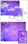
(A) Shown is a field of normal lobules (terminal duct lobular units), each composed of multiple acini. (B) Complete regression (involution) of these lobules has occurred, leaving small residual structures largely depleted of acini. Reprinted with permission.
Fig 2.
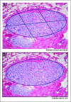
(A) We subdivided an intact lobule to facilitate counting of individual acini. (B) Delineation of the circumference of the lobule for calculation of lobule area by the computer software is demonstrated.
Fig 3.
Whole-breast mounts from (A) preinvolutional and (B) postinvolutional women. Reprinted with permission.
Similar articles
- Assessment of the accuracy of the Gail model in women with atypical hyperplasia.
Pankratz VS, Hartmann LC, Degnim AC, Vierkant RA, Ghosh K, Vachon CM, Frost MH, Maloney SD, Reynolds C, Boughey JC. Pankratz VS, et al. J Clin Oncol. 2008 Nov 20;26(33):5374-9. doi: 10.1200/JCO.2007.14.8833. Epub 2008 Oct 14. J Clin Oncol. 2008. PMID: 18854574 Free PMC article. - Assessment of clinical validity of a breast cancer risk model combining genetic and clinical information.
Mealiffe ME, Stokowski RP, Rhees BK, Prentice RL, Pettinger M, Hinds DA. Mealiffe ME, et al. J Natl Cancer Inst. 2010 Nov 3;102(21):1618-27. doi: 10.1093/jnci/djq388. Epub 2010 Oct 18. J Natl Cancer Inst. 2010. PMID: 20956782 Free PMC article. - Standardized measures of lobular involution and subsequent breast cancer risk among women with benign breast disease: a nested case-control study.
Figueroa JD, Pfeiffer RM, Brinton LA, Palakal MM, Degnim AC, Radisky D, Hartmann LC, Frost MH, Stallings Mann ML, Papathomas D, Gierach GL, Hewitt SM, Duggan MA, Visscher D, Sherman ME. Figueroa JD, et al. Breast Cancer Res Treat. 2016 Aug;159(1):163-72. doi: 10.1007/s10549-016-3908-7. Epub 2016 Aug 3. Breast Cancer Res Treat. 2016. PMID: 27488681 Free PMC article. - Reassessing risk models for atypical hyperplasia: age may not matter.
Mazzola E, Coopey SB, Griffin M, Polubriaginof F, Buckley JM, Parmigiani G, Garber JE, Smith BL, Gadd MA, Specht MC, Guidi A, Hughes KS. Mazzola E, et al. Breast Cancer Res Treat. 2017 Sep;165(2):285-291. doi: 10.1007/s10549-017-4320-7. Epub 2017 Jun 6. Breast Cancer Res Treat. 2017. PMID: 28589368 Free PMC article. Review. - Atypia in the assessment of breast cancer risk: implications for management.
Vogel VG. Vogel VG. Diagn Cytopathol. 2004 Mar;30(3):151-7. doi: 10.1002/dc.20004. Diagn Cytopathol. 2004. PMID: 14986294 Review.
Cited by
- A genetic mosaic mouse model illuminates the pre-malignant progression of basal-like breast cancer.
Zeng J, Singh S, Zhou X, Jiang Y, Casarez E, Atkins KA, Janes KA, Zong H. Zeng J, et al. Dis Model Mech. 2023 Nov 1;16(11):dmm050219. doi: 10.1242/dmm.050219. Epub 2023 Nov 13. Dis Model Mech. 2023. PMID: 37815460 Free PMC article. - Host, reproductive, and lifestyle factors in relation to quantitative histologic metrics of the normal breast.
Abubakar M, Klein A, Fan S, Lawrence S, Mutreja K, Henry JE, Pfeiffer RM, Duggan MA, Gierach GL. Abubakar M, et al. Breast Cancer Res. 2023 Aug 15;25(1):97. doi: 10.1186/s13058-023-01692-7. Breast Cancer Res. 2023. PMID: 37582731 Free PMC article. - Beyond Breast Density: Risk Measures for Breast Cancer in Multiple Imaging Modalities.
Acciavatti RJ, Lee SH, Reig B, Moy L, Conant EF, Kontos D, Moon WK. Acciavatti RJ, et al. Radiology. 2023 Mar;306(3):e222575. doi: 10.1148/radiol.222575. Epub 2023 Feb 7. Radiology. 2023. PMID: 36749212 Free PMC article. Review. - Glandular Tissue Component on Breast Ultrasound in Dense Breasts: A New Imaging Biomarker for Breast Cancer Risk.
Lee SH, Moon WK. Lee SH, et al. Korean J Radiol. 2022 Jun;23(6):574-580. doi: 10.3348/kjr.2022.0099. Korean J Radiol. 2022. PMID: 35617993 Free PMC article. Review. No abstract available. - Association of Genetic Ancestry With Terminal Duct Lobular Unit Involution Among Healthy Women.
Sung H, Koka H, Marino N, Pfeiffer RM, Cora R, Figueroa JD, Sherman ME, Gierach GL, Yang XR. Sung H, et al. J Natl Cancer Inst. 2022 Oct 6;114(10):1420-1424. doi: 10.1093/jnci/djac063. J Natl Cancer Inst. 2022. PMID: 35333343 Free PMC article.
References
- Freedman AN, Seminara D, Gail MH, et al. Cancer risk prediction models: A workshop on development, evaluation, and application. J Natl Cancer Inst. 2005;97:715–723. - PubMed
- Elmore JG, Fletcher SW. The risk of cancer risk prediction: “What is my risk of getting breast cancer?”. J Natl Cancer Inst. 2006;98:1673–1675. - PubMed
- Hartmann LC, Sellers TA, Frost MH, et al. Benign breast disease and the risk of breast cancer. N Engl J Med. 2005;353:229–237. - PubMed
- Dupont WD, Page DL. Risk factors for breast cancer in women with proliferative breast disease. N Engl J Med. 1985;312:146–151. - PubMed
Publication types
MeSH terms
LinkOut - more resources
Full Text Sources
Medical
Research Materials


